Here we are providing Class 11 Biology Important Extra Questions and Answers Chapter 18 Body Fluids and Circulation. Important Questions for Class 11 Biology are the best resource for students which helps in Class 11 board exams.
Class 11 Biology Chapter 18 Important Extra Questions Body Fluids and Circulation
Body Fluids and Circulation Important Extra Questions Very Short Answer Type
Question 1.
What is systole?
Answer:
The contraction phase of the cardiac chamber (s) is called systole.
Question 2.
What is diastole?
Answer:
The relaxation phase of the cardiac chamber is called diastole.
Question 3.
Where is SA-node located?
Answer:
It is located at the place of merge of sinus venous at the right wall of the right atrium.
Question 4.
In which animals we can find the sinus Venosus?
Answer:
Sinus venosus can be found in fishes, amphibians and reptiles.
Question 5.
What is the ‘lubb’ sound?
Answer:
It is the first sound produced by the heart due to the sharp closure of AV valves at the start of ventricular systole.
Question 6.
What is AV-node?
Answer:
It is a node of specialised fibres, located at the junction of the right atrium and right ventricle.
Question 7.
What is hemolymph?
Answer:
The blood of insects that lack haemoglobin is called haemolymph.
Question 8.
What is a sphygmomanometer?
Answer:
The instrument which measures blood pressure is called a sphygmomanometer.
Question 9.
What is the “dup” sound?
Answer:
It is the second sound produced by the heart due to the sharp closure of semilunar valves at the start of ventricular systole.
Question 10.
What is the role of AV-node?
Answer:
It collects the wave contraction generated by SA-node and passes down to a bundle of His and Purkinje fibres.
Question 11.
Where is RBCs (red blood cells) produced?
Answer:
RBCs are produced in the bone marrow.
Question 12.
Name different types of blood groups present in human beings.
Answer:
Types of blood group: O, A, B, AB group.
Question 13.
Name various parts of the circulation.
Answer:
Heart, blood, gases and same waste materials.
Question 14.
Name the blood vessel which carries impure blood to the heart.
Answer:
Vena cava.
Question 15.
The transport system in animals is called
Answer:
Circulatory system.
Question 16.
What is the difference between artery and vein?
Answer:
- Artery: Thin-walled
- Vein: thick-walled.
Question 17.
What are the different components of blood?
Answer:
Plasma, RBC, WBC, Sugar, salt, waste products.
Question 18.
What is the heartbeat rate/minute?
Answer:
72 beat/minute.
Question 19.
What are circulatory fluid and its component?
Answer:
Circulatory fluid is blood and its components are haemoglobin, RBC, WBC.
Question 20.
Give the function of blood platelets.
Answer:
They help in the clotting of blood.
Question 21.
Which blood group is known as a universal donor?
Answer:
‘O’ group is known as a universal donor.
Body Fluids and Circulation Important Extra Questions Short Answer Type
Question 1.
Why does the ventricle contract as a closed chamber in the early phase of its systole?
Answer:
In the early phases of systole the ventricle contracts as a closed chamber, so to increase the pressure in the atrium. Backflow of blood into atria is prevented by closure of AV-valves. In a closed chamber, the ventricles contract and increased pressure cause the opening of semi-lunar valves. The blood is passed into arteries with great force.
Question 2.
The blood vascular system is considered efficient than the water circulatory system in animals, why?
Answer:
The dissolved oxygen and nutrient present in water are in fewer amounts. Oxygen is supplied through oxygen carrier molecule (haemoglobin) present in plasma or cells, in higher animals. Oxygen and nutrients are supplied quickly and in fairly large amounts in animals with a blood vascular system, so considered comparatively efficient.
Question 3.
Mention, in brief, the important events that happen during the cardiac cycle.
Answer:
The cardiac cycle comprises the following three phases.
(a) Atrial systole
(b) Ventricular systole
(c) Joint diastole.
(a) Atrial systole: The atria contract from anterior to posterior and the blood is sent to respective ventricles. Time taken is 0.1 sec.
(b) Ventricular systole: The ventricles contract, deoxygenated blood is sent to lungs for oxygenation, while oxygenated blood received from lungs is sent to different parts of the body through the aortic arch. Time taken is 0.3 sec.
(c) Joint diastole: All the chambers are in systole, called joint diastole. Time taken is 0.4 sec.
Question 4
Describe in brief the types of valves present in the heart.
Answer:
Different valves present in the heart are described as under:
(a) Tricuspid valve: The valve present at the right atrioventricular aperture formed of three flaps.
(b) Bicuspid valves: The valve present at the left atrioventricular aperture, formed of two flaps, also called the mitral valve.
(c) Semi-lunar valve: Two sets of half-moon shaped (3) valves, present at openings of pulmonary aorta and aorta, present here to check backflow of blood.
Question 5.
What is hypertension? What are causative factors?
Answer:
It is a manifestation of an increase in blood pressure. A continuous or sustained rise in arterial blood pressure is known as hypertension.
The normal systolic and diastolic pressure of a healthy human is 120m and 80mm Hg respectively. Under various physiological conditions, a moderate level of fluctuation may occur and blood pressure increase. It is referred to as high blood pressure.
Question 6.
What is heart rhythm? Discuss.
Answer:
Heart muscles continuously generate impulses in a manner that causes rhythmic contraction and relaxation of the heart chambers. For the purpose of maintenance of the heart rhythm, a highly excitatory’ and conductive system is present, which includes sino-atrial node (SA) inter- nodal pathways, the atria-ventricular node (Avnode) AV bundle and the bundle of Purkinje fibres.
In normal conduction of impulse, the atrial contraction precedes that of the ventricle.
The automatic rhythmicity of the heart is its ability to contract spon¬taneously at a regular rate.
Question 7.
Define the terms:
(a) SA node,
Answer:
SA: It is a self-excitatory node that initiates excitatory waves at the highest rate. SA-node normally generates the cardiac impulse and working by knowing the rate of discharge of cardiac impulse. It can determine the rate of heartbeat. The SA-node works as the pacemaker of the heart.
(b) Systole,
Answer:
Systole: The pumping out of blood due to contraction of the heart is called systole.
(c) Diastole,
Answer:
Diastole: The relaxation of the heart is called diastole. During diastole, blood flows into the auricles to their ventricles through the open auriculoventricular valve.
(d) Pulmonary circulation.
Answer:
Pulmonary circulation: The circulatory system which is associated with the lungs through arteries and veins, is known as pulmonary circulation.
Question 8.
Fill in the blanks:
Answer:
(a) Eosinophil is a bilobed nucleus
(b) Pulmonary artery carries deoxygenated blood.
(c) Heart of cockroach is present in the pericardial sinus.
(d) Platelets helps in coagulation
(e) Haemoglobin is present in RBCs due to which its colour is red.
Question 9.
What is an artificial pacemaker? Explain.
Answer:
A pacemaker is a rhythmic centre that establishes a pace of activity. Sometimes the component of the impulse conduction system is disrupted, causing irregularity in the heart rhythm like heart failure. Such types of patients are provided with an artificial electronic device, which regularly sends a small amount of electrical charge for maintaining the rhythmicity of the heart.
The device is known as an artificial pacemaker. It is implanted subcu¬taneously in the upper thoracic region having a connection with the heart.
If patients having symptoms of ventricular escape, in which atrial impulse suddenly fails, the artificial pacemaker is connected to the right ventricle for controlling its rhythm. The artificial pacemaker consists of a pulse generator containing a cell to produce electric impulse.
Question 10.
How arteriosclerosis is different from atherosclerosis? Discuss.
Answer:
Arteriosclerosis: It is the hardening of the arteries due to deposition and thickening. In the case of arteriosclerosis, calcium salts precipitate with the cholesterol and plague. The affected artery loses the property of distension and its walls may rupture. The blood leaking from the ruptured walls may clot and block the pathway of blood flow. This may lead to a heart attack and even death.
Atherosclerosis: It is the deposition of lipids (cholesterol) on the wall lining the lumen of large and medium-sized arteries. This type of deposition is called atherosclerosis. Its formation starts with the deposition of cholesterol particles/crystals in the tunica intima and smooth muscles. This results in the reduction of the lumen size of the artery and the flow of the blood also reduce.
In extreme circumstances, these plaques may completely block the artery. The proliferation of smooth muscles occurs because these plaques provide a rough surface to the platelets causing the release of platelet, derived from growth factor (PDGF). Such plaques reduce the blood supply to the heart or may stop the supply due to complete blockage. This may result in a heart attack or stroke.
Question 11.
What is heart rhythm? Discuss.
Answer:
Heart muscles continuously generate impulses in a manner that causes rhythmic contraction and relaxation of the heart chambers. For the purpose of maintenance of the heart rhythm, a highly excitatory and conductive system is present, which includes sino-atrial node (SA) intermodal pathways, the atrioventricular node (Av node) AV bundle and the bundle of Purkinje fibres.
In normal conduction of impulse, the atrial contraction precedes that of the ventricle.
The automatic rhythmicity of the heart is its ability to contract spontaneously at a regular rate. ,
Question 12.
What is an electrocardiogram? Write about its significance.
Answer:
The passage of cardiac impulse through the heart spreads electric current into the tissues around the heart and a small portion spreads throughout the surface of the body.
These electrical changes can be recorded along the cardiac cycle. The recording of electrical potential generated by the spreading of cardiac impulse is called an electrocardiogram (ECG). The electrical activity of the heart can be graphically represented by ECG, which shows different waves.
Question 13.
Match column I with column II.
| Column I | Column II |
| (a) Haemolymph | (i) Coagulation |
| (b) RBC | (ii) Immunity |
| (c) Antibody | (iii) Cockroach |
| (d) Platelets | (iv) Contraction |
| (e) Systole | (v) Gas transport |
Answer:
| Column I | Column II |
| (a) Haemolymph | (iii) Cockroach |
| (b) RBC | (v) Gas transport |
| (c) Antibody | (ii) Immunity |
| (d) Platelets | (i) Coagulation |
| (e) Systole | (iv) Contraction |
Question 14.
What is blood? Describe its components.
Answer:
Blood is a complex connective tissue, which consists of two components:
- Plasma: It is the extracellular fluid of blood constituting about 55% of the blood volume. Plasma contains 91 – 92% of water, 7% of proteins, 0.9% of inorganic constituents, 0.1% of glucose and the rest includes various organic and inorganic substances. Proteins are the second-largest constituents of plasma.
- Blood corpuscles: Nearly 45% volume of blood consists of corpuscles or blood cells. Blood corpuscles are of three types: erythrocytes or red blood corpuscles (RBC), leucocytes or white blood corpuscles (WBC) and thrombocytes or blood platelets.
Question 15.
What is hypertension? What are different contributory fac¬tors?
Answer:
An abnormal rise in arterial blood pressure is called hypertension.
The contributory factors of this disease are:
(a) Hypercholesterolemia,
(b) Nervous strain,
(c) Renal disorder/impaired functioning.
(d) Arteriosclerosis.
Question 16.
What is the main advancement in avian and mammalian heart over the amphibians and reptiles heart?
Answer:
Avian and mammalian heart show complete double circulation and in them, oxygenated blood remains completely separate from deoxygenated blood. There are no accessory chambers in them. There is a single aorta in aves and mammals and double aorta in reptiles and amphibians which quickens the supply of O2 and food to release the energy and aids in the removal of wastes from the body.
Question 17.
What is the base efficiency of the heart?
Answer:
The efficiency of the heart on beating throughout life without fatigue is based upon the principle that it rests double the time it works i.e. contraction of the heart is followed immediately by relaxation but relaxation is not followed by the next contraction. The beating of the heart is 72 times per minute. It is indicated by pulse rate per minute. The amount of blood pumped out per minutes is called heart output.
Question 18.
What does ECG stand for? Define it.
Answer:
ECG stands for electrocardiograph. It is defined as a permanent record of electrical events (depolarization and ventricles) during a cardiac cycle made on graph paper with the help of an instalment called an electrocardiogram.
It shows 5 waves i.e., P, G, R, S, T. In this P, R, T are positive waves that lie above the baseline whereas Q and S are negative waves that lie below the baseline. The part of the baseline between two deflections is called an interval.
Question 19.
What are the advantages of physical exercise?
Answer:
Any physical exercise initiates an impulse from the nervous system which causes the release of adrenaline hormone from adrenal glands into the blood.
Advantages of physical exercise:
- Exercise increases heartbeat which reduces the workload of the heart. It increases blood circulation so that every tissue gets a supply of nutrients and oxygen.
- It maintains normal blood pressure by lowering the chances of developing atherosclerosis- a disease, which causes narrowing of arteries.
- Exercise increases the level of high-density lipoproteins (HDLS) which help in reducing the harmful cholesterol from arteries.
- Exercise prevents heart attack with the development of additional or collateral blood vessels which provide alternative routes for blood supply to the muscles of the heart.
- Exercise adds more number RBCs
- Haemoglobin of blood increases which helps in supplying more O, to the tissues.
Question 20.
What is blood pressure? What do you mean by diastolic and systolic pressure?
Answer:
It is the pressure against the walls of blood vessels produced by the discharge of blood from the heart. It is high in arteries and Ipw in veins. It rises and falls in arteries due to heartbeat.
The temporary rise in B.P. during contraction of the heart is called systolic pressure. The temporary fall in the B.P during relaxation of the heart is called diastolic pressure.
Question 21.
What is a lymph node?
Answer:
The lymphatic vessels at intervals bear small lymph nodes which act as the filters of poisonous and foreign substances such as dust, debris, bacteria and other injurious substances. A lymph node has an inner border called the hilum. These are the masses of dense connective tissue, rich in phagocytic white corpuscles and macrophages.
Lymph nodes also produce lymphocytes, monocyte and antibodies into the lymph which are carried to the blood. These lymph nodes help in detecting and destroying cancer cells at the initial stages.
Question 22.
What is systemic circulation?
Answer:
The system of blood vessels that ensures the supply of oxygenated blood from the left ventricle to all the body organs and the return of deoxygenated blood to the right atrium is called systematic circulation.
Question 23.
Describe the importance of pulmonary circulation.
Answer:
The circulatory system associated with the lungs through arteries and veins forms the pathway of the circulatory system.
The flow of blood from the heart to the lungs and back to the heart is called pulmonary circulation. The regular oxygenated blood is returned to the heart and the pulmonary artery supplies deoxygenated blood from the right ventricle to the lungs. It is the pathway of the blood through arteries from t aorta arising from the heart and returning to the right atrium.
Question 24.
What is an electrocardiogram? Write about its significance.
Answer:
The passage of cardiac impulse .through the heart spreads electric current into the tissues around the heart and a small portion spreads throughout the surface of the body.
These electrical changes can be recorded along the cardiac cycle. The recording of electrical potential generated by the spreading of cardiac impulse is called an electrocardiogram (ECG). The electrical activity of the heart can be graphically represented by ECG, which shows different waves.
Question 25.
Write true or false:
(a) Atrio-ventricular node is the natural pacemaker of the heart.
Answer:
False
(b) Human heart has inter-auricular foramen.
Answer:
True
(c) Heart of the cockroach is segmentally arranged.
Answer:
True
(d) Right atriventricular valve is a semilunar valve
Answer:
True
(e) Normal systolic and diastolic pressure of humans is 120 and 60 mm Hg, respectively.
Answer:
False
Body Fluids and Circulation Important Extra Questions Long Answer Type
Question 1.
(a) Why is the AV bundle essential for the conduction of cardiac muscles? Explain.
Answer:
The sinoatrial node (pacemaker) of the heart spreads the cardiac impulse over the two atria to bring about their systole. It, however, cannot spread along the common cardiac muscle fibres from the atria to the ventricles. It is because, in the mammalian heart, there is no continuity between the cardiac muscle fibres of the atria and those of the ventricles.
Although the fibres of each individual chamber exist in a functional syncytium (not separatable). It is because a bond of specialized cardiac muscle fibres exists on the interatrial septum called the ATRIOVENTICULAR BUNDLE (AV bundle).
AV bundle forms the only muscular continuity between atrial and ventricular muscles. The AV bundle descends from the AV node along the interatrial septum and the interventricular septum. It branches into right and left bundle branches as it enters the ventricle.
From each AV bundle branch Purkinje which fibres spread out and connect with the common ventricular muscle fibre. Thus, the cardiac impulse spreads over the atria to reach the AV node; the AV bundle is necessary for the conductions of impulse through its two branches and the Purkinje fibres to reach the ventricular muscle fibres causing contraction of ventricles.
(b) Make a graphic representation of double circulation in the mammalian heart.
Answer:
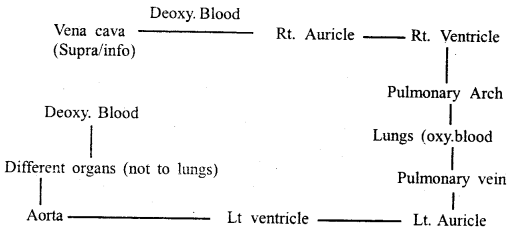
Representation of double circulation in animals.
Question 2.
Define portal system. How is the hepatic portal system useful to our body?
Answer:
Portal system: A portal system is the circulatory system in which blood collected from one set of organs or tissues is conveyed to another organ through capillaries before entering the heart.
For example, in the hepatic portal system, blood collected from the alimentary -canal is first conveyed to the liver by the hepatic portal vein. After passing through the capillaries of the liver, it passes directly to the posterior vena cave by the hepatic vein.
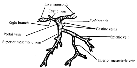
Hepatic portal circulation
Use of hepatic portal system:
(i) Veins coming from the various parts of the alimentary canal carry deoxygenated and food-laden blood. Through the portal vein, it reaches the network of portal vein capillaries in the liver. The excess of food is filtered and stored in the liver as glycogen. Thus, heavily food loaded blood is not allowed to go to the heart, which may have to work more in pumping the blood.
(ii) Liver also consumes drugs and toxins present in the blood coming from the intestine. So, that heart can be saved from their harmful effects.
Question 3.
What is the lymphatic system? Discuss its importance.
Answer:
The lymphatic system consists of vessels and lymph organs, like lymph nodes, bone marrows, spleen and thymus. The fluid present in the lymphatic system is called lymph. This fluid has a composition similar to that of plasma except it is low in protein.
Fine channels present in the tissue are called lymph vessels, these are similar to veins. Besides these, a number of lymph nodes are present. The lymphatic vessels are distributed in the limbs, abdomen, thorax and neck.
The lymphatic system provides an accessory route for the flow of interstitial fluid into the blood. The lymphocytes present in the lymphatic system play important role in the defence against foreign agents or microbes.
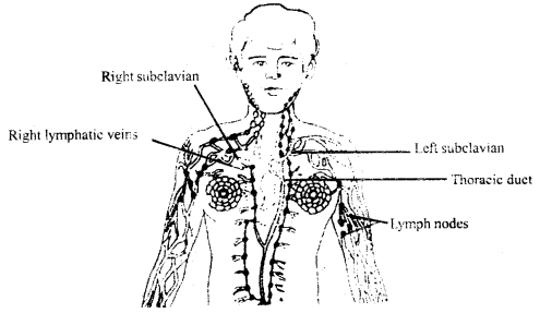
Shows the various lymphatic organs in the human body
Question 4.
Describe the structure of the human heart.
Answer:
The human heart consists of four chambers: Two upper thin layered (Atrium) and two lower thick layered ventricles. The atria are situated at the broader end while the ventricles are situated at the lower conical end. Externally a transverse groove is present between the atria and ventricles, called the anterior interventricular sulcus and posterior intraventricular sulcus.
The right and left atria receive blood from different body parts. The right atrium receives deoxygenated blood from all parts of the body except the lungs, through the superior and inferior vena cava. Pulmonary veins bring oxygenated blood to the left atrium horn of the lungs.
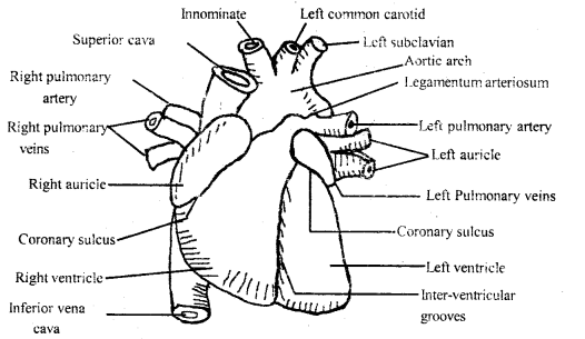
Extma1 features of the human heart
The right and left atria pump their blood into the right and left ventricles. From the right ventricle, the pulmonary trunk arises which bifurcates into right and left pulmonary arteries, which supply deoxygenated blood to the lungs. The left ventricle gives rise to an ascending aorta from ‘which the oxygenated blood is supplied to the coronary- arteries and the systemic circulation of the body occurs.
Question 5.
Write the differences between
(a) Blood and haemolymph
Answer:
| Blood | Haemolymph |
| I. It is composed of blood plasma RBCs, WBCs and platelets. | 1. It is composed of watery fluid, lymphocytes. few monocytes, but no RBCs. |
| 2. It is red in colour | 2. It is a watery fluid. |
| 3. Blood contains more plasma proteins, calcium and phosphorus ions. | 3. Lymph contains fewer proteins, less calcium and phosphorus ions |
| 4. Flow of blood is fairly rapid. | 4. Haemolymph flow is very slow. |
| 5. Blood is pumped out from the heart, flow through the arteries, capillaries and veins. | 5. Haemolymph flow begins from the tissue spaces, goes to lymph capillaries. |
| 6. Glucose concentration is comparatively low. | 6. Glucose Concentration is comparatively high. |
(b) Open and closed system of circulation
Answer:
Open type of circulatory System and Closed system of circulation:
Open type of circulatory System: It mainly occurs in arthropods and molluscs, such as insects, prawns, spiders, oysters, etc. In these colourless blood coming through blood vessels, flows through open spaces and channels called lacunae and sinuses.
Lacunae and sinuses together called haemocoel, which carries colourless blood called haemolymph. The oxygen-carrying pigments are generally dissolved in the plasma of blood. Deoxygenated blood from these goes to the gills for oxygenation after oxygenation blood is returned to the sinus surrounding the heart.
Closed type of circulatory system: It mainly occurs in annelids and chordates. Heart and blood vessels together constitute the closed circulatory system. The heart is a pumping organ that is provided with valves. It pumps the blood into arteries, which earn’ the blood to different organs to the body.
Question 6.
Describe the circulatory system of cockroach.
Answer:
The circulatory system is of open type. It consists of the heart and dorsal blood vessel, sinuses and haemolymph. The haemocoel is divided into three chambers or sinuses dorsal, middle and ventral. The dorsal chamber is called the pericardial sinus, the middle is the perivisceral sinus whereas the ventral is the perineural sinus. The dorsal and ventral diaphragms bear a number of pores through which haemolymph flows.
The heart of a cockroach is an elongated tubular structure, closed behind and open in front. It has thirteen funnel-shaped and segmentally arranged chambers i.e. three in the thoracic segment and ten in the abdominal segment. Valves, ensuring the unidirectional flow of blood, guard the passage of each heart chamber.
Blood flows from the posterior to the anterior end and is discharged into the tissue space of the head. Laterally each heart chamber bears a pair of apertures called Ostia which communicates with the pericardial sinus.
In each segment, a pair of triangular alary muscles is present on either side of the heart.
The blood of the cockroach is not responsible for the transportation of respiratory gases but serves for
(1) the transportation of nutrients.
(2) maintains hydrostatic pressure and
(3) acts as a reservoir of water.
The haemolymph of cockroach circulates due to contraction and relaxation of the heart and ciliary muscles,
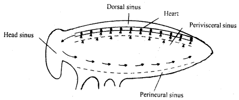
Open type of circulatory system in cockroach
Must Read: