Here we are providing Class 11 Biology Important Extra Questions and Answers Chapter 21 Neural Control and Coordination. Important Questions for Class 11 Biology are the best resource for students which helps in Class 11 board exams.
Class 11 Biology Chapter 21 Important Extra Questions Neural Control and Coordination
Neural Control and Coordination Important Extra Questions Very Short Answer Type
Question 1.
What are the major divisions of the forebrain?
Answer:
Cerebrum, Thalamus, Hypothalamus.
Question 2.
Which parts of the central nervous system constitute the grey matter?
Answer:
Areas that contain cell bodies of the neurons.
Question 3.
Name the major lobes of the cerebral hemisphere?
Answer:
Frontal, parietal, temporal and occipital.
Question 4.
What is the function of cerebral spinal fluid?
Answer:
It maintains a constant pressure inside the cranium.
Question 5.
What is the junction between two neurons known as?
Answer:
Synapse.
Question 6.
What is the polarized state of the nerve membrane?
Answer:
It is the state of the nerve membrane when its inner side is electronegative to its outer side.
Question 7.
Give two examples of unconditioned reflexes.
Answer:
(i) Salivation on tasting food.
(ii) Constriction of the pupil on the illumination of the eye.
Question 8.
Name the types of cells present in the retina.
Answer:
Rods, cones, bipolar neurons, ganglion cells, supporting cells.
Question 9.
Where is iodopsin present in the eye?
Answer:
In the cone cells of the retina
Question 10.
Where are taste buds located?
Answer:
In the mucous membrane over the papillae on the tongue.
Question 11.
Name the main parts of the human brain.
Answer:
Cerebrum, cerebellum, medulla, thalamus, and hypothalamus.
Question 12.
How many cranial nerves and spinal nerves do we possess?
Answer:
12 pairs of cranial nerves and 31 pairs of spinal nerves.
Question 13.
Which part of the brain controls posture and equilibrium?
Answer:
Cerebellum.
Question 14.
What is a polarised membrane?
Answer:
It is electrically positive outside and negative inside.
Question 15.
Compare rods and cones.
Answer:
Rods work in dim light and dark. Cones work in bright light.
Question 16.
What is the blind spot?
Answer:
The point of the retina from where the optic nerve starts and receptor cells are absent.
Neural Control and Coordination Important Extra Questions Short Answer Type
Question 1.
What are receptors?
Answer:
Receptors are peripheral nerve endings in the skin or special sense organs. They collect information from the external or internal environment of the body; transform them into electrical potential changes, which then pass along the afferent neurons to CNS.
Question 2.
Why does vitamin A deficiency produce night blindness?
Answer:
Vitamin ‘A’ is the constituent of rhodopsin, a pigment present in the photoreceptor cells of the eye. Rhodopsin breaks up into opsin and rode to visualize things in bright and dim light. There is constant consumption of vitamin A in rod cells. Deficiency of vitamin A causes impairment of synthesis of rhodopsin leading to night blindness, i.e., inability to see in the dark.
Question 3.
Why does the nerve impulse flow more rapidly in myelinated nerve fibers than in the non-myelinated fibers?
Answer:
Due to the following reasons nerve impulse flows more rapidly in myelinated nerve fibers:
- Myelin sheath provides insulation to the nerve fibers from electrical disturbances between the neighboring fibers.
- Myelin sheath is impermeable to free ions present in the extracellular fluid. So, it prevents the exchange of ions between the extracellular fluid and the interior of the myelinated axon.
- The myelin sheath is absent at the Nodes of Ranvier, so, action potential jumps from one Node of Ranvier to the next. Thus, the nerve impulse flows in the form of leaps or jumps. This is known as the saltatory conduction of impulse.
- It is more rapid than the smooth flow of impulse.
Question 4.
What is a synapse?
Answer:
It is the junction between axon terminals of a neuron and dendrites or the cell body of another neuron. There is a narrow fluid-filled space, called Synaptic Cleft separating axon terminals and dendrites at the synaptic junction. So, the two-neurons forming synapse does not form actual continuity between the neurons.
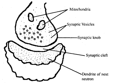
Structure of Synapse
Question 5.
Draw a labeled diagram of a section of the retina to illustrate its structure.
Answer:
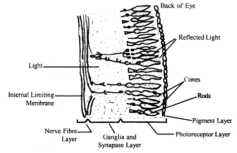
Diagrammatic representation of the sectional view of the retina.
Question 6.
What functions does the hypothalamus serve in coordinating the various activities of the body?
Answer:
- It contains nerve centers for temperature regulation, hunger, thirst, and emotional reactions.
- It secretes neurohormones, which control the secretion of anterior pituitary hormones.
- It synthesizes the posterior pituitary hormones and controls their release into the blood.
Question 7.
What is a nerve fiber? How is it classified according to myelin sheath?
Answer:
A nerve fiber is a long axon or dendrite of a neuron. According to the presence and absence of myelin sheath around the fibers.
These are classified as:
- Myelinated nerve fiber (i.e., presence of myelin sheath) and
- Non-myelinated nerve fiber (i.e., absence of myelin sheath).
Question 8.
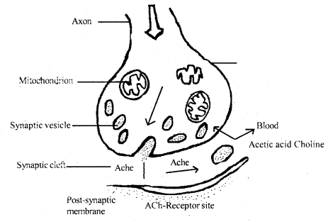
Explain Motor-end plate.
Answer:
A Motor-end plate is a specialized structure formed by the muscle fiber at the point where the axon terminal is applied to it. The axon of the motor neuron is divided into branches near the muscle fibers. Each branch loses its myelin sheath near its termination and ends in an expanded foot-like form which is supplied closely to a muscle fiber.
There is no actual continuity between the neuron and muscle fiber. The membranes of the two are separated from each other by a narrow cleft-like fluid-filled space.
Question 9.
What are the biological functions of Dorsal and Ventral spinal nerve roots fibers?
Answer:
Dorsal spinal nerve root fibers bring impulses from the peripheral tissues, giving rise to sensations like touch, temperature, and pain, or to involuntary spontaneous activities called Reflexes.
Ventral spinal nerve root fibers: Some of the root fibers go to skeletal muscle fibers directly to stimulate or inhibit their activities; many others go to autonomic ganglia and end in them.
Question 10.
Our rods and cones evenly distributed over the entire surface of the retina? Why or not? At which point on the retina is a point-to-point image formed?
Answer:
The retina is composed of several layers of cells. First, there are the photoreceptor cells, the rods, and cones, partially embedded in the microvilli of pigmented epithelium cells of the choroids. The rod cells are present on the periphery of the retina in the human eye. The total number of rod cells has been estimated to be between 110 – 125 million. They contain a visual pigment called Rhodopsin.
The cone cells are shorter, thicker, and conical in shape. Cone cells are responsible for the perception of different colors. The total number of cone cells is 6.36 – 6.8 million. Cones are abundant on the rear wall and fovea centralis of the retina.
The point-to-point image is formed on the blind spot. From it, the optic nerve and blood vessels exit the retina and join the diencephalon of the brain.
Question 11.
Blindspot in the eye is devoid of the ability of vision. Why is it so?
Answer:
It is devoid of rods and cone cells. It is unstable to light rays.
Question 12.
If a strong odor is smelled continuously for some time, the sensation of that weakens. Justify.
Answer:
When a person continuously inhales the fumes in the air of a strong-smelling substance the sense of smell progressively and rapidly declines and finally disappears. This is because the olfactory cells get fatigued rapidly due to overstimulation. This is called olfactory adaptation, which develops from various changes in the olfactory epithelium and olfactory centers of the brain.
Question 13.
Which part of the nervous system participates in the maintenance of balance and co-ordinate body movements?
Answer:
The cerebellum process all the data and co-ordinates muscle movement in conjunction with the cortex and sends signals to the muscles to adjust.
Question 14.
What is a reflex action? What units of the nervous systems are involved with a typical vertebrate reflex arc?
Answer:
It is a spontaneous, automatic, mechanical, nerve-mediated response evoked at the unconscious level by the stimulation of any specific receptor without exercising the will of an organism.
There, are more than 200 reflexes “wired” into our nervous system all following the sequence from stimulus to reflex along the specific neural pathway that makes up the reflex arc. The simplest reflex arc involves some specific receptor, afferent sensory neuron towards an aggregation of nervous tissue which may be ganglion or the spinal cord.
Question 15.
Which nerve tract connects the right and left hemispheres of the cerebrum? Into what four lobes in each hemisphere divided?
Answer:
A longitudinal fissure splits the brain into two halves, the left, and right cerebral hemispheres. Other grooves divide the surface of each cerebral hemisphere into four lobes. The frontal lobe, temporal lobe at the front, parietal lobe, and occipital lobe at the back.
Question 16.
What is the primary function of neuroglia cells? What special structure is produced by Schwann cells?
Answer:
The neuroglia cells perform many house-keeping functions, provide nutritional support to the neurons and consume waste products. They also insulate, separating each neuron from the others.
Schwann cells, a type of neuroglial, wrap around the axon with concentric layers of the insulating plasma membrane.
Question 17.
How does a wave of depolarization spread along with a nerve fiber?
Answer:
Nerve cells have polarized membranes, having an electrical potential difference across the membrane. The trigger zone for a particular neuron is the place on the membrane where voltage-gated channels are clustered most densely. When stimulated opening of voltage-gated Na+ ion channel brings Na+ ions into the cell, a temporary, very localized, but rapid inflow of Na+ ions into the cells occurs, wiping out the local electrical potential difference in the immediate vicinity. This is called depolarization.
When the site of stimulation has less charge difference than the membrane surface surrounding it, this potential difference establishes a small, localized current in the immediate vicinity, which influences the nearby Na+ channels to open and depolarizing these cells.
The depolarization thus spreads, producing a local current, which induces passive Na+ channels to open and so to depolarize the near % site. In this way, initial depolarization passes outward over the membrane and spreads out in all directions along with the nerve fiber, from dendron to axon.
Question 18.
What is a synapse? How does the nerve cell across the synapse?
Answer:
A nerve signal travels from neuron to neuron all around the body. These associations are called Synapse.
There are mainly two types of synapses:
- Electrical and
- Chemical depending upon the nature of the transfer of information across the synapse.
In electrical synapses, cells are separated by a gap, the synaptic cleft, of only 0.2 mm. So that an action potential arriving at the presynaptic side of the cleft can sufficiently depolarize the postsynaptic membrane to directly trigger its action potential.
Chemical synapses are the common type of synapse consists of a bulbous expansion of a nerve terminal called a synaptic knob. The cytoplasm of the synaptic knob contains numerous tiny round sacs synaptic vesicles. Each vesicle contains a neurotransmitter substance responsible for the transmission of nerve impulses across the synapse.
Question 19.
What is the action potential of a neuron? Do all neurons possess the same action potential?
Answer:
Depolarisation is caused by rapid change in membrane permeability and a corresponding shift in the balance of ions. If the shift of ions and consequent shift in electrical charges is sufficient, it will trigger a wave of transient membrane depolarisation known as nerve impulse or Action potential. Different neurons possess different densities of Na+ ion channels, different neurons exhibit different action potentials. However, for anyone neuron, the action potential is always the same.
Question 20.
Why is the mode of conduction of electrical impulse along the myelinated neuron is advantageous to a non-myelinated neuron? What is this type of conduction called?
Answer:
The myelinated nerve fibers carry impulses nearly 20 times faster than the non-myelinated nerve fibers. These avoid the dissipation of impulses into adjacent fibers. The myelin sheath serves as a highly insulating layer that prevents the flow of ions between the fluid external to the myelin sheath and within the axon.
In non-myelinated fiber, ionic charges and depolarization are repeated over the membrane along the length of the fiber and action potential flow over the entire length of the fiber. But in myelinated fibers, ionic changes and depolarization are repeated only at the nodes. Thus the impulse is more rapid in myelinated fibers and requires less energy. This jumping of depolarization from node to node is called saltatory conduction of nerve impulse.
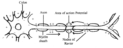
Saltatory conduction
Question 21.
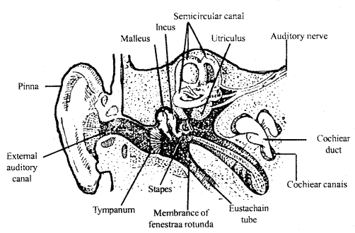
(a) Make a clearly labeled diagram of the inner ear of a human being.
Answer:
Diagrams showing the inner ear
(b) Describe how each of the following is achieved in us
(i) hearing
(ii) balance.
Answer:
Neural Control and Coordination Important Extra Questions Long Answer Type
Question 1.
(a) Describe the reflex arc with a diagram.
Answer:
The neurons forming the pathway taken by the nerve impulses in the reflex action form the Reflex Arc.
The reflex arc consists of
- Receptor,
- An afferent neuron or sensory neurons from receptor to CN system,
- The efferent neuron of motor neurons from CN system to specific muscle fibers or gland cells,
- a number of connectors or intermediate neurons conducting impulses form the afferent to the efferent neurons.
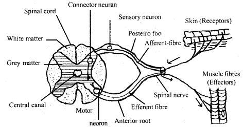
Reflex Arc
When a specific stimulus is applied to a specific group of receptors, it stimulates the receptor to initiate a nerve impulse along the afferent neurons. This impulse travels along with the afferent connector and efferent neurons to reach an effector-muscle or gland for that reflex. Thus the flow of impulse can only be in a single direction in a reflex arc, i.e.,
Stimulus → receptor → afferent neuron → CN system efferent neuron ← (connector neuron)
(b) Distinguish between conditioned reflex and unconditioned reflex.
Answer:
Differences between conditioned reflex and unconditioned reflex:
| Conditioned Reflex | Unconditioned Reflex |
| It is a reflex, acquired after birth by applying an indifferent stimulus before or along with the stimulus for an inborn reflex. | It is a reflex, which can be evoked even immediately after birth and needs no previous encounter with the stimulus exciting it. |
Question 2.
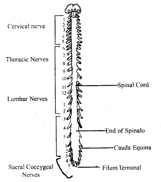
(a) Give an account of spinal nerves in man.
Answer:
There are 31 pairs of a spinal nerve in man. From each segment of the spinal cord, there are two spinal nerves. Each spinal nerve is a mixed nerve, containing both sensory and motor nerve fibers. It runs between the spinal cord and peripheral tissue. The two roots, i.e., motor or ventral and sensory or dorsal connect the spinal nerve to the spinal cord.
The DORSAL ROOT carries sensory or afferent fiber and has a dorsal root ganglion at its middle. The VENTRAL ROOT contains a motor or efferent nerve fibers. The dorsal root fibers bring impulses from the peripheral tissue and give rise to sensations like touch, temperature, and pain.
The ventral nerve root fibers pass impulses to muscles and glands in the peripheral tissues. The spinal nerve has been named according to its relation with the vertebral column.
These are
- Eight pairs of cervical
- 12 pairs of thoracic
- 5 pairs of lumber,
- 5 pairs of sacral and
- a pair of coccygeal or caudal.
(b) What biological functions are served by the skeletal system?
Answer:
- The skeletal system forms the rigid structural framework of the body and supports the weight of the body along with its limbs.
- It affords protection to the internal organs against mechanical injury by forming cage-like compartments, e.g., skull.
- It serves as a storage depot for calcium and phosphate, which are released for a number of functions of the body.
- It participates in movement and locomotion.
The spinal nerve in man
Question 3.
Distinguish between:
(a) Afferent neurons and efferent neurons.
Answer:
Afferent neurons and efferent neurons:
Afferent neurons: These conduct sensory impulses from the receptors present in the peripheral organs and tissues towards the central nervous system. Their bodies are called afferent neurons.
Efferent neurons: These conduct motor impulses from the central nervous system to the peripheral organs and tissues serving as effectors. Their cell bodies are called efferent neurons.
(b) Rods and cones
Answer:
Rods: Rod cells are rod-like, elongated cells, bearing long, thin cylinders, containing a visual pigment called Rhodopsin. Rod cells are present on the periphery of the retina in the human eye. These cells do not form color vision.
Cones: Cone cells are shorter, thicker, and conical in shape. These are highly sensitive to bright light and colors. They contain a violet color pigment called rhodopsin. Cone cells are responsible for the perception of different colors. Cones are abundant on the rear wall and fovea centralis of the retina.
(c) Resting membrane potential and action potential.
Answer:
Resting membrane potential: The surface of the axon carries a positive charge relative to its interior and this electrical potential difference across the plasma membrane is called resting membrane potential.
Action potential: The shift of ions and consequents shift in electrical charges is sufficient enough; it will trigger a wave of transient membrane depolarization known as nerve impulse or Action potential.
(d) Impulse conduction in myelinated nerve fiber and unmyelinated nerve fiber.
Answer:
Impulse conduction in myelinated nerve fiber: The myelinated fibers carry impulses nearly 20 times faster than the non-myelinated nerve fibers. These avoid dissipation of impulse into adjusting fibers. The myelin sheath serves as a highly insulating layer that prevents the flow of ions. Impulses are rapid.
Non-myelinated nerve fiber: Ionic changes and depolarization are repeated over the membrane all along with the fiber. Impulse requires less energy and does not need to run all along with the fiber.
(e) Aqueous humor and vitreous humor.
Answer:
Aqueous humor: The chamber between the cornea and lens is filled with a clear watery fluid, the aqueous humor.
Vitreous humor; The chamber behind the lens is filled with a semisolid gelatinous material the vitreous humor.
(f) Blindspot and yellow spot.
Answer:
Blindspot: It s a small insensitive light area of about 0.5 cm. in diameter. It is devoid of rod and cone cells. It is unable to receive light rays.
Yellow spot: A tiny circular area, about 6 mm in diameter in the retina is a yellow spot. Here the vision is sharpest. It has rod and cone cells.
(g) Cranial nerve and spinal nerves.
Answer:
Cranial nerve: There are 12 pairs of cranial nerves, 10 originate from the brain stem, but all pass through the foramina of the skull. Cranial nerves contain only sensory fibers. The remainder contains both sensory and motor fibers.
Spinal Nerve: They arise from the cord. 31 pairs of segmental spinal nerves arise from the cord. They contain both receptor neurons and effectors neurons.
Must Read: