By going through these CBSE Class 12 Biology Notes Chapter 6 Molecular Basis of Inheritance, students can recall all the concepts quickly.
Molecular Basis of Inheritance Notes Class 12 Biology Chapter 6
→ Gene is a macromolecule attached to an undifferentiated protein thread (chromonema) that can pass from one cell to another and can be transmitted from one generation to another without being changed at all.
→ Gene is a unit of heredity and responsible for inheritance.
→ Gene is a part of DNA and is made up of nucleotides. It can grow, reproduce and mutate. Gene is the unit of recombination. Genetic information is conveyed from DNA to mRNA.
→ Genes mainly act by producing proteins or enzymes which are required at various levels of metabolism. Proteins are made up of polypeptides which are formed from amino acids. Genes determine the physical and physiological characteristics of living beings.
→ DNA and RNA are two types of nucleic acids, which are polymers of nucleotides. DNA is the genetic material in most organisms. RNA is found only in some viruses as genetic material, it acts as a messenger to transfer the genetic information from DNA to proteins. It also functions as an adapter, structural and catalytic molecule.
→ DNA (deoxyribonucleic acid) is the polymer of deoxyribonucleotides. The haploid content of human DNA is 3.3 × 10⁹ bp. DNA is chemically and structurally more stable than RNA.
→ Nucleic acids were first isolated by Friedrich Miescher (1869) from pus cells. These were named nuclein. Due to their acidic nature, they were named nucleic acid, and Altmann (1899) named them nucleic acids. Fisher (1880s) discovered the presence of purine and pyrimidine bases. Levene (1910) found deoxyribose nucleic acid contains phosphoric acid and deoxyribose sugar. He characterized four types of nucleotides.
Chargaff (1950) found that purines are equal to pyrimidines in DNA, and adenine = thymine, and guanine = cytosine. W.T. Astbury discovered through X-ray diffraction that DNA is a polynucleotide with nucleotides arranged perpendicular to the long axis of the molecule and are 0.34nm away from one another.
Wilkins and Franklin (1953) through X-ray photographs found that DNA was a helix with 2.0nm width. One turn of the helix was 3.4 nm with 10 layers of bases stacked in it. Through these photographs, Watson and Crick (1 953) built a three-dimensional model of DNA. They were awarded Nobel Prize in 1962. They purposed that DNA consists of a double helix with two chains having sugar-phosphate as the backbone and nitrogen bases on the inner sides.
The nitrogen bases of two chains form complementary pairs with purines of one and pyrimidine of the other held together by hydrogen bonds. The two chains are antiparallel with 5′ → 3′ orientation and 3′ → 5′ orientation. The two chains are twisted helically as a rope ladder with rigid steps twisted into a spiral. Each turn contains 10 nucleotides. In Vitro, DNA synthesis was carried out by Komberg (l 959). DNA has got an implicit mechanism for replication and copying.
→ A nucleotide is made up of three components: a nitrogenous base, a pentose sugar (ribose in RNA, deoxyribose in DNA), and a phosphate group. Nitrogenous bases are of two types: purines and pyrimidines. Purines are Adenine and Granite and pyrimidines are Cytosine, Uracil, and Thymine. Thymine is present in DNA. Thymine is replaced by Uracil in RNA.
→ A nucleoside is formed by a nitrogenous base linked to pentose sugar by an N-glycosidic bond, e.g. deoxyadenosine or adenosine, deoxycytidine, or cytidine.
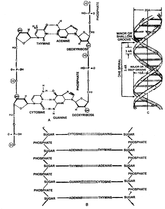
Structure of DNA.
A. Chemical Structure and bonding of different constituents of DNA in the two chains.
B. sequence of nucleotides in a part of the double helix of DNA. C. coiling in double helix or duplex of DNA.
→ Two nucleotides are bound to each other through a phosphodiester bond. The phosphate group provides acidity to the nucleic acids. In Bacteria nucleotide, the DNA is covalently closed at its two ends. It’s called circular DNA. Its also present in mitochondria, plastids, and some viruses. Eukaryotic cells have free linear DNA. Both types of DNA are coiled and supercoiled to get into small space.
→ DNA replication is autocatalytic, it occurs during the S-phase of the cell cycle. Watson and Crick proposed a semiconservative model of replication for DNA. During which one strand of the daughter strand is derived from the parental duplex and the other strand is formed a new. Taylor et al (1957) worked with Broad Bean root tips, using
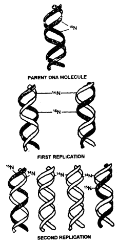
The experiment of Mcselson and Stahi (i958) to prove semi-conservative replication of DNA
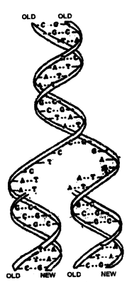
Semi-conservative replication of DNA showing Zipper duplication
radiolabelled thymine and showed DNA replication is semi-conservative. In 1958 Nesselson and Stahl proved the semi-conservative mode of replication. They worked with E.Coli, using a heavy isotope of nitrogen, N15. E.Coli was grown with N15 for several generations till the bacteria became completely labeled with N15. They were then shifted to medium with normal N14 nitrogen.
The sample was taken after every generation and tested for the presence of N15 and N14 using density gradient centrifugation with CsCl. The first generation was found to be a hybrid between N115 and N14. The second-generation contained two types of DNA, 50% light (N14) and 50% intermediate. This is only possible if two strands separate during replication and act as a template for the synthesis of new complementary strands. Thus proving a semiconservative mode of replication.
→ The process of replication starts at a particular spot known as the origin of replication. The deoxyribonucleotides are first phosphorylated and activated. Energy and phosphorylase enzymes are required for activation. Enzymes’ topoisomerases are specialized to break and reseal the DNA strands. Enzyme helicases unwind the DNA helix and separate the two strands. DNA binding proteins bind on the separated strands. The whole of DNA does not open up for replication, the point of separation proceeds from one end to another. The replication fork appears Y- Y-shaped during the process of replication.
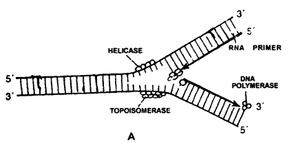
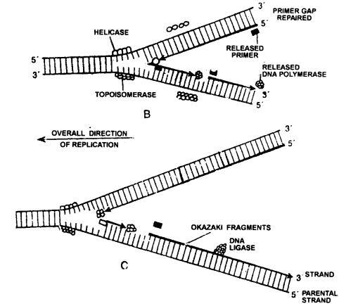
Replication of DNA continuous over one strand and discontinuous over other strands
DNA polymerase enzyme requires an RNA primer to initiate the process/ RAN primer is synthesized at the 5′ end of new DNA strand by enzyme primase. The two separated strands act as templates. The new nucleotides are added according to the law of base pairing i.e. A pairs with T and G pairs with C.

Energy is utilized in forming hydrogen bonds between the free nucleotides and nitrogen bases of templates. Elongation of the DNA chain requires enzyme DNA polymerase III in the presence of Mg2+ and ATP. The adjacent nucleotides attach to one another by phosphodiester bonds. As replication proceeds, the strands unwind and at the end, the RNA primer is removed and the gap is filled with the help of DNA polymerase 1. This is also called Zipper duplication.
DNA polymerase can act only in the 5′ → 3′ direction so replication is continuous on the strand (3′ → 5′) known as the leading strand. The other strand called lagging strand 1 (5′ → 3′) replicates itself in small stretches called Okazaki fragments. These fragments are joined by the DNA ligase enzyme. Any mismatch or mutation if occurs during replication can be corrected by proofreading and DNA repair mechanisms
→ Frederick Griffith (1928) by doing transforming experiments on mice with Streptococcus pneumonia found out the transfer of genetic material from a heat-killed infectious stain to the normal non-infectious stain.
→ Earlier the genetic material was thought to be protein. In 1933 – 44 Oswald Avery, Colin Macleod, and Maclyn McCarty discovered that the transforming biochemical in Griffith’s experiments was DNA.
→ Alfred Hershey and Martha Chase (1952) proved that DNA is the hereditary material. They worked with bacteriophage T, which infects E.Coli.
→ Francis Crick proposed the central dogma in molecular biology which explains the flow of genetic information
i.e. DNA → RNA → Protein.
→ The formation of RNA over a DNA template is called transcription. Transcription is meant for taking the coded information from DNA to the site where it is required for protein synthesis. Only one DNA strand transcribes RNA, it is called a sense strand. Transcription requires enzyme RNA polymerase. Prokaryotes have only one RNA polymerase. Eukaryotes have three RNA polymerases.
RNA polymerase 1 synthesizes rRNA, 28S, 18S, and 5.8S, RNA polymerase II synthesizes hnRNA, mRNA, and snRNAs, and RNA polymerase III synthesizes tRNA, 5SRNA, and siRNAs. A transcription unit consists of a promoter, the structural gene, and a terminator.

Schematic Structure of a Transcription Unit
→ In eukaryotes, the primary transcript contains exons and introns so it is non-functional. Exons are coding sequences whereas introns are intervening sequences. These introns are removed by splicing. It is followed by capping, methyl guanosine triphosphate addition to 5′ end and of hnRNA and tailing around 200 – 300 adenyl residues are added at 3′ end. The fully processed hnRNA is called mRNA and is transported out of the nucleus.
→ The relationship between the sequence of amino acids in a polypeptide and nucleotides sequence of DNA or mRNA is called genetic code.
→ George Gamow proposed that the genetic code is a triplet in nature. Marshall and Nirenberg’s method for cell-free protein synthesis helped to decipher the code. The chemical method developed by Har Gobind Khorana helped in synthesizing RNA molecules with defined base combinations. Severo Ochoa’s enzyme polynucleotide phosphorylase helped to polymerize RNA with defined sequences. All these methods helped in deciphering the genetic code.
→ The translation is the process of polymerization of amino acids to form a polypeptide. mRNA sequence decides the sequence of amino acids. Amino acids are joined through peptide bonds. Ribosomes are considered as the protein-synthesizing cellular factory. It requires amino acids, mRNA, tRNA, aminoacyl tRNA synthetase, and various enzymes along with ribosomes to complete the process of protein synthesis.
The process of translation occurs in the cytoplasm. The process takes place in several steps like activation of amino acids, initiation, elongation, and termination. Polyribosomes or polysomes help to produce a number of copies of the same polypeptide. In it, different ribosomes are held together by a strand of messenger RNA. These ribosomes usually form rosette or helical groups during active protein synthesis and are known as polyribosomes.
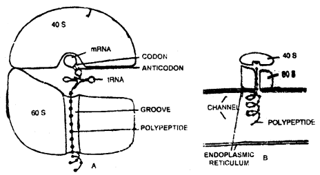
Ribosomes as a protein factory. A relationship between the various components. B. synthesis of the polypeptide on ribosome connected with the endoplasmic reticulum.
Cistron is a segment of DNA consisting of a stretch of base sequences that codes for one polypeptide chain, one transfer RNA (tRNA), ribosomal RNA tRNA) a molecule or performs any other specific function in connection with transcription, including controlling the functioning of other cistrons (operon model of gene action).
Regulation of gene expression may occur at various steps like
- transcriptional level,
- processing level (splicing),
- transport of mRNA from the nucleus to the cytoplasm.
- translational level.
An operon is a part of genetic material (or DNA) which acts a: a single regulated unit having one or more structural genes, an operator gene, a promoter gene, a regulator gene, a repressor, and an inducer or corepressor (from outside). Operons are of two types, inducible and repressive.
Lac operon is an example of an inducible operon system. In E.Col; the enzyme beta-galactosidase catalyzes the hydrolysis of lactose into glucose and galactose, which is used as a source of energy. If the bacteria do not have lactose in the medium they won’t need beta-galactosidase enzyme. Thus the production of an enzyme (protein) is regulated by the presence of lactose (inducer).
→ Anticodon: A triplet of bases present on tRN A complementing with mRNA codon is called the anticodon.
→ A-site: It’s the site where the second and next amino-acyl-tRNA enters the ribosome.
→ Codon: Triplet of bases on mRNA, which codes for one amino acid.
→ Central dogma: Unidirectional flow of information from DNA to RNA to protein.
→ Cistron: A segment of DNA that determines a single polypeptide chain.
→ DNA polymerase: The enzyme playing a pivotal role in adding the building blocks to the primer in a sequence as guided by the DNA template. It can polymerize nucleotides only in the 5’ → ‘3’ direction.
→ Frameshift mutation: The mutations which are caused by shifting the entire reading frame by addition or deletion on the segment of DNA.
→ Genetic code: The genetic presentation of codon through which the information in RNA is decoded in a polypeptide chain.
→ Glycosylation: The addition of sugar residues by modification of certain proteins, which are released in the lumen and are trapped in Golgi vesicles.
→ Hydrogen bond: The bond between nitrogenous bases of DNA binding two nucleotide chains. It is a weak bond.
→ Inducer: An effector molecule responsible for the induction of enzyme synthesis at recognition sites, to prevent self-cleavage by a modified enzyme that recognizes the sites and methylates specific nucleotides at each site.
→ Jumping genes: These are genes that shuffle from one location to another.
→ Phosphodiester bond: The bond between two adjacent nucleotides of two adjacent sugar moieties at 3’ and 5’ positions with phosphoric acid.
→ Phenocopy: When a normal gene under a different set of environmental conditions copies down the phenotypic characters of a mutant.
→ Restriction enzyme: An endonuclease that recognizes specific nucleotide sequences in DNA and makes a double-strand cleavage of DNA molecule.
→ Regulatory gene: Any gene which regulates or modifies the activity of other genes.
→ Repression: The phenomenon in which the synthesis of a set of enzymes leading to a product is shut down if the product is present in plentiful amounts.
→ Rho-factor: The factor which is required for termination of RNA synthesis at some sites.
→ Structural gene: A gene that codes for a polypeptide.
→ Silent mutation: This kind of mutation does not cause any change in the protein.
→ Thalassemia: Haemoglobin-based genetic disorder which involves frameshift mutation in β-chain of hemoglobin.
→ Transformation: The process in which the cell takes up the segment of the naked DNA from its surroundings and incorporates it in its hereditary material and ultimately expresses the character specified by incoming DNA.
→ Wobble position: The position on the codon where mutation occurs at 3rd base of triplet which still permits the normal interaction with anticodon.