Learninsta presents the core concepts of Biology with high-quality research papers and topical review articles.
Detection of Foetal Disorders During Early Pregnancy
Ultrasound scanning Ultrasound has no known risks other than mild discomfort due to pressure from the transducer on the abdomen or vagina. No radiation is used during this procedure. Ultrasonography is usually performed in the first trimester for dating, determination of the number of foetuses, and for assessment of early pregnancy complications.
Amniocentesis
Amniocentesis involves taking a small sample of the amniotic fluid that surrounds the foetus to diagnose for chromosomal abnormalities (Fig. 3.1).
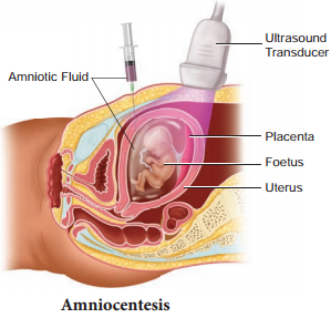
Amniocentesis is generally performed in a pregnant woman between the 15th and 20th weeks of pregnancy by inserting a long, thin needle through the abdomen into the amniotic sac to withdraw a small sample of amniotic fluid. The amniotic fluid contains cells shed from the foetus.
Chorionic villus sampling (CVS)
CVS is a prenatal test that involves taking a sample of the placental tissue to test for chromosomal abnormalities.
Foetoscope
Foetoscope is used to monitor the foetal heart rate and other functions during late pregnancy and labour. The average foetal heart rate is between 120 and 160 beats per minute. An abnormal foetal heart rate or pattern may mean that the foetus is not getting enough oxygen and it indicates other problems.
A hand-held doppler device is often used during prenatal visits to count the foetal heart rate. During labour, continuous electronic foetal monitoring is often used.