Learninsta presents the core concepts of Biology with high-quality research papers and topical review articles.
Flagella – Definition Structure and its Types
Prokaryotic Flagellum
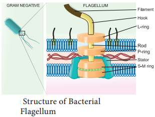
Bacterial flagella are helical appendages helps in motility. They are much thinner than flagella or cilia of eukaryotes. The filament contains a protein called flagellin. The structure consists of a basal body associated with cytoplasmic membrane and cell wall with short hook and helical filament. Bacteria rotates their helical flagella and propels rings present in the basal body which are involved in the rotary motor that spins the flagellum.
Structure of Flagella in Bacteria
The gram positive bacteria contain only two basal rings. S-ring is attached to the inside of peptidoglycan and M-ring is attached to the cell membrane. In Gram negative bacteria two pairs of rings proximal and distal ring are connected by a central rod.
They are L-Lipopolysaccharide ring, P-Peptidoglycan ring, S-Super membrane ring and M-membrane ring. The outer pair L and P rings is attached to cell wall and the inner pair S and M rings attached to cell membrane (Figure 6.27).
Mechanism of Flagellar Movement – Proton Motive Force
In flagellar rotation only proton movements are involved and not ATP. Protons flowing back into the cell through the basal body rings of each flagellum drives it to rotate. These rings constitute the rotary motor.The proton motive force (The force derived from the electrical potential and the hydrogen ion gradient across the cytoplasmic membrane) drives the flagellar motor.
For the rotation of flagellum the energy is derived from proton gradient across the plasma membrane generated by oxidative phosphorylation. In bacteria flagellar motor is located in the plasma membrane where the oxidative phosphorylation takes place. Therefore, plasma membrane is a site of generation of proton motive force.
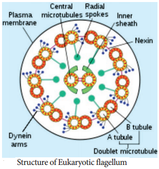
Eukaryotic Flagellum – Cell Motility Structure
Eukaryotic Flagella are enclosed by unit membrane and it arises from a basal body. Flagella is composed of outer nine pairs of microtubules with two microtubules in its centre (9+2 arrangement). Flagella are microtubule projection of the plasma membrane. Flagellum is longer than cilium (as long as 200µm). The structure of flagellum has an axoneme made up microtubules and protein tubulin (Figure 6.28)
Movement
Outer microtubule doublet is associated with axonemal dynein which generates force for movement. The movement is ATP driven. The interaction between tubulin and dynein is the mechanism for the contraction of cilia and flagella. Dynein molecules uses energy from ATP to shift the adjacent microtubules. This movement bends the cilium or flagellum.
Cilia
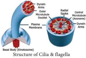
Cilia (plural) are short cellular, numerous microtubule bound projections of plasma membrane. Cilium (singular) is membrane bound structure made up of basal body, rootlets, basal plate and shaft.
The shaft or axoneme consists of nine pairs of microtubule doublets, arranged in a circle along the periphery with a two central tubules, (9+2) arrangement of microtubules is present. Microtubules are made up of tubulin. The motor protein dynein connects the outer microtubule pair and links them to the central pair. Nexin links the peripheral doublets of microtubules (Figure 6.29).