Learninsta presents the core concepts of Biology with high-quality research papers and topical review articles.
Human Reproductive Gametogenesis
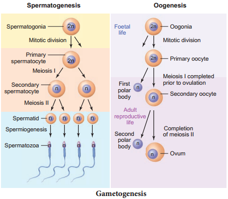
Gametogenesis is the process of formation of gametes i.e., sperms and ovary from the primary sex organs in all sexually reproducing organisms. Meiosis plays the most significant role in the process of gametogenesis (Fig. 2.5).
Spermatogenesis
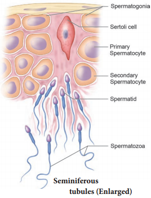
Spermatogenesis is the sequence of events in the seminiferous tubules of the testes that produce the male gametes, the sperms. During development, the primordial germ cells migrate into the testes and become immature germ cells called sperm mother cells or spermatogonia in the inner surfaces of the seminiferous tubules (Fig. 2.6 a).
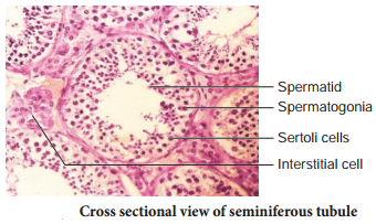
The spermatogonia begin to undergo mitotic division at puberty and continue throughout life. In the first stage of spermatogenesis, the spermatogonia migrate among sertoli cells towards the central lumen of the seminiferous tubule and become modified and enlarged to form primary spermatocytes which are diploid with 23 pairs i.e., 46 chromosomes.
Some of the primary spermatocytes undergo first meiotic division to form two secondary spermatocytes which are haploid with 23 chromosomes each. The secondary spermatocytes undergo second meiotic division to produce four haploid spermatids. The spermatids are transformed into mature spermatozoa (sperms) by the process called spermiogenesis. Sperms are finally released into the cavity of seminiferous tubules by a process called spermiation.
The whole process of spermatogenesis takes about 64 days. At any given time, different regions of the seminiferous tubules contain spermatocytes in different stages of development (Fig. 2.6 b). The sperm production remains nearly constant at a rate of about 200 million sperms per day.
Spermatogenesis starts at the age of puberty and is initiated due to the increase in the release of Gonadotropin Releasing Hormone (GnRH) by the hypothalamus. GnRH acts on the anterior pituitary gland and stimulates the secretion of two gonadotropins namely Follicle Stimulating Hormone (FSH) and Lutenizing Hormone (LH).
FSH stimulates testicular growth and enhances the production of Androgen Binding Protein (ABP) by the sertoli cells and helps in the process of spermiogenesis. LH acts on the Leydig cells and stimulates the synthesis of testosterone which in turn stimulates the process of spermatogenesis.
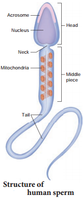
Structure of human spermatozoan The human sperm is a microscopic, flagellated and motile gamete (Fig. 2.7). The whole body of the sperm is enveloped by plasma membrane and is composed of a head, neck and a tail. The head comprises of two parts namely acrosome and nucleus.
Acrosome is a small cap like pointed structure present at the tip of the nucleus and is formed mainly from the Golgi body of the spermatid. It contains hyaluronidase, a proteolytic enzyme, popularly known as sperm lysin which helps to penetrate the ovum during Fertilization. The nucleus is flat and oval. The neck is very short and is present between the head and the middle piece.
It contains the proximal centriole towards the nucleus which plays a role in the first division of the zygote and the distal centriole gives rise to the axial filament of the sperm. The middle piece possesses mitochondria spirally twisted around the axial filament called mitochondrial spiral or nebenkern. It produces energy in the form of ATP molecules for the movement of sperms. The tail is the longest part of the sperm and is slender and tapering.
It is formed of a central axial filament or axoneme and an outer protoplasmic sheath. The lashing movements of the tail push the sperm forward. The human male ejaculates about 200 to 300 million sperms during coitus. It is estimated that around 60 percent of sperms must have normal shape of which at least 40 per cent must show vigorous motility for normal fertility.
Oogenesis
Oogenesis is the process of development of the female gamete or ovum or egg in the ovaries. During foetal development, certain cells in the germinal epithelium of the foetal ovary divide by mitosis and produce millions of egg mother cells or oogonia.
No more oogonia are formed or added after birth. The oogonial cells start dividing and enter into Prophase I of meiotic division I to form the primary oocytes which are temporarily arrested at this stage. The primary oocytes then get surrounded by a single layer of granulosa cells to form the primordial or primary follicles (Fig. 2.8 a). A large number of follicles degenerate during the period from birth to puberty, so at puberty only
60,000 to 80,000 follicles are lef in each ovary.
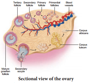
The primary follicle gets surrounded by many layers of granulosa cells and a new theca layer to form the secondary follicle. A fluid filed space, the antrum develops in the follicle and gets transformed into a tertiary follicle. The theca layer gets organized into an inner theca interna and an outer theca externa. At this time, the primary oocyte within the tertiary follicle grows in size and completes its first meiotic division and forms the secondary oocyte.
It is an unequal division resulting in the formation of a large haploid secondary oocyte and a first polar body. The first polar body disintegrates. During Fertilization, the secondary oocyte undergoes second meiotic
division and produces a large cell, the ovum and a second polar body.
The second polar body also degenerates. The tertiary follicle eventually becomes a mature follicle or Graafin follicle. If Fertilization does not take place, second meiotic division is never completed and the egg disintegrates. At the end of gametogenesis in females, each primary oocyte gives rise to only one haploid ovum.
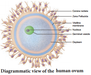
Structure of ovum
Human ovum is non-cleidoic, alecithal and microscopic in nature. (Fig. 2.8 (b)). Its cytoplasm called ooplasm contains a large nucleus called the germinal vesicle. The ovum is surrounded by three coverings namely an inner thin transparent vitelline membrane, middle thick zona pellucida and outer thick coat of follicular cells called corona radiata. Between the vitelline membrane and zona pellucida is a narrow perivitelline space.