Learninsta presents the core concepts of Biology with high-quality research papers and topical review articles.
Microscopy – Bright Field Microscope and Electron Microscope
Microscope is an inevitable instrument in studying the cell and subcellular structures. It offers scope in studying microscopic organisms therefore it is named as microscope (mikros – small; skipein – to see) in Greek terminology. Compound microscope was invented by Z. Jansen.
Microscope basically works on the lens system and its properties of light and lens such as reflection, magnification and numerical aperture. The common light microscope which has many lenses are called as compound microscope. The microscope transmits visible light from sources to eye or camera through sample.
Bright Field Microscope
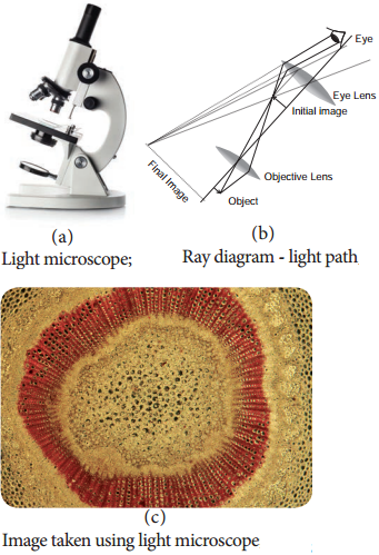
Bright field microscope is the routinely used microscope in studying various aspects of cells. It allows light to pass directly through specimen and shows a well distinguished image from different portions of the specimen. The contrast can be increased by staining the specimen with reagent that reacts with cells and tissue components of the object.
The light rays are focused by condenser on to the specimen on a microslide placed upon the adjustable platform called stage. Light comes from the Compact Flourescent Lamp (CFL) or Light Emitting Diode (LED). Then it passes through two lens systems namely objective lens (closer to the object) and the eye piece (closer to eye).
There are four objective lenses (5X, 10X, 45X and 100X) which can be rotated and fixed at certain point to get required magnification. It works on the principle of numerical aperture value and its own resolving power.
The first magnification of the microscope is done by the objective lens which is called primary magnification and it is real, inverted image. The second magnification of the microscope is obtained through eye piece lens called as secondary magnification and it is virtual and inverted image (Figure 6.2 a, b and c).
Electron Microscope
Electron Microscope was first introduced by Ernest Ruska (1931) and developed by G Binning and H Roher (1981). It is used to analyse the fine details of cell and organelles called ultrastructure. It uses beam of accelerated electrons as source of illumination and therefore the resolving power is 1,00,000 times greater than that of light microscope.
The specimen to be viewed under electron microscope is dehydrated and impregnated with electron opaque chemicals like gold or palladium. This is essential for withstanding electrons and also for contrast of the image.
There are two kinds of electron microscopes namely:
- Transmission Electron Microscope (TEM)
- Scanning Electron Microscope (SEM)
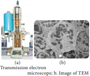
1. Transmission Electron Microscope:
This is the most commonly used electron microscope which provides two dimensional image. The components of the microscope are as follows:
- Electron generating system
- Electron condensor
- Specimen objective
- Tube lens
- Projector
A beam of electron passes through the specimen to form an image on fluorescent screen. The magnification is 1-3 lakhs times and resolving power is 2-10 Å. It is used for studying detailed structrue of viruses, mycoplasma, cellular organelles, etc (Figure 6.3 a and b).
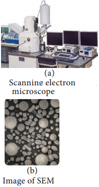
2. Scanning Electron Microscope:
This is used to obtain three dimensional image and has a lower resolving power than TEM. In this, electrons are focused by means of lenses into a very fine point.
The interaction of electrons with the specimen results in the release of different forms of radiation (such as auger electrons, secondary electrons, back scattered electrons) from the surface of the specimen. These radiations are then captured by an appropriate detector, amplified and then imaged on fluorescent screen. The magnification is 2,00,000 times and resolution is 5-20 nm (Figure 6.4 a and b).