Learninsta presents the core concepts of Biology with high-quality research papers and topical review articles.
Post Fertilization Structure and Events
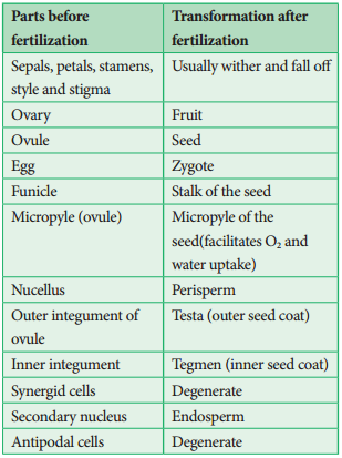
After fertilization, several changes take place in the floral parts up to the formation of the seed (Figure 1.20).
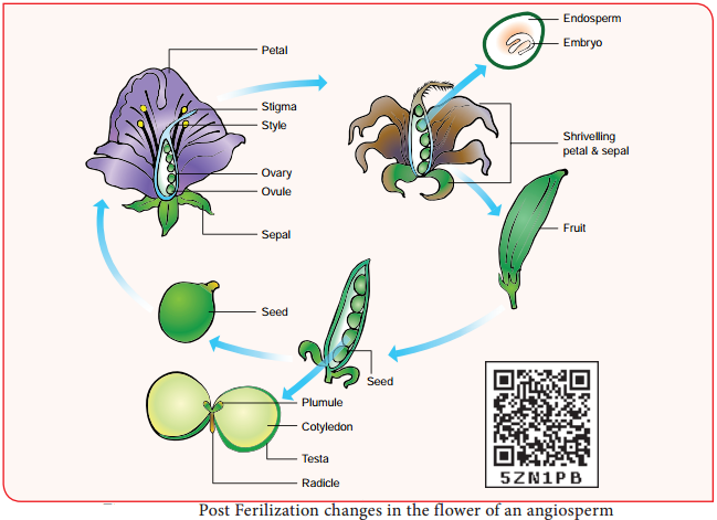
The events after fertilization (endosperm, embryo development, formation of seed, fruits) are called post fertilization changes.
Endosperm
The primary endosperm nucleus (PEN) divides immediately after fertilization but before the zygote starts to divide, to form the endosperm. The primary endosperm nucleus is the result of triple fusion (two polar nuclei and one sperm nucleus) and thus has 3n number of chromosomes. It is a nutritive tissue and regulatory structure that nourishes the developing embryo.
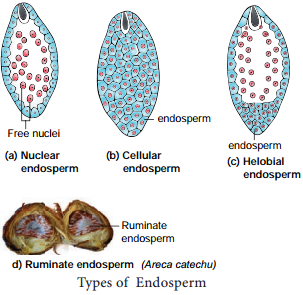
Depending upon the mode of development three types of endosperm are recognized in angiosperms. They are nuclear endosperm, cellular endosperm and helobial endosperm (Figure 1.21).
Nuclear endosperm:
Primary Endosperm Nucleus undergoes several mitotic divisions without cell wall formation thus a free nuclear condition exists in the endosperm. Examples: Coccinia, Capsella and Arachis
Cellular endosperm:
Primary endosperm nucleus divides into 2 nuclei and it is immediately followed by wall formation. Subsequent divisions also follow cell wall formation. Examples: Adoxa, Helianthus and Scoparia
Helobial endosperm:
Primary Endosperm Nucleus moves towards base of embryo sac and divides into two nuclei. Cell wall formation takes place leading to the formation of a large micropylar and small chalazal chamber. The nucleus of the micropylar chamber undergoes several free nuclear division whereas that of chalazal chamber may or may not divide. Examples: Hydrilla and Vallisneria.
The endosperms may either be completely consumed by the developing embryo or it may persist in the mature seeds. These seeds without endosperms are called non-endospermous or ex-albuminous seeds. Examples: Pea, Groundnut and Beans. These seeds with endosperms are called endospermous or albuminous seeds. The endosperms in these seeds supply nutrition to the embryo during seed germination.
Examples: Paddy, Coconut and Castor.
Ruminate endosperm:
The endosperm with irregularity and unevenness in its surface forms ruminate endosperm. Examples: Areca catechu, Passiflra and Myristica
Functions of endosperm:
- It is the nutritive tissue for the developing embryo.
- In majority of angiosperms, the zygote divides only after the development of endosperm.
- Endosperm regulates the precise mode of embryo development.
Development of Dicot embryo
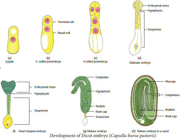
The Stages involved in the development of Dicot embryo (Capsella bursa-pastoris – Onagrad or crucifer type) is given in Figure 1.22. The embryo develops at micropylar end of embryo sac. The zygote undergoes transverse division to form upper or terminal cell and lower or basal cell.
Further divisions in the zygote during the development lead to the formation of embryo. Embryo undergoes globular, heart shaped stages before reaching a mature stage. Th mature embryo has a radicle, two cotyledons and a plumule.
Seed
The fertilized ovule is called seed and possesses an embryo, endosperm and a protective coat. Seeds may be endospermous (wheat, maize, barley and sunflower) or non endospermous. (Bean, Mango, Orchids and cucurbits).
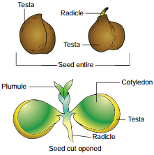
Cicer seed (example for Dicot seed)
The mature seeds are attached to the fruit wall by a stalk called funiculus. The funiculus disappears leaving a scar called hilum. Below the hilum a small pore called micropyle is present. It facilitates entry of oxygen and water into the seeds during germination.
Each seed has a thick outer covering called seed coat. The seed coat is developed from integuments of the ovule. The outer coat is called testa and is hard whereas the inner coat is thin, membranous and is called tegmen.
In Pea plant the tegmen and testa are fused. Two cotyledons laterally attached to the embryonic axis and store the food materials in pea whereas in other seeds like castor the endosperm contains reserve food and the Cotyledons are thin. The portion of embryonal axis projecting beyond the cotyledons is called radicle or embryonic root.
The other end of the axis called embryonic shoot is the plumule. Embryonal axis above the level of cotyledon is called epicotyl whereas the cylindical region between the level of cotyledon is called hypocotyl (Figure 1.23 a).
Oryza seed (example for Monocot seed)
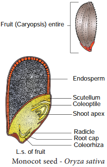
The seed of paddy is one seeded and is called Caryopsis. Each seed remains enclosed by a brownish husk which consists of glumes arranged in two rows. The seed coat is a brownish, membranous layer closely adhered to the grain.
Endosperm forms the bulk of the grain and is the storage tissue. It is separated from embryo by a defiite layer called epithelium. The embryo is small and consists of one shieldshaped cotyledon known as scutellum present towards lateral side of embryonal axis.
A short axis with plumule and radicle protected by the root cap is present. The plumule is surrounded by a protective sheath called coleoptile. The radicle including root cap is also covered by a protective sheath called coleorhiza. The scutellum supplies the growing embryo with food material absorbed from the endosperm with the help of the epithelium (Figure 1.23 b).
