Learninsta presents the core concepts of Biology with high-quality research papers and topical review articles.
Structure of Contractile Proteins
Contraction of the muscle depends on the presence of contractile proteins (Figure 9.2) such as actin and myosin in the myofilaments. The thick filaments are composed of the protein myosin which are bundled together whose heads produce at opposite ends of the filament.
Each myosin molecule is made up of a monomer called meromyosin. The meromyosin has two regions, a globular head with a short arm and a tail. The short arm constitutes the heavy meromyosin (HMM).
The tail portion forms the light meromyosin (LMM). The head bears an actin-binding site and an ATP – binding site. It also contains ATP case enzyme that split ATP to generate energy for the contraction of muscle. The thin filaments are composed of two interwined actin molecules. Actin has polypeptide subunits called globular actin or G-actin and filamentous form or F-actin.
Each thin filament is made of two F-actins helically wound to each other. Each F-actin is a polymer of monomeric G-actins. It also contains a binding site for myosin. The thin filaments also contain several regulatory proteins like tropomyosin and troponin which help in regulating the contraction of muscles along with actin and myosin.
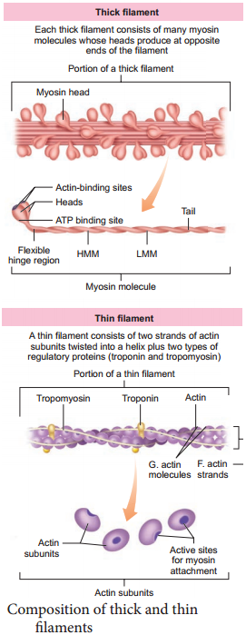
Structure of Contractile Proteins. Each actin (thin) filament is made of two ‘F’ (filamentous) actins helically wound to each other. Each ‘F’ actin is a polymer of monomeric ‘G’ (Globular) actins. Two filaments of another protein, tropomyosin also run close to the ‘F’ actins throughout its length.
The contractile proteins are myosin, the principal component of thick myofilaments, and actin, which is the principal component of thin myofilaments.
Contractile fibers are intracellular protein filament-based structures that are primarily composed of actin, myosin and tropomyosin.
Contractile proteins are proteins that mediate sliding of contractile fibres (contraction) of a cell’s cytoskeleton, and of cardiac and skeletal muscle.
Thick filaments contain myosin, thin filaments contain actin , troponin and tropomyosin. Scientists think that muscles contract by the two types of filament sliding over each other so that they overlap more.
Contractile function is a fundamental part of the CMR examination. Contractile function imaging is used for global and regional wall motion assessment and has been demonstrated to be highly accurate and reproducible for LV and right ventricular (RV) volume, ejection fraction, and mass measurements. Non-contractile (inert) tissues – joint capsules, ligaments, nerves and their sheaths, bursae, and cartilages.
Fiber-tracheids are intermediate forms between tracheids and libriform fibers. Tracheids are not fibers, as their major function is conducting water and the cell shape is not typical of a fiber, though they have relatively thick cell walls.
ATP is a nucleotide that consists of three main structures: the nitrogenous base, adenine; the sugar, ribose; and a chain of three phosphate groups bound to ribose. The phosphate tail of ATP is the actual power source which the cell taps.
Any metabolic process that requires oxygen to occur is referred to as aerobic. Humans, most other multicellular organisms, and some microorganisms require oxygen for the efficient capture of the chemical energy from food and its transformation into the cellular energy form known as ATP.