Theories Of Biological Evolution

Learninsta presents the core concepts of Biology with high-quality research papers and topical review articles.

Theories Of Biological Evolution

Lamarck’s theory

Jean Baptiste de Lamarck, was the first to postulate the theory of evolution in his famous book ‘Philosophie Zoologique’ in the year 1809. The two principles of Lamarckian theory are:

(i) The theory of use and disuse – Organs that are used often will increase in size and those that are not used will degenerate. Neck in giraffe is an example of use and absence of limbs in snakes is an example for disuse theory.

(ii) The theory of inheritance of acquired characters – Characters that are developed during the life time of an organism are called acquired characters and these are then inherited.

The main objection to Lamarckism

Lamarck’s “Thory of Acquired characters” was disproved by August Weismann who conducted experiments on mice for twenty generations by cutting their tails and breeding them. All mice born were with tail. Weismann proved his germplasm theory that change in the somatoplasm will not be transferred to the next generation but changes in the germplasm will be inherited.

Neo-Lamarckism

The followers of Lamarck (Neo-Lamarckists) like Cope, Osborn, Packard and Spencer tried to explain Lamarck’s theory on a more scientific basis. They considered that adaptations are universal. Organisms acquire new structures due to their adaptations to the changed environmental conditions. They argued that external conditions stimulate the somatic cells to produce certain ‘secretions’ which reach the sex cells through the blood and bring about variations in the offspring.

Darwin’s theory of Natural Selection

Charles Darwin explained the theory of evolution in his book ‘The Origin of Species by Natural Selection’. During his journey around the Earth, he made extensive observations of plants and animals. He noted a huge variety and remarkable similarities among organisms and their adaptive features to cope up to their environment. He proved that fitest organisms can survive and leave more progenies than the unfit ones through natural selection.

Darwin’s theory was based on several facts, observations and influences. They are:

1. Over production (or) prodigality of production:

All living organisms increase their population in larger number. For example, Salmon fish produces about 28 million eggs during breeding season and if all of them hatch, the seas would be filed with salmon in few generations. Elephant, the slowest breeder that can produce six young ones in its life time can produce 6 million descendants at the end of 750 years in the absence of any check.

2. Struggle for existence:

Organisms struggle for food, space and mate. As these become a limiting factor, competition exists among the members of the population. Darwin denoted struggle for existence in three ways – Intra specific struggle between the same species for food, space and mate. Inter specific struggle with different species for food and space. Struggle with the environment to cope with the climatic variations, flood, earthquakes, drought, etc.,

3. Universal occurrence of variations

No two individuals are alike. There are variations even in identical twins. Even the children born of the same parents differ in colour, height, behavior, etc., The useful variations found in an organism help them to overcome struggle and such variations are passed on to the next generation.

4. Origin of species by Natural Selection

According to Darwin, nature is the most powerful selective force. He compared origin of species by natural selection to a small isolated group. Darwin believed that the struggle for existence resulted in the survival of the fittest. Such organisms become better adapted to the changed environment.

Objections to Darwinism

Some objections raised against Darwinism were –

  • Darwin failed to explain the mechanism of variation.
  • Darwinism explains the survival of the fittest but not the arrival of the fittest.
  • He focused on small fluctuating variations that are mostly non-heritable.
  • He did not distinguish between somatic and germinal variations.
  • He could not explain the occurrence of vestigial organs, over specialization of some organs like large tusks in extinct mammoths, oversized antlers in the extinct Irish deer, etc.,

Neo Darwinism

Neo Darwinism is the interpretation of Darwinian evolution through Natural Selection as it has been modified since it was proposed. New facts and discoveries about evolution have led to modifications of Darwinism and is supported by Wallace, Heinrich, Haeckel, Weismann and Mendel. This theory emphasizes the change in the frequency of genes in population arises due to mutation, variation, isolation and Natural
selection.

Mutation theory

Hugo de Vries put forth the Mutation theory. Mutations are sudden random changes that occur in an organism that is not heritable. De Vries carried out his experiments in the Evening Primrose plant (Oenothera lamarckiana) and observed variations in them due to mutation.

According to de Vries, sudden and large variations were responsible for the origin of new species whereas Lamarck and Darwin believed in gradual accumulation of all variations as the causative factors in the origin of new species. Hugo de Vries believed that Mutations are random and directionless, but Darwinian variations are small and directional.

Salient features of Mutation Theory

  • Mutations or discontinuous variation are transmitted to other generations.
  • In naturally breeding populations, mutations occur from time to time.
  • There are no intermediate forms, as they are fully fledged.
  • They are strictly subjected to natural selection.

Modern synthetic theory

Sewell Wright, Fisher, Mayer, Huxley, Dobzhansky, Simpson and Haeckel explained Natural Selection in the light of Post-Darwinian discoveries. According to this theory gene mutations, chromosomal mutations, genetic recombinations, natural selection and reproductive isolation are the fie basic factors involved in the process of organic evolution.

(i) Gene mutation

Refers to the changes in the structure of the gene. It is also called gene/point mutation. It alters the phenotype of an organism and produces variations in their offspring.

(ii) Chromosomal mutation

Refers to the changes in the structure of chromosomes due to deletion, addition, duplication, inversion or translocation. This too alters the phenotype of an organism and produces variations in their offspring.

(iii) Genetic recombination

Is due to crossing over of genes during meiosis. This brings about genetic variations in the individuals of the same species and leads to heritable variations.

(iv) Natural selection

Does not produce any genetic variations but once such variations occur it favours some genetic changes while rejecting others (driving force of evolution).

(v) Reproductive Isolation

Helps in preventing interbreeding between related organisms.

Evolution by anthropogenic sources

Natural Selection (Industrial melanism)

Natural selection can be explained clearly through industrial melanism. Industrial melanism is a classical case of Natural selection exhibited by the peppered month, Biston betularia. These were available in two colours, white and black. Before industrialization peppered moth both white and black coloured were common in England. Pre-industrialization witnessed white coloured background of the wall of the buildings hence the white coloured months escaped from their predators.

Post industrialization, the tree trunks became dark due to smoke and soot let out from the industries. The black moths camouflaged on the dark bark of the trees and the white moths were easily identified by their predators. Hence the dark coloured month population was selected and their number increased when compared to the white months. Nature offered positive selection pressure to the black coloured months. The above proof shows that in a population, organisms that can adapt will survive and produce more progenies
resulting in increase in population through natural selection.

Artificial selection is a byproduct of human exploitation of forests, oceans and fisheries or the use of pesticides, herbicides or drugs. For hundreds of years humans have selected various types of dogs, all of which are variants of the single species of dog. If human beings can produce new varieties in short period, then “nature” with its vast resources and long duration can easily produce new species by selection.

Adaptive Radiation

The evolutionary process which produces new species diverged from a single ancestral form becomes adapted to newly invaded habitats is called adaptive radiation. Adaptive radiations are best exemplified in closely related groups that have evolved in relatively short time.

Darwin’s finches and Australian marsupials are best examples for adaptive radiation. When more than one adaptive radiation occurs in an isolated geographical area, having the same structural and functional similarity is referred to as convergent evolution.
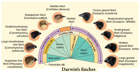
Darwin’s fiches

Their common ancestor arrived on the Galapagos about 2 million years ago. During that time, Darwin’s finches have evolved into 14 recognized species differing in body size, beak shape and feeding behavior. Changes in the size and form of the beak have enabled different species to utilize different food resources such as insects, seeds, nectar from cactus flowers and blood from iguanas, all driven by Natural selection. Fig. 6.5 represents some of the finches observed by Darwin.
Theories Of Biological Evolution img 1

Genetic variation in the ALX1 gene in the DNA of Darwin finches is associated with variation in the beak shape. Mild mutation in the ALX1 gene leads to phenotypic change in the shape of the beak of the Darwin finches. Marsupials in Australia and placental mammals in North America are two subclasses of mammals they have adapted in similar way to a particular food resource, locomotory skill or climate.

They were separated from the common ancestor more than 100 million year ago and each lineage continued to evolve independently. Despite temporal and geographical separation, marsupials in Australia and placental mammals in North America have produced varieties of species living in similar habitats with similar ways of life. Their overall resemblance in shape, locomotory mode, feeding and foraging are superimposed upon different modes of reproduction. This feature reflects their distinctive evolutionary relationships.

Over 200 species of marsupials live in Australia along with many fewer species of placental mammals. The marsupials have undergone adaptive radiation to occupy the diverse habitats in Australia, just as the placental mammals have radiated across North America.

Evidences For Biological Evolution

Learninsta presents the core concepts of Biology with high-quality research papers and topical review articles.

Evidences For Biological Evolution

Paleontological evidences

Paleontology is the study of prehistoric life through fossils. Fossils are described as the true witnesses of evolution or documents of various geological strata of evolution. Fossilization is the process by which plant and animal remains are preserved in sedimentary rocks. They fall under three main categories.

(i) Actual remains:

The original hard parts such as bones, teeth or shells are preserved as such in the earth’s atmosphere. This is the most common method of fossilization. When marine animals die, their hard parts such as bones, shells, etc., are covered with sediments and are protected from further deterioration. They get preserved as such as they are preserved in vast ocean; the salinity in them prevents decay.

The sediments become hardened to form definite layers or strata. For example, Woolly Mammoth that lived 22 thousand years ago were preserved in the frozen coast of Siberia as such. Several human beings and animals living in the ancient city of Pompeii were preserved intact by volcanic ash which gushed out from Mount Vesuvius.

(ii) Petrifaction:

When animals die the original portion of their body may be replaced molecule for molecule by minerals and the original substance being lost through disintegration. This method of fossilization is called petrifaction. The principle minerals involved in this type fossilization are iron pyrites, silica, calcium carbonate and bicarbonates of calcium and magnesium.

(iii) Natural moulds and casts:

Even after disintegration, the body of an animal might leave indelible impression on the soft mud which later becomes hardened into stones. Such impressions are called moulds. The cavities of the moulds may get filled up by hard minerals and get fossilized, which are called casts.

Hardened faecal matter termed as coprolites occur as tiny pellets. Analysis of the coprolites enables us to understand the nature of diet the pre-historic animals thrived on.

Evidences from comparative anatomy

Similarities in structure between groups of organisms are accepted as indicators of relationship. For example, a comparative study of the forelimbs of different vertebrates exhibits a fundamental plan of similarity in structure. These relationships can be studied under homologous organs, analogous organs, vestigial organs, connecting links and atavistic organs.

Homologous structures

In vertebrates, comparative anatomical studies reveal a basic plan in various structures such as fore limbs and hind limbs. Fore limbs of vertebrates exhibit anatomical similarity with each other and is made of similar bones such as humerus, radius, ulna, carpals, metacarpals and phalanges.

Structures which are similar in origin but perform different functions are called homologous structures that brings about divergent evolution (Fig 6.2).
Evidences For Biological Evolution img 1

Similarly the thorn of Bougainvillea and the tendrils of Curcurbita and Pisum sativum represent homology. The thorn in former is used as a defence mechanism from grazing animals and the tendrils of latter is used as a support for climbing.

Analogous structures

Organs having different structural patterns but similar function are termed as analogous structures. For example, the wings of birds and insects are different structurally but perform the same function of flight that brings about convergent evolution (Fig. 6.3).
Evidences For Biological Evolution img 2

Other examples of analogous organs include the eyes of the Octopus and of mammals and the flippers of Penguins and Dolphins. Root modification in sweet potato and stem modification in potato are considered as analogous organs. Both of these plants have a common function of storage of food.

Vestigial organs

Structures that are of no use to the possessor, and are not necessary for their existence are called vestigial organs. Vestigial organs may be considered as remnants of structures which were well developed and functional in the ancestors, but disappeared in course of evolution due to their nonutilization. Human appendix is the remnant of caecum which is functional in the digestive tract of herbivorous animals like rabbit.

Cellulose digestion takes place in the caecum of these animals. Due to change in the diet containing less cellulose, caecum in human became functionless and is reduced to a vermiform appendix, which is vestigial. Other examples of vestigial organs in human beings include coccyx, wisdom teeth, ear muscles, body hair, mammae in male, nictitating membrane of the eye, etc.

Connecting link

The organisms which possess the characters of two different groups (transitional stage) are called connecting links. Example Peripatus (connecting link between Annelida and Arthropoda), Archeopteryx (connecting link between Reptiles and Aves).

Atavistic organs

Sudden appearance of vestigial organs in highly evolved organisms is called atavistic organs. Example, presence of tail in a human baby is an atavistic organ.

Embryological evidences

Embryology deals with the study of the development of individual from the egg to the adult stage. A detailed study of the embryonic development of different forms makes us to think that there is a close resemblance during development.

The development of heart in all vertebrates follows the same pattern of development as a pair of tubular structures that later develop into two chambered heart in fishes, three chambered in amphibians and in most reptiles and four chambered in crocodiles, birds and mammals; indicating a common ancestry for all the vertebrates, Hence scientists in the 19th century concluded that higher animals during their embryonic development pass through stages of lower animals (ancestors). Ernst Von Haeckel, propounded the “biogenetic law or theory of recapitulation” which states that the life history of an individual (ontogeny) briefly repeats or recapitulates the evolutionary history of the race (phylogeny).

In other words “Ontogeny recapitulates Phylogeny”. The embryonic stages of a higher animal resemble the adult stage of its ancestors. Appearance of pharyngeal gill slits, yolk sac and the appearance of tail in human embryos are some of the examples (Fig. 6.4). The biogenetic law is not universal and it is now thought that animals do not recapitulate the adult stage of any ancestors. The human embryo recapitulates the embryonic history and not the adult history of the organisms.
Evidences For Biological Evolution img 3

The comparative study of the embryo of different animals shows structural similarities among themselves. The embryos of fish, salamander, tortoise, chick and human start life as a single cell, the zygote, and undergo cleavage to produce the blastula, change to gastrula and are triploblastic. This indicates that all the above said animals have evolved from a common ancestor.

Molecular evidences

Molecular evolution is the process of change in the sequence composition of molecules such as DNA, RNA and proteins across generations. It uses principles of evolutionary biology and population genetics to explain patterns in the changes of molecules.

One of the most useful advancement in the development of molecular biology is proteins and other molecules that control life processes are conserved among species. A slight change that occurs over time in these conserved molecules (DNA, RNA and protein) are often called molecular clocks. Molecules that have been used to study evolution are cytochrome c (respiratory pathway) and rRNA (protein synthesis).

Biological Evolution

Learninsta presents the core concepts of Biology with high-quality research papers and topical review articles.

Biological Evolution

Formation of protobionts

Abiotically produced molecules can spontaneously self assemble into droplets that enclose a watery solution and maintain a chemical environment different from their surroundings. Scientists call these spheres as ‘protobionts’. Liposomes are lipids in a solution that can self assemble into a lipid bilayer. Some of the proteins inside the liposomes acquired the properties of enzymes resulting in fast multiplication of molecules.

The coacervates with nucleoprotein and nutrients had a limiting surface membrane that had the characters of a virus or free living genes. Sub sequently number of genes united to form ‘proto viruses’ somewhat similar to present day viruses. Two major cell types that appeared during this time were significant. One form of the earliest cell contained clumps of nucleoproteins embedded in the cell substance.

Such cells were similar to the Monera. They are considered as ancestral to the modern bacteria and blue green algae. The other form of earliest cells contained nucleoprotein clumps that condensed into a central mass surrounded by a thin membrane.

This membrane separated nucleoproteins from the cell substances. Such cells were referred to as Protista. When the natural sources of food in the ocean declined in course of time the ancestors of Monera and Protista had to evolve different methods for food procurement. These may be summarized as parasitism, saprophytism, predator or animalism and chemosynthesis or photosynthesis. When the number of photosynthetic organisms increased there was an increase in the free O2 in the sea and atmosphere.

CH4 + 2O2 → CO2 + 2H2O
4NH3 + 3O2 → 2N2 + 6H2O

The atmospheric oxygen combined with methane and ammonia to form CO2 and free nitrogen. The presence of the free O2 brought about the evolution of aerobic respiration which could yield large amounts of energy by oxidation of food stuffs. Thus Prokaryotes and Eukaryotes evolved.

Experimental approach to the origin of life

Urey and Miller (1953), paved way for understanding the possible synthesis of organic compounds that led to the appearance of living organisms is depicted in the Fig. 6.1. In their experiment, a mixture of gases was allowed to circulate over electric discharge from an tungsten electrode. A small flask was kept boiling and the steam emanating from it was made to mix with the mixture of gases (ammonia, methane and hydrogen) in the large chamber that was connected to the boiling water.

The steam condensed to form water which ran down the ‘U’ tube. Experiment was conducted continuously for a week and the liquid was analysed. Glycine, alanine, beta alanine and aspartic acid were identified. This Miller’s experiments had an insight as to the possibility of abiogenetic synthesis of large amount of variety of organic compounds in nature from a mixture of sample gases in which the only source of carbon was methane. Later in similar experiments, formation of all types of amino acids, and nitrogen bases were noticed.
Biological Evolution img 1

Geological Time Scale

Learninsta presents the core concepts of Biology with high-quality research papers and topical review articles.

Geological Time Scale

The duration of the earth’s history has been divided into eras that include the Paleozoic, Mesozoic, and Cenozoic. Recent eras are further divided into periods, which are split into epochs. The geological time scale with the duration of the eras and periods with the dominant forms of life is shown in Table 6.1.

The Paleozoic era is characterized by abundance of fossils of marine invertebrates. Towards the later half, other vertebrates (marine and terrestrial) except birds and mammals appeared.

The six periods of Paleozoic era in order from oldest to the youngest are Cambrian (Age of invertebrates), Ordovician (fresh water fihes, Ostracoderms, various types of Molluscs), Silurian (origin of fishes), Devonian (Age of fihes, many types of fishes such as lung fishes, lobe fined fishes and ray fined fihes), Mississippian (earliest amphibians, Echinoderms), Pennsylvanian (earliest reptiles), Permian (mammal like reptiles).

Geological Time Scale img 1

Mesozoic era (dominance of reptiles) called the Golden age of reptiles, is divided into three periods namely, Triassic (origin of egg laying mammals), Jurassic (Dinosaurs were dominant on the earth, fossil bird – Archaeopteryx) and Cretaceous (extinction of toothed birds and dinosaurs, emergence of modern birds).
Cenozoic era (Age of mammals) is sub divided into two periods namely Tertiary and Quaternary.

Tertiary period is characterized by abundant mammalian fauna. This period is subdivided into five epochs namely, Paleocene (placental mammals, Eocene (Monotremes except duck billed Platypus and Echidna, hoofed mammals and carnivores), Oligocene (higher placental mammals appeared), Miocene (origin of first man like apes) and Pliocene (origin of man from man like apes).

Quaternary period witnessed decline of mammals and beginning of human social life. The age of fossils can be determined using two methods namely, relative dating and absolute dating. Relative dating is used to determine a fossil by comparing it to similar rocks and fossils of known age. Absolute dating is used to determine the precise age of a fossil by using radiometric dating to measure the decay of isotopes.

Origin Of Life – Evolution Of Life Forms

Learninsta presents the core concepts of Biology with high-quality research papers and topical review articles.

Origin Of Life – Evolution Of Life Forms

Theory of special creation states that life was created by a supernatural power, respectfully referred to as “God”. According to Hinduism, Lord Brahma created the Earth. Christianity, Islam and most religions believe that God created the universe, the plants and the animals.

According to the theory of spontaneous generation or Abiogenesis, living organisms originated from non-living materials and occurred through stepwise chemical and molecular evolution over millions of years. Thomas Huxley coined the term abiogenesis.

Big bang theory explains the origin of universe as a singular huge explosion in physical terms. The primitive earth had no proper atmosphere, but consisted of ammonia, methane, hydrogen and water vapour. The temperature of the earth was extremely high.

UV rays from the sun split up water molecules into hydrogen and oxygen. Gradually the temperature cooled and the water vapour condensed to form rain. Rain water filled all the depressions to form water bodies. Ammonia and methane in the atmosphere combined with oxygen to form carbon-dioxide and other gases.

According to the theory of biogenesis life arose from pre-existing life. The term biogenesis also refers to the biochemical process of production of living organisms This term was coined by Henry Bastian.

According to the theory of chemical evolution primitive organisms in the primordial environment of the earth evolved spontaneously from inorganic substances and physical forces such, as lightning, UV radiations, volcanic activities, etc.,., Oparin (1924) suggested that the organic compounds could have undergone a series of reactions leading to more complex molecules. He proposed that the molecules formed colloidal aggregates or ‘coacervates’ in an aqueous environment.

The coacervates were able to absorb and assimilate organic compounds from the environment. Haldane (1929) proposed that the primordial sea served as a vast chemical laboratory powered by solar energy. The atmosphere was oxygen free and the combination of CO2, NH3 and UV radiations gave rise to organic compounds. The sea became a ‘hot’ dilute soup containing large populations of organic monomers and polymers.

They envisaged that groups of monomers and polymers acquired lipid membranes and further developed into the first living cell. Haldane coined the term prebiotic soup and this became the powerful symbol of the Oparin-Haldane view on the origin of life (1924-1929). Oparin and Haldane independently suggested that if the primitive atmosphere was reducing and if there was appropriate supply of energy such as lightning or UV light then a wide range of organic compounds can be synthesized.

DNA Finger Printing Technique

Learninsta presents the core concepts of Biology with high-quality research papers and topical review articles.

DNA Finger Printing Technique

The DNA fingerprinting technique was first developed by Alec Jeffreys in 1985 (Recipient of the Royal Society’s Copley Medal in 2014). Each of us have the same chemical structure of DNA. But there are millions of differences in the DNA sequence of base pairs. This makes the uniqueness among us so that each of us except identical twins is different from each other genetically.

The DNA of a person and finger prints are unique. There are 23 pairs of human chromosomes with 1.5 million pairs of genes. It is a well known fact that genes are segments of DNA which differ in the sequence of their nucleotides.

Not all segments of DNA code for proteins, some DNA segments have a regulatory function, while others are intervening sequences (introns) and still others are repeated DNA sequences. In DNA fingerprinting, short repetitive nucleotide sequences are specific for a person. These nucleotide sequences are called as variable number tandem repeats (VNTR).The VNTRs of two persons generally show variations and are useful as genetic markers.

DNA figer printing involves identifying diffrences in some specific regions in DNA sequence called repetitive DNA, because in these sequences, a small stretch of DNA is repeated many times. These repetitive DNA are separated from bulk genomic DNA as different peaks during density gradient centrifugation. The bulk DNA forms a major peak and the other small peaks are referred to as satellite DNA.

Depending on base composition (A : T rich or G : C rich), length of segment and number of repetitive units, the satellite DNA is classified into many sub categories such as micro-satellites, minisatellites, etc., These sequences do not code for any proteins, but they form a large portion of human genome.

These sequences show high degree of polymorphism and form the basis of DNA figerprinting (Fig. 5.15). DNA isolated from blood, hair, skin cells, or other genetic evidences lef at the scene of a crime can be compared through VNTR patterns, with the DNA of a criminal suspect to determine guilt or innocence. VNTR patterns are also useful in establishing the identity of a homicide victim, either from DNA found as evidence or from the body itself.
DNA Finger Printing Technique img 1

The Steps in DNA Fingerprinting technique is depicted in Fig. 5.16.

1. Extraction of DNA

The process of DNA fingerprinting starts with obtaining a sample of DNA from blood, semen, vaginal fluids, hair roots, teeth, bones, etc.,

2. Polymerase chain reaction (PCR)

In many situations, there is only a small amount of DNA available for DNA figerprinting. If needed many copies of the DNA can be produced by PCR (DNA amplifiation).

3. Fragmenting DNA

DNA is treated with restriction enzymes which cut the DNA into smaller fragments at specific sites.
DNA Finger Printing Technique img 2

4. Separation of DNA by electrophoresis

During electrophoresis in an agarose gel, the DNA fragments are separated into bands of different sizes. The bands of separated DNA are sieved out of the gel using a nylon membrane (treated with chemicals that allow for it to break the hydrogen bonds of DNA so there are single strands).

5. Denaturing DNA

The DNA on gels is denatured by using alkaline chemicals or by heating.

6. Blotting

The DNA band pattern in the gel is transferred to a thin nylon membrane placed over the ‘size fractionated DNA strand’ by Southern blotting.

7. Using probes to identify specific DNA

A radioactive probe (DNA labeled with a radioactive substance) is added to the DNA bands. The probe attaches by base pairing to those restriction fragments that are complementary to its sequence. The probes can also be prepared by using either ‘florescent substance’ or ‘radioactive isotopes’.

8. Hybridization with probe

After the probe hybridizes and the excess probe washed off a photographic film is placed on the membrane containing ‘DNA hybrids’.

9. Exposure on fim to make a genetic/DNA Fingerprint

The radioactive label exposes the film to form an image (image of bands) corresponding to specific DNA bands. The thick and thin dark bands form a pattern of bars which constitutes a genetic fingerprint.

Application of DNA figer printing

Forensic analysis:

1. It can be used in the identification of a person involved in criminal activities, for settling paternity or maternity disputes, and in determining relationships for immigration purposes.

2. Pedigree analysis – inheritance pattern of genes through generations and for detecting inherited diseases.

3. Conservation of wild life – protection of endangered species. By maintaining DNA records for identification of tissues of the dead endangered organisms.

4. Anthropological studies – It is useful in determining the origin and migration of human populations and genetic diversities.

Human Genome Project (HGP)

Learninsta presents the core concepts of Biology with high-quality research papers and topical review articles.

Human Genome Project (HGP)

The international human genome project was launched in the year 1990. It was a mega project and took 13 years to complete. The human genome is about 25 times larger than the genome of any organism sequenced to date and is the first vertebrate genome to be completed. Human genome is said to have approximately 3 × 10⁹ bp. HGP was closely associated with the rapid development of a new area in biology called bioinformatics.

Goals and methodologies of Human Genome Project

The main goals of Human Genome Project are as follows

  • Identify all the genes (approximately 30000) in human DNA.
  • Determine the sequence of the three billion chemical base pairs that makeup the human DNA.
  • To store this information in databases.
  • Improve tools for data analysis.
  • Transfer related technologies to other sectors, such as industries.
  • Address the ethical, legal and social issues (ELSI) that may arise from the project.

The methodologies of the Human Genome Project involved two major approaches. One approach was focused on identifying all the genes that are expressed as RNA (ESTS – Expressed Sequence Tags). The other approach was sequence annotation. Here, sequencing the whole set of genome was taken, that contains all the coding and non-coding sequences and later assigning different regions in the sequences with functions.

For sequencing, the total DNA from a cell is isolated and converted into random fragments of relatively smaller sizes and cloned in suitable hosts using specialized vectors. This cloning results in amplification of pieces of DNA fragments so that it could subsequently be sequenced with ease.

Bacteria and yeast are two commonly used hosts and these vectors are called as BAC (Bacterial Artificial Chromosomes) and YAC (Yeast Artificial Chromosomes). The fragments are sequenced using automated DNA sequencers (developed by Frederick Sanger).

The sequences are then arranged based on few overlapping regions, using specialized computer based programs. These sequences were subsequently annotated and are assigned to each chromosome. The genetic and physical maps on the genome are assigned using information on polymorphism of restriction endonuclease recognition sites and some repetitive DNA sequences, called microsatellites.

The latest method of sequencing even longer fragments is by a method called Shotgun sequencing using super computers, which has replaced the traditional sequencing methods.

Salient features of Human Genome Project:

  • The human genome contains 3 billion nucleotide bases.
  • An average gene consists of 3000 bases, the largest known human gene being dystrophin with 2.4 million bases.
  • Genes are distributed over 24 chromosomes. Chromosome 19 has the highest gene density. Chromosome 13 and Y chromosome have lowest gene densities.
  • The chromosomal organization of human genes shows diversity.
  • There may be 35000-40000 genes in the genome and almost 99.9 nucleotide bases are exactly the same in all people.
  • Functions for over 50 percent of the discovered genes are unknown.
  • Less than 2 percent of the genome codes for proteins.
  • Repeated sequences make up very large portion of the human genome. Repetitive sequences have no direct coding functions but they shed light on chromosome structure, dynamics and evolution (genetic diversity).
  • Chromosome 1 has 2968 genes whereas chromosome ’Y’ has 231 genes.
  • Scientists have identified about 1.4 million locations where single base DNA differences (SNPs – Single nucleotidepolymorphism – pronounce as ‘snips’) occur in humans.
  • Identification of ‘SNIPS’ is helpful in finding chromosomal locations for disease associated sequences and tracing human history.

Applications and future challenges

The mapping of human chromosomes is possible to examine a person’s DNA and to identify genetic abnormalities. This is extremely useful in diagnosing diseases and to provide genetic counselling to those planning to have children.

This kind of information would also create possibilities for new gene therapies. Besides providing clues to understand human biology, learning about non-human organisms, DNA sequences can lead to an understanding of their natural capabilities that can be applied towards solving challenges in healthcare, agriculture, energy production and environmental remediation.

A new era of molecular medicine, characterized by looking into the most fundamental causes of disease than treating the symptoms will be an important advantage.

  • Once genetic sequence becomes easier to determine, some people may attempt to use this information for profit or for political power.
  • Insurance companies may refuse to insure people at ‘genetic risk’ and this would save the companies the expense of future medical bills incurred by ‘less than perfect’ people.
  • Another fear is that attempts are being made to “breed out” certain genes of people from the human population in order to create a ‘perfect race’.

Regulation Of Gene Expression

Learninsta presents the core concepts of Biology with high-quality research papers and topical review articles.

Regulation Of Gene Expression

We have previously established how DNA is organized into genes, how genes store genetic information, and how this information is expressed. We now consider the most fundamental issues in molecular genetics. How is genetic expression regulated? Evidence in support of the idea that genes can be turned on and of is very convincing. Regulation of gene expression has been extensively studied in prokaryotes, especially in E. coli.

Gene expression can be controlled or regulated at transcriptional or post transcriptional or translational level. Here, we are going to discuss regulation of gene expression at transcriptional level. Usually, small extracellular or intracellular metabolites trigger initiation or inhibition of gene expression. The clusters of gene with related functions are called operons. They usually transcribe single mRNA molecules. In E.coli, nearly 260 genes are grouped into 75 different operons.

Structure of the operon:

Each operon is a unit of gene expression and regulation and consists of one or more structural genes and an adjacent operator gene that controls transcriptional activity of the structural gene.

  • The structural gene codes for proteins, rRNA and tRNA required by the cell.
  • Promoters are the signal sequences in DNA that initiate RNA synthesis. RNA polymerase binds to the promoter prior to the initiation of transcription.
  • The operators are present between the promoters and structural genes. The repressor protein binds to the operator region of the operon.

The Lac (Lactose) operon:

The metabolism of lactose in E.coli requires three enzymes – permease, β-galactosidase (β-gal) and transacetylase. The enzyme permease is needed for entry of lactose into the cell, β-galactosidase brings about hydrolysis of lactose to glucose and galactose, while transacetylase transfers acetyl group from acetyl Co A to β-galactosidase.

The lac operon consists of one regulator gene (‘i’ gene refers to inhibitor) promoter sites (p), and operator site (o). Besides these, it has three structural genes namely lac z, y and lac a. The lac ‘z’ gene codes for β-galactosidase, lac ‘y’ gene codes for permease and ‘a’ gene codes for transacetylase.

Jacob and Monod proposed the classical model of Lac operon to explain gene expression and regulation in E.coli. In lac operon, a polycistronic structural gene is regulated by a common promoter and regulatory gene. When the cell is using its normal energy source as glucose, the ‘i’ gene transcribes a repressor mRNA and after its translation, a repressor protein is produced.

It binds to the operator region of the operon and prevents translation, as a result, β-galactosidase is not produced. In the absence of preferred carbon source such as glucose, if lactose is available as an energy source for the bacteria then lactose enters the cell as a result of permease enzyme. Lactose acts as an inducer and interacts with the repressor to inactivate it.

The repressor protein binds to the operator of the operon and prevents RNA polymerase from transcribing the operon. In the presence of inducer, such as lactose or allolactose, the repressor is inactivated by interaction with the inducer.

This allows RNA polymerase to bind to the promotor site and transcribe the operon to produce lac mRNA which enables formation of all the required enzymes needed for lactose metabolism (Fig. 5.14). This regulation of lac operon by the repressor is an example of negative control of transcription initiation. Lac operon is also under the control of positive regulation as well.
Regulation Of Gene Expression img 1

Rna – The Adapter Molecule

Learninsta presents the core concepts of Biology with high-quality research papers and topical review articles.

Rna – The Adapter Molecule

The transfer RNA, (tRNA) molecule of a cell acts as a vehicle that picks up the amino acids scattered through the cytoplasm and also reads specific codes of mRNA molecules. Hence it is called an adapter molecule. This term was postulated by Francis Crick.

The two dimensional clover leaf model of tRNA was proposed by Robert Holley. The secondary structure of tRNA depicted in Fig. 5.11 looks like a clover leaf. In actual structure, the tRNA is a compact molecule which looks like an inverted L. The clover leaf model of tRNA shows the presence of three arms namely DHU arm, middle arm and TΨC arm. These arms have loops such as amino acyl binding loop, anticodon loop and ribosomal binding loop at their ends.
Rna - The Adapter Molecule img 1

In addition it also shows a small lump called variable loop or extra arm. The amino acid is attached to one end (amino acid acceptor end) and the other end consists of three anticodon nucleotides. The anticodon pairs with a codon in mRNA ensuring that the correct amino acid is incorporated into the growing polypeptide chain.

Four different regions of double-stranded RNA are formed during the folding process. Modified bases are especially common in tRNA. Wobbling between anticodon and codon allows some tRNA molecules to read more than one codon.

The process of addition of amino acid to tRNA is known as aminoacylation or charging and the resultant product is called aminoacyl – tRNA (charged tRNA). Without aminoacylation tRNA is known as uncharged tRNA (Fig. 5.12).

If two such tRNAs are brought together peptide bond formation is favoured energetically. Numbers of amino acids are joined by peptide bonds to form a polypeptide chain. This aminoacylation is catalyzed by an enzyme aminoacyl – tRNA synthetase. This is an endothermic reaction and is associated with ATP hydrolysis. 20 different aminoacyl – tRNA synthetases are known. The power to recognize codon on the mRNA lies in the tRNA and not in the attached amino acid molecule.
Rna - The Adapter Molecule img 2

Translation and Its Mechansim

Learninsta presents the core concepts of Biology with high-quality research papers and topical review articles.

Translation and Its Mechansim

Translation refers to the process of polymerization of amino acids to form poly peptide chain. The decoding process is carried out by ribosomes that bind mRNA and charged tRNA molecules. The mRNA is translated, starting at the 5′ end.

After binding to mRNA, the ribosomes move along it, adding new amino acids to the growing polypeptide chain each time it reads a codon. Each codon is read by an anticodon on the corresponding tRNA. Hence the order and sequence of amino acids are defined by the sequence of bases in the mRNA.

Mechanism of Translation

The cellular factory responsible for synthesizing protein is the ribosome. The ribosome consists of structural RNAs and about 80 different proteins. In inactive state, it exists as two subunits; large subunit and small subunit.

When the subunit encounters an mRNA, the process of translation of the mRNA to protein begins. The prokaryotic ribosome (70 S) consists of two subunits, the larger subunit (50 S) and smaller subunit (30 S). The ribosomes of eukaryotes (80 S) are larger, consisting of 60 S and 40 S sub units. ‘S’ denotes the sedimentation coefficient which is expressed as Svedberg unit (S).

One of the alternative ways of dividing up a sequence of bases in DNA or RNA into codons is called reading frame. Any sequence of DNA or RNA, beginning with a start codon and which can be translated into a protein is known as an Open Reading Frame (ORF). A translational unit in mRNA is the sequence of RNA that is flanked by the start codon (AUG) and the stop codon and codes for polypeptides.

mRNA also have some additional sequences that are not translated and are referred to as Untranslated Regions (UTR). UTRs are present at both 5′ end (before start codon) and at 3′ end (after stop codon). The start codon (AUG) begins the coding sequence and is read by a special tRNA that carries methionine (met).

The initiator tRNA charged with methionine binds to the AUG start codon. In prokaryotes, N – formyl methionine (f met) is attached to the initiator tRNA whereas in eukaryotes unmodified methionine is used. The 5′ end of the mRNA of prokaryotes has a special sequence which precedes the initial AUG start codon of mRNA.

This ribosome binding site is called the Shine – Dalgarno sequence or S-D sequence. This sequences base-pairs with a region of the 16Sr RNA of the small ribosomal subunit facilitating initiation. The subunits of the ribosomes (30 S and 50 S) are usually dissociated from each other when not involved in translation (Fig. 5.13 a).
Translation and Its Mechansim img 1

Initiation of translation in E. coli begins with the formation of an initiation complex, consisting of the 30S subunits of the ribosome, a messenger RNA and the charged N-formyl methionine tRNA (fmet – t RNA fmet), three proteinaceous initiation factors (IF1, IF2, IF3), GTP (Guanine Tri Phosphate) and Mg2+.

The components that form the initiation complex interact in a series of steps. IF3 binds to the 30S and allows the 30S subunit to bind to mRNA. Another initiation protein (IF2) then enhances the binding of charged formyl methionine tRNA to the small subunit in response to the AUG triplet. This step ‘sets’ the reading frame so that all subsequent groups of three ribonucleotides are translated accurately.

The assembly of ribosomal subunits, mRNA and tRNA represent the initiation complex. Once initiation complex has been assembled, IF3 is released and allows the initiation complex to combine with the 50S ribosomal subunit to form the complete ribosome (70S). In this process a molecule of GTP is hydrolyzed providing the required energy and the initiation factors (IF1 and IF2 and GDP) are released (Fig. 5.13 b).
Translation and Its Mechansim img 2

Elongation is the second phase of translation. Once both subunits of the ribosomes are assembled with the mRNA, binding sites for two charged tRNA molecules are formed. The sites in the ribosome are referred to as the aminoacyl site (A site), the peptidyl site (P site) and the exit site (E site). The charged initiator tRNA binds to the P site.

The next step in prokaryotic translation is to position the second tRNA at the ‘A’ site of the ribosome to form hydrogen bonds between its anticodon and the second codon on the mRNA (step1). This step requires the correct transfer RNA, another GTP and two proteins called elongation factors (EF-Ts and EF-Tu).

Once the charged tRNA molecule is positioned at the A site, the enzyme peptidyl transferase catalyses the formation of peptide bonds that link the two amino acids together (step 2). At the same time, the covalent bond between the amino acid and tRNA occupying the P site is hydrolyzed (broken).

The product of this reaction is a dipeptide which is attached to the 3′ end of tRNA still residing in the A site. For elongation to be repeated, the tRNA attached to the P site, which is now uncharged is released from the large subunit. The uncharged tRNA moves through the ‘E’ site on the ribosome.

The entire mRNA-tRNA-aa1-aa2 complex shift in the direction of the ‘P’ site by a distance of three nucleotides (step 3). This step requires several elongation factors (EFs) and the energy derived from hydrolysis of GTP. This results in the third triplet of mRNA to accept another charged tRNA into the A site (step 4).

The sequence of elongation is repeated over and over (step 5 and step 6). An additional amino acid is added to the growing polypeptide, each time mRNA advances through the ribosome. Once a polypeptide chain is assembled, it emerges out from the base of the large subunit (Fig. 5.13 c).
Translation and Its Mechansim img 3

Termination is the third phase of translation. Termination of protein synthesis occurs when one of the three stop codons appears in the ‘A’ site of the ribosome. The terminal codon signals the action of GTP – dependent release factor, which cleaves the polypeptide chain from the terminal tRNA releasing it from the translational complex (step 1). The tRNA is then released from the ribosome, which then dissociates into its subunits (step 2) (Fig. 5.13 d).
Translation and Its Mechansim img 4

Genetic Code and Its Mutation

Learninsta presents the core concepts of Biology with high-quality research papers and topical review articles.

Genetic Code and Its Mutation

DNA is the genetic material that carries genetic information in a cell and from generation to generation. At this stage, an attempt will be made to determine in what manner the genetic information exists in DNA molecule? Are they written in coded language on a DNA molecule? If they occur in the language of codes what is the nature of genetic code?

The translation of proteins follows the triplet rule; a sequence of three mRNA base (a codon) designates one of the 20 different kinds of amino acids used in protein synthesis. Genetic code is the sequence relationship between nucleotide in genes (or mRNA) and the amino acids in the proteins they encode.

There are 64 possible triplets, and 61 of them are used to represent amino acids. The remaining three triplet codons are termination signals for polypeptide chains. Since there are only 20 amino acids involved in protein synthesis, most of them are encoded by more than one triplet. Two things make this multiple (degenerate) coding possible. First, there is more than one tRNA for most amino acids.

Each tRNA has a different anticodon. Second, this pairing is highly specific for the first two portions on the codon, permitting Watson and Crick base pairs (A – U and G – C) to be formed. But at the third position there is a great deal of flexibility as to which base pairs are acceptable. Most part of the genetic code is universal, being the same in prokaryotes and eukaryotes.

The order of base pairs along DNA molecule controls the kind and order of amino acids found in the proteins of an organism. This specific order of base pairs is called genetic code, the blue print establishing the kinds of proteins to be synthesized which makes and organism unique.

Marshall Nirenberg, Severo Ochoa (enzyme polynucleotide phosphorylase called Ochoa’s enzyme), Hargobind Khorana, Francis Crick and many others have contributed signifiantly to decipher the genetic code. The order in which bases are arranged in mRNA decides the order in which amino acids are arranged in proteins. Finally a checker board for genetic code was prepared (table 5.1). The salient features of genetic code are as follows:
Genetic Code img 1

1. The genetic codon is a triplet code and 61 codons code for amino acids and 3 codons do not code for any amino acid and function as stop codon (Termination).

2. The genetic code is universal. It means that all known living systems use nucleic acids and the same three base codons (triplet codon) direct the synthesis of protein from amino acids. For example, the mRNA (UUU) codon codes for phenylalanine in all cells of all organisms. Some exceptions are reported in prokaryotic, mitochondrial and chloroplast genomes. However similarities are more common than differences.

3. A non-overlapping codon means that the same letter is not used for two different codons. For instance, the nucleotide sequence GUU GUC represents only two codons.

4. It is comma less, which means that the message would be read directly from one end to the other i.e., no punctuation are needed between two codes.

5. A degenerate code means that more than one triplet codon could code for a specific amino acid. For example, codons GUU, GUC, GUA and GUG code for valine.

6. Non-ambiguous code means that one codon will code for one amino acid.

7. The code is always read in a fixed direction i.e. from 5′ → 3′ direction called polarity.

8. AUG has dual functions. It acts as a initiator codon and also codes for the amino acid methionine.

9. UAA, UAG and UGA) codons are designated as termination (stop) codons and also are known as “non sense” codons.

Mutation and genetic code

Comparative studies of mutations (sudden change in a gene) and corresponding alteration in amino acid sequence of specific protein have confirmed the validity of the genetic code. The relationship between genes and DNA are best understood by mutation studies.

The simplest type of mutation at the molecular level is a change in nucleotide that substitutes one base for another. Such changes are known as base substitutions which may occur spontaneously or due to the action of mutagens. A well studied example is sickle cell anaemia in humans which results from a point mutation of an allele of β-haemoglobin gene (βHb).

A haemoglobin molecule consists of four polypeptide chains of two types, two α chains and two β-chains. Each chain has a heme group on its surface. The heme groups are involved in the binding of oxygen. The human blood disease, sickle cell anaemia is due to abnormal haemoglobin. This abnormality in haemoglobin is due to a single base substitution at the sixth codon of the beta globin gene from GAG to GTG in β – chain of haemoglobin. It results in a change of amino acid glutamic acid to valine at the 6th position of the β – chain.

This is the classical example of point mutation that results in the change of amino acid residue glutamic acid to valine (Fig. 5.10). The mutant haemoglobin undergoes polymerisation under oxygen tension causing the change in the shape of the RBC from biconcave to a sickle shaped structure. The effect of point mutation can be understood by the following example.
Genetic Code img 2

ABC DEF GHI JKL

If we insert a letter O between DEF and GHI the arrangement would be

ABC DEF OGH IJK L

If we insert OQ at the same place the arrangement would be

ABC DEF OQG HIJ KL

The above information shows that insertion or deletion of one or two bases, changes the reading frame from the point of insertions or deletions. Such mutations are referred to as frame shift insertion or deletion mutations. This forms the genetic basis of proof that codon is a triplet and is read in a continuous manner.