Common Diseases In Human Beings

Learninsta presents the core concepts of Biology with high-quality research papers and topical review articles.

Common Diseases In Human Beings

Disease can be defined as a disorder or malfunction of the mind or body. It involves morphological, physiological and psychological disturbances which may be due to environmental factors or pathogens or genetic anomalies or life style changes. Diseases can be broadly grouped into infectious and non infectious types.
Common Diseases In Human Beings img 1

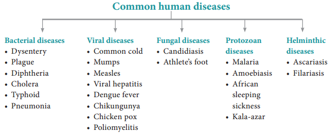
Diseases which are transmitted from one person to another are called infectious diseases or communicable diseases. Such disease causing organisms are called pathogens and are transmitted through air, water, food, physical contact and vectors.

The disease causing pathogen may be virus, bacteria, fungi, protozoan parasites, helminthic parasites, etc., Infectious diseases are common and everyone suffers from such diseases at some time or the other. Most of the bacterial diseases are curable but all viral diseases are not. Some infectious disease like AIDS may be fatal.

Non-infectious diseases are not transmitted from an infected person to a healthy person. In origin they may be genetic (cystic fibrosis), nutritional (vitamin deficiency diseases) and degenerative (arthritis, heart attack, stroke). Among non – infectious diseases, cancer is one of the major causes of death.

Bacterial and viral diseases

Bacterial diseases

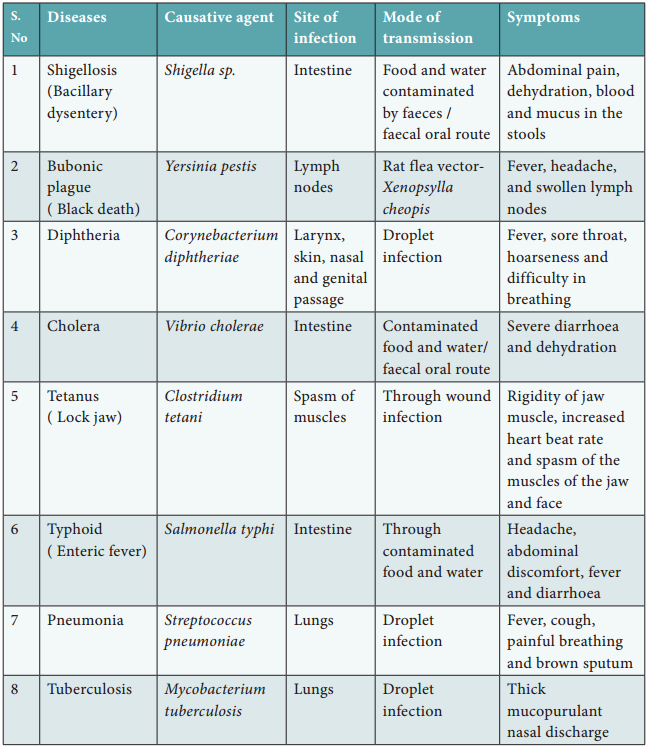
Though the number of bacterial species is very high, only a few bacteria are associated with human diseases and are called pathogenic bacteria. Such pathogens may emit toxins and affects the body. Common pathogenic bacteria and the bacterial diseases are given in table 7.1.
Common Diseases In Human Beings img 2

Bacteria spread through air, water or by inhaling the droplets/aerosols or even by sharing utensils, dresses with an infected person. Typhoid fever can be confirmed by Widal test.

Viral diseases

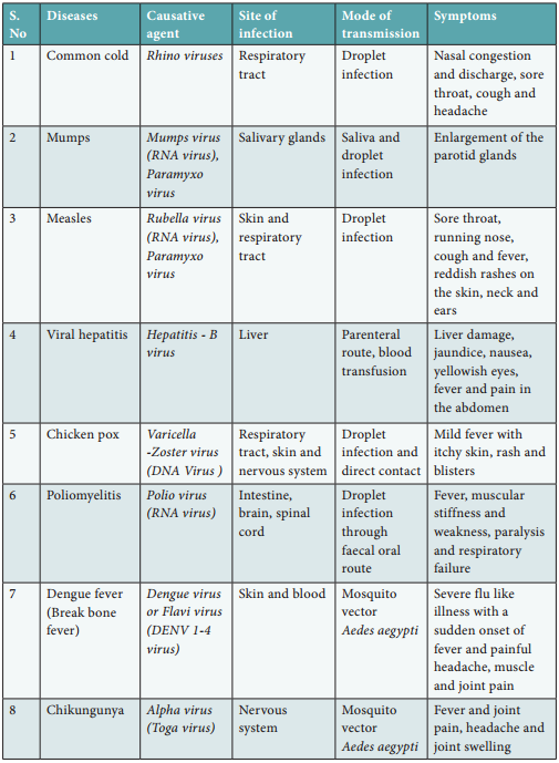
Viruses are the smallest intracellular obligate parasites, which multiply within living cells. Outside the living cells they cannot carry out the characteristics of a living organism. Viruses invade living cells, forcing the cells to create new viruses. The new viruses break out of the cell, killing it and invade other cells in the body, causing diseases in human beings. Rhino viruses cause one of the most infectious human ailment called the “Common cold”.

Viral diseases are generally grouped into four types on the basis of the symptoms produced in the body organs.

  • Pneumotropic diseases (respiratory tract infected by influenza)
  • Dermotropic diseases (skin and subcutaneous tissues affected by chicken pox and measles)
  • Viscerotropic diseases (blood and visceral organs affcted by yellow fever and dengue fever)
  • Neurotropic diseases (central nervous system affcted by rabies and polio).

Some common viral diseases of human beings are given in table 7.2.
Common Diseases In Human Beings img 3

Protozoan diseases

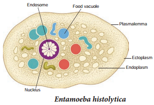
About 15 genera of protozoans live as parasites within the human body and cause diseases. Amoebiasis also called amoebic dysentery or amoebic colitis is caused by Entamoeba histolytica, which lives in the human large intestine and feeds on mucus and bacteria (Fig. 7.1).

Infective stage of this parasite is the trophozoite, which penetrates the walls of the host intestine (colon) and secretes histolytic enzymes causing ulceration, bleeding, abdominal pain and stools with excess mucus. Symptoms of amoebiasis can range from diarrhoea to dysentery with blood and mucus in the stool. House flies (Musca domestica) acts as a carrier for transmitting the parasite from contaminated faeces and water.
Common Diseases In Human Beings img 4

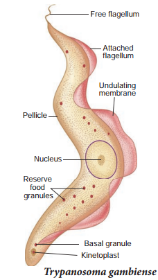
African sleeping sickness is caused by Trypanosoma species. Trypanosoma is generally transmitted by the blood sucking Tsetse flies. Three species of Trypanosoma cause sleeping sickness in man.

1. T. gambiense is transmitted by Glossina palpalis (Tsetse fly) and causes Gambian or Central African sleeping sickness (Fig. 7.2).
Common Diseases In Human Beings img 5

2. T. rhodesiense is transmitted by Glossina morsitans causing Rhodesian or East African sleeping sickness.

3. T. cruzi is transmitted by a bug called Triatoma megista and causes Chagas disease or American trypanosomiasis.

Kala – azar or visceral leishmaniasis is caused by Leishmania donovani, which is transmitted by the vector Phlebotomus (sand fly). Infection may occur in the endothelial cells, bone marrow, liver, lymph glands and blood vessels of the spleen. Symptoms of Kala azar are weight loss, anaemia, fever, enlargement of spleen and liver.

Malaria is caused by different types of Plasmodium species such as P. vivax, P. ovale, P. malariae and P. falciparum (Table 7.3). Plasmodium lives in the RBC of human in its mature condition it is called as trophozoite. It is transmited from one person to another by the bite of the infected female Anopheles mosquito.
Common Diseases In Human Beings img 6

Life cycle of Plasmodium

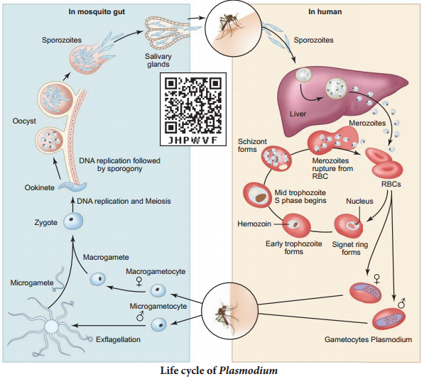
Plasmodium vivax is a digenic parasite, involving two hosts, man as the secondary host and female Anopheles mosquito as the primary host. The life cycle of Plasmodium involves three phases namely schizogony, gamogony and sporogony (Fig. 7.3).
Common Diseases In Human Beings img 7

The parasite first enters the human blood stream through the bite of an infected female Anopheles mosquito. As it feeds, the mosquito injects the saliva containing the sporozoites. The sporozoite within the blood stream immediately enters the hepatic cells of the liver. Further in the liver they undergo multiple asexual fission (schizogony) and produce merozoites. After being released from liver cells, the merozoites penetrate the RBC’s.

Inside the RBC, the merozoite begins to develop as unicellular trophozoites. The trophozoite grows in size and a central vacuole develops pushing them to one side of cytoplasm and becomes the signet ring stage. The trophozoite nucleus then divides asexually to produce the schizont. The large schizont shows yellowish – brown pigmented granules called Schuffers granules. The schizont divides and produces mononucleated merozoites.

Eventually the erythrocyte lyses, releasing the merozoites and haemozoin toxin into the blood stream to infect other erythrocytes. Lysis of red blood cells results in cycles of fever and other symptoms. This erythrocytic stage is cyclic and repeats itself approximately every 48 to 72 hours or longer depending on the species of Plasmodium involved. The sudden release of merozoites triggers an attack on the RBCs.

Occasionally, merozoites diffrentiate into macrogametocytes and microgametocytes. When these are ingested by a mosquito, they develop into male and female gametes respectively.

In the mosquito’s gut, the infected erythrocytes lyse and male and female gametes fertilize to form a diploid zygote called ookinete. The ookinete migrates to the mosquito’s gut wall and develop into an oocyte. The oocyte undergoes meiosis by a process called sporogony to form sporozoites. These sporozoites migrate to the salivary glands of the mosquito. The cycle is now completed and when the mosquito bites another human host, the sporozoites are injected and the cycle begins a new.

The pathological changes caused by malaria, affects not only the erythrocytes but also the spleen and other visceral organs. Incubation period of malaria is about 12 days.

The early symptoms of malaria are headache, nausea and muscular pain. The classic symptoms first develop with the synchronized release of merozoites, haemozoin toxin and erythrocyte debris into the blood stream resulting in malarial paroxysms – shivering chills, high fever followed by sweating. Fever and chills are caused partly by malarial toxins that induce macrophages to release tumour necrosis factor (TNF-α) and interleukin.

Prevention

It is possible to break the transmission cycle by killing the insect vector. Mosquitoes lay their eggs in water. Larvae hatch and develop in water but breathe air by moving to the surface. Oil can be sprayed over the water surface, to make it impossible for mosquito larvae and pupae to breathe.

Ponds, drainage ditches and other permanent bodies of water can be stocked with fishes such as Gambusia which feed on mosquito larvae. Preparations containing Bacillus thuringiensis can be sprayed to kill the mosquito larvae since it is not toxic to other forms of life. The best protection against malaria is to avoid being bitten by mosquito. People are advised to use mosquito nets, wire gauging of windows and doors to prevent mosquito bites.

In the 1950’s the World Health Organisation (WHO) introduced the Malaria eradication programme. This programme was not successful due to the resistance of Plasmodium to the drugs used to treat it and resistance of mosquitoes to DDT and other insecticides.

Fungal diseases

Fungi was recognized as a causative agent of human diseases much earlier than bacteria. Dermatomycosis is a cutaneous infection caused by fungi belonging to the genera Trichophyton, Microsporum and Epidermophyton.

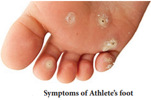
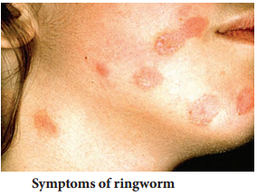
Ringworm is one of the most common fungal disease in humans (Fig. 7.4). Appearance of dry, scaly lesions on the skin, nails and scalp are the main symptoms of the disease. Heat and moisture help these fungi to grow and makes them to thrive in skin folds such as those in the groin or between the toes. Ringworms of the feet is known as Athlete’s foot caused by Tinea pedis (Fig. 7.5). Ringworms are generally acquired from soil or by using clothes, towels and comb used by infected persons.
Common Diseases In Human Beings img 8
Common Diseases In Human Beings img 9

Helminthic diseases

Helminthes are mostly endoparasitic in the gut and blood of human beings and cause diseases called helminthiasis. The two most prevalent helminthic diseases are Ascariasis and Filariasis.

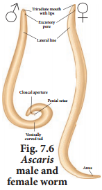
Ascaris is a monogenic parasite and exhibits sexual dimorphism. Ascariasis is a disease caused by the intestinal endoparasite Ascaris lumbricoides commonly called the round worms (Fig. 7.6). It is transmitted through ingestion of embryonated eggs through contaminated food and water.

Children playing in contaminated soils are also prone to have a chance of transfer of eggs from hand to mouth. The symptoms of the disease are abdominal pain, vomiting, headache, anaemia, irritability and diarrhoea. A heavy infection can cause nutritional deficiency and severe abdominal pain and causes stunted growth in children. It may also cause enteritis, hepatitis and bronchitis.
Common Diseases In Human Beings img 10

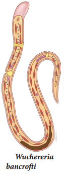
Filariasis is caused by Wuchereria bancroft, commonly called fiarial worm. It is found in the lymph vessels and lymph nodes of man (Fig. 7.7). Wuchereria bancroft is sexually dimorphic, viviparous and digenic. The life cycle is completed in two hosts, man and the female Culex mosquito The female fiarial worm gives rise to juveniles called microfiariae larvae.

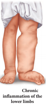
In the lymph glands, the juveniles develop into adults. The accumulation of the worms block the lymphatic system resulting in inflammation of the lymph nodes. In some cases, the obstruction of lymph vessels causes elephantiasis or fiariasis of the limbs, scrotum and mammary glands (Fig. 7.8).
Common Diseases In Human Beings img 11
Common Diseases In Human Beings img 12

Origin And Evolution Of Man

Learninsta presents the core concepts of Biology with high-quality research papers and topical review articles.

Origin And Evolution Of Man

Mammals evolved in the early Jurassic period, about 210 million years ago (mya). Hominid evolution occurred in Asia and Africa. Hominids proved that human beings are superior to other animals and efficient in making tools and culture.

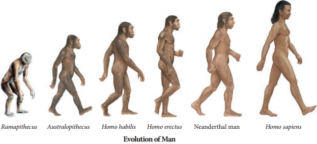
The earliest fossils of the prehistoric man like Ramapithecus and Sivapithecus lived some 14 mya and were derived from ape like Dryopithecus. Dryopithecus and Ramapithecus were hairy and walked like gorillas and chimpanzees. Ramapithecus is regarded as a possible ancester of Australopithecus and therefore of modern humans. They were vegetarians (Fig 6.10).
Origin And Evolution Of Man img 1

Australopithecus lived in East African grasslands about 5 mya and was called the Australian ape man. He was about 1.5 meters tall with bipedal locomotion, omnivorous, semi erect, and lived in caves. Low forehead, brow ridges over the eyes, protruding face, lack of chin, low brain capacity of about 350 – 450 cc, human like dentition, lumbar curve in the vertebral column were his distinguishing features.

Homo habilis lived about 2 mya. Their brain capacity was between 650 – 800cc, and was probably vegetarian. They had bipedal locomotion and used tools made of chipped stones.

Homo erectus the fist human like being was around 1.7 mya and was much closer to human in looks, skull was flitter and thicker than the modern man and had a large brain capacity of around 900 cc. Homo erectus probably ate meat Homo ergaster and Homo erectus were the first to leave Africa.

Neanderthal human was found in Neander Valley, Germany with a brain size of 1400 cc and lived between 34,000 – 1,00,000 years ago. They differ from the modern human in having semierect posture, flat cranium, sloping forehead, thin large orbits, heavy brow ridges, protruding jaws and no chin. They used animal hides to protect their bodies, knew the use of fie and buried their dead. They did not practice agriculture and animal domestication.

Cro-Magnon was one of the most talked forms of modern human found from the rocks of Cro-Magnon, France and is considered as the ancestor of modern Europeans. They were not only adapted to various environmental conditions, but were also known for their cave paintings, failures on flours and walls. Homo sapiens or modern human arose in Africa some 25,000 years ago and moved to other continents and developed into distinct races. They had a brain capacity of 1300 – 1600 cc. They started cultivating crops and domesticating animals.

Hardy Weinberg Principle

Learninsta presents the core concepts of Biology with high-quality research papers and topical review articles.

Hardy Weinberg Principle

In nature, populations are usually evolving such as the grass in an open meadow, wolves in a forest and bacteria in a person’s body are all natural populations. All of these populations are likely to be evolving some of their genes.

Evolution does not mean that the population is moving towards perfection rather the population is changing its genetic makeup over generations. For example in a wolf population, there may be a shift in the frequency of a gene variant for black fur than grey fur. Sometimes, this type of change is due to natural selection or due to migration or due to random events.

First we will see the set of conditions required for a population not to evolve. Hardy of UK and Weinberg of Germany stated that the allele frequencies in a population are stable and are constant from generation to generation in the absence of gene flow, genetic drift, mutation, recombination and natural selection. If a population is in a state of Hardy Weinberg equilibrium, the frequencies of alleles and genotypes or sets of alleles in that population will remain same over generations.

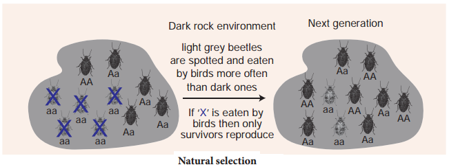
Evolution is a change in the allele frequencies in a population over time. Hence population in Hardy Weinberg is not evolving. Suppose we have a large population of beetles, (infinitely large) and appear in two colours dark grey (black) and light grey, and their colour is determined by ‘A’ gene. ‘AA’ and ‘Aa’ beetles are dark grey and ‘aa’ beetles are light grey. In a population let’s say that ‘A’ allele has frequency (p) of 0.3 and ‘a’ allele has a frequency (q ) of 0.7. Then p + q = 1.

If a population is in Hardy Weinberg equilibrium, the genotype frequency can be estimated by Hardy Weinberg equation.

(p + q)2 = p2 + 2pq + q2
p2 = frequency of AA
2pq = frequency of Aa
q2 = frequency of aa
p = 0.3, q = 0.7 then,
p3 = (0.3)2 = 0.09 = 9 % AA
2pq = 2(0.3) (0.7) = 0.42 = 42 % Aa
q2 = (0.7)2 0.49 = 49 % aa

Hence the beetle population appears to be in Hardy – Weinberg equilibrium. When the beetles in Hardy – Weinberg equilibrium reproduce, the allele and genotype frequency in the next generation would be: Let’s assume that the frequency of ‘A’ and ‘a’ allele in the pool of gametes that make the next generation would be the same, then there would be no variation in the progeny.

The genotype frequencies of the parent appears in the next generation. (i.e. 9% AA, 42% Aa and 49% aa). If we assume that the beetles mate randomly (selection of male gamete and female gamete in the pool of gametes), the probability of getting the offspring genotype depends on the genotype of the combining parental gametes.

Hardy Weinberg’s assumptions include No mutation – No new alleles are generated by mutation nor the genes get duplicated or deleted.

Random mating:
Every organism gets a chance to mate and the mating is random with each other with no preferences for a particular genotype.

No gene flow:
Neither individuals nor their gametes enter (immigration) or exit (emigration) the population.

Very large population size:
The population should be infinite in size.

No natural selection:
All alleles are fit to survive and reproduce. If any one of these assumptions were not met, the population will not be in HardyWeinberg equilibrium. Only if the allele frequencies changes from one generation to the other, evolution will take place.

Mechanism Of Evolution

Learninsta presents the core concepts of Biology with high-quality research papers and topical review articles.

Mechanism Of Evolution

Mechanism of evolution

Microevolution (evolution on a small scale) refers to the changes in allele frequencies within a population. Allele frequencies in a population may change due to four fundamental forces of evolution such as natural selection, genetic drift mutation and gene flow.

Natural selection

It occurs when one allele (or combination of alleles of differences) makes an organism more or less fit to survive and reproduce in a given environment. If an allele reduces fitness, its frequencies tend to drop from one generation to the next.

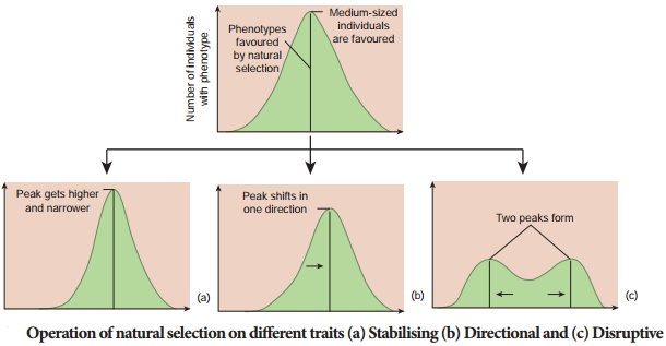
The evolutionary path of a given gene i.e., how its allele’s change in frequency in the population across generation, may result from several evolutionary mechanisms acting at once. For example, one gene’s allele frequencies might be modified by both gene flow and genetic drift for another gene, mutation may produce a new allele, that is favoured by natural selection (Fig. 6.6).
Mechanism Of Evolution img 1

Selection

There are mainly three types of natural selection

(i) Stabilising Selection (centipetal selection):

This type of selection operates in a stable environment (Fig. 6.7 a). The organisms with average phenotypes survive whereas the extreme individuals from both the ends are eliminated. There is no speciation but the phenotypic stability is maintained within the population over generation.

For example, measurements of sparrows that survived the storm clustered around the mean, and the sparrows that failed to survive the storm clustered around the extremes of the variation showing stabilizing selection.
Mechanism Of Evolution img 2

(ii) Directional Selection:

The environment which undergoes gradual change is subjected to directional selection (Fig. 6.7 b). This type of selection removes the individuals from one end towards the other end of phenotypic distribution. For example, size differences between male and female sparrows. Both male and female look alike externally but differ in body weight. Females show directional selection in relation to body weight.

(iii) Disruptive Selection (centrifugal selection):

When homogenous environment changes into heterogenous environment this type of selection is operational (Fig. 6.7 c). The organisms of both the extreme phenotypes are selected whereas individuals with average phenotype are eliminated.

This results in splitting of the population into sub population/species. This is a rare form of selection but leads to formation of two or more different species. It is also called adaptive radiation. E.g. Darwin’s fichesbeak size in relation to seed size inhabiting Galapagos islands.

Group selection and sexual selection are other types of selection. The two major group selections are Altrusim and Kin selection.

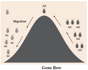
Gene flow

Movement of genes through gametes or movement of individuals in (immigration) and out (emigration) of a population is referred to as gene flow. Organisms and gametes that enter the population may have new alleles or may bring in existing alleles but in different proportions than those already in the population. Gene flow can be a strong agent of evolution (Fig 6.8).
Mechanism Of Evolution img 3

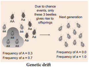
Genetic drift / Sewall Wright Effect

Genetic drift is a mechanism of evolution in which allele frequencies of a population change over generation due to chance (sampling error). Genetic drift occurs in all population sizes, but its effects are strong in a small population (Fig. 6.9).

It may result in a loss of some alleles (including benefiial ones) and fitation of other alleles. Genetic drift can have major effects, when the population is reduced in size by natural disaster due to bottle neck effect or when a small group of population splits from the main population to form a new colony due to founder’s effect.
Mechanism Of Evolution img 4

Mutation

Although mutation is the original source of all genetic variation, mutation rate for most organisms is low. Hence new mutations on an allele frequencies from one generation to the next is usually not large.

Theories Of Biological Evolution

Learninsta presents the core concepts of Biology with high-quality research papers and topical review articles.

Theories Of Biological Evolution

Lamarck’s theory

Jean Baptiste de Lamarck, was the first to postulate the theory of evolution in his famous book ‘Philosophie Zoologique’ in the year 1809. The two principles of Lamarckian theory are:

(i) The theory of use and disuse – Organs that are used often will increase in size and those that are not used will degenerate. Neck in giraffe is an example of use and absence of limbs in snakes is an example for disuse theory.

(ii) The theory of inheritance of acquired characters – Characters that are developed during the life time of an organism are called acquired characters and these are then inherited.

The main objection to Lamarckism

Lamarck’s “Thory of Acquired characters” was disproved by August Weismann who conducted experiments on mice for twenty generations by cutting their tails and breeding them. All mice born were with tail. Weismann proved his germplasm theory that change in the somatoplasm will not be transferred to the next generation but changes in the germplasm will be inherited.

Neo-Lamarckism

The followers of Lamarck (Neo-Lamarckists) like Cope, Osborn, Packard and Spencer tried to explain Lamarck’s theory on a more scientific basis. They considered that adaptations are universal. Organisms acquire new structures due to their adaptations to the changed environmental conditions. They argued that external conditions stimulate the somatic cells to produce certain ‘secretions’ which reach the sex cells through the blood and bring about variations in the offspring.

Darwin’s theory of Natural Selection

Charles Darwin explained the theory of evolution in his book ‘The Origin of Species by Natural Selection’. During his journey around the Earth, he made extensive observations of plants and animals. He noted a huge variety and remarkable similarities among organisms and their adaptive features to cope up to their environment. He proved that fitest organisms can survive and leave more progenies than the unfit ones through natural selection.

Darwin’s theory was based on several facts, observations and influences. They are:

1. Over production (or) prodigality of production:

All living organisms increase their population in larger number. For example, Salmon fish produces about 28 million eggs during breeding season and if all of them hatch, the seas would be filed with salmon in few generations. Elephant, the slowest breeder that can produce six young ones in its life time can produce 6 million descendants at the end of 750 years in the absence of any check.

2. Struggle for existence:

Organisms struggle for food, space and mate. As these become a limiting factor, competition exists among the members of the population. Darwin denoted struggle for existence in three ways – Intra specific struggle between the same species for food, space and mate. Inter specific struggle with different species for food and space. Struggle with the environment to cope with the climatic variations, flood, earthquakes, drought, etc.,

3. Universal occurrence of variations

No two individuals are alike. There are variations even in identical twins. Even the children born of the same parents differ in colour, height, behavior, etc., The useful variations found in an organism help them to overcome struggle and such variations are passed on to the next generation.

4. Origin of species by Natural Selection

According to Darwin, nature is the most powerful selective force. He compared origin of species by natural selection to a small isolated group. Darwin believed that the struggle for existence resulted in the survival of the fittest. Such organisms become better adapted to the changed environment.

Objections to Darwinism

Some objections raised against Darwinism were –

  • Darwin failed to explain the mechanism of variation.
  • Darwinism explains the survival of the fittest but not the arrival of the fittest.
  • He focused on small fluctuating variations that are mostly non-heritable.
  • He did not distinguish between somatic and germinal variations.
  • He could not explain the occurrence of vestigial organs, over specialization of some organs like large tusks in extinct mammoths, oversized antlers in the extinct Irish deer, etc.,

Neo Darwinism

Neo Darwinism is the interpretation of Darwinian evolution through Natural Selection as it has been modified since it was proposed. New facts and discoveries about evolution have led to modifications of Darwinism and is supported by Wallace, Heinrich, Haeckel, Weismann and Mendel. This theory emphasizes the change in the frequency of genes in population arises due to mutation, variation, isolation and Natural
selection.

Mutation theory

Hugo de Vries put forth the Mutation theory. Mutations are sudden random changes that occur in an organism that is not heritable. De Vries carried out his experiments in the Evening Primrose plant (Oenothera lamarckiana) and observed variations in them due to mutation.

According to de Vries, sudden and large variations were responsible for the origin of new species whereas Lamarck and Darwin believed in gradual accumulation of all variations as the causative factors in the origin of new species. Hugo de Vries believed that Mutations are random and directionless, but Darwinian variations are small and directional.

Salient features of Mutation Theory

  • Mutations or discontinuous variation are transmitted to other generations.
  • In naturally breeding populations, mutations occur from time to time.
  • There are no intermediate forms, as they are fully fledged.
  • They are strictly subjected to natural selection.

Modern synthetic theory

Sewell Wright, Fisher, Mayer, Huxley, Dobzhansky, Simpson and Haeckel explained Natural Selection in the light of Post-Darwinian discoveries. According to this theory gene mutations, chromosomal mutations, genetic recombinations, natural selection and reproductive isolation are the fie basic factors involved in the process of organic evolution.

(i) Gene mutation

Refers to the changes in the structure of the gene. It is also called gene/point mutation. It alters the phenotype of an organism and produces variations in their offspring.

(ii) Chromosomal mutation

Refers to the changes in the structure of chromosomes due to deletion, addition, duplication, inversion or translocation. This too alters the phenotype of an organism and produces variations in their offspring.

(iii) Genetic recombination

Is due to crossing over of genes during meiosis. This brings about genetic variations in the individuals of the same species and leads to heritable variations.

(iv) Natural selection

Does not produce any genetic variations but once such variations occur it favours some genetic changes while rejecting others (driving force of evolution).

(v) Reproductive Isolation

Helps in preventing interbreeding between related organisms.

Evolution by anthropogenic sources

Natural Selection (Industrial melanism)

Natural selection can be explained clearly through industrial melanism. Industrial melanism is a classical case of Natural selection exhibited by the peppered month, Biston betularia. These were available in two colours, white and black. Before industrialization peppered moth both white and black coloured were common in England. Pre-industrialization witnessed white coloured background of the wall of the buildings hence the white coloured months escaped from their predators.

Post industrialization, the tree trunks became dark due to smoke and soot let out from the industries. The black moths camouflaged on the dark bark of the trees and the white moths were easily identified by their predators. Hence the dark coloured month population was selected and their number increased when compared to the white months. Nature offered positive selection pressure to the black coloured months. The above proof shows that in a population, organisms that can adapt will survive and produce more progenies
resulting in increase in population through natural selection.

Artificial selection is a byproduct of human exploitation of forests, oceans and fisheries or the use of pesticides, herbicides or drugs. For hundreds of years humans have selected various types of dogs, all of which are variants of the single species of dog. If human beings can produce new varieties in short period, then “nature” with its vast resources and long duration can easily produce new species by selection.

Adaptive Radiation

The evolutionary process which produces new species diverged from a single ancestral form becomes adapted to newly invaded habitats is called adaptive radiation. Adaptive radiations are best exemplified in closely related groups that have evolved in relatively short time.

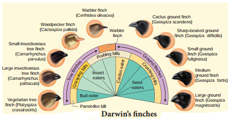
Darwin’s finches and Australian marsupials are best examples for adaptive radiation. When more than one adaptive radiation occurs in an isolated geographical area, having the same structural and functional similarity is referred to as convergent evolution.

Darwin’s fiches

Their common ancestor arrived on the Galapagos about 2 million years ago. During that time, Darwin’s finches have evolved into 14 recognized species differing in body size, beak shape and feeding behavior. Changes in the size and form of the beak have enabled different species to utilize different food resources such as insects, seeds, nectar from cactus flowers and blood from iguanas, all driven by Natural selection. Fig. 6.5 represents some of the finches observed by Darwin.
Theories Of Biological Evolution img 1

Genetic variation in the ALX1 gene in the DNA of Darwin finches is associated with variation in the beak shape. Mild mutation in the ALX1 gene leads to phenotypic change in the shape of the beak of the Darwin finches. Marsupials in Australia and placental mammals in North America are two subclasses of mammals they have adapted in similar way to a particular food resource, locomotory skill or climate.

They were separated from the common ancestor more than 100 million year ago and each lineage continued to evolve independently. Despite temporal and geographical separation, marsupials in Australia and placental mammals in North America have produced varieties of species living in similar habitats with similar ways of life. Their overall resemblance in shape, locomotory mode, feeding and foraging are superimposed upon different modes of reproduction. This feature reflects their distinctive evolutionary relationships.

Over 200 species of marsupials live in Australia along with many fewer species of placental mammals. The marsupials have undergone adaptive radiation to occupy the diverse habitats in Australia, just as the placental mammals have radiated across North America.

Evidences For Biological Evolution

Learninsta presents the core concepts of Biology with high-quality research papers and topical review articles.

Evidences For Biological Evolution

Paleontological evidences

Paleontology is the study of prehistoric life through fossils. Fossils are described as the true witnesses of evolution or documents of various geological strata of evolution. Fossilization is the process by which plant and animal remains are preserved in sedimentary rocks. They fall under three main categories.

(i) Actual remains:

The original hard parts such as bones, teeth or shells are preserved as such in the earth’s atmosphere. This is the most common method of fossilization. When marine animals die, their hard parts such as bones, shells, etc., are covered with sediments and are protected from further deterioration. They get preserved as such as they are preserved in vast ocean; the salinity in them prevents decay.

The sediments become hardened to form definite layers or strata. For example, Woolly Mammoth that lived 22 thousand years ago were preserved in the frozen coast of Siberia as such. Several human beings and animals living in the ancient city of Pompeii were preserved intact by volcanic ash which gushed out from Mount Vesuvius.

(ii) Petrifaction:

When animals die the original portion of their body may be replaced molecule for molecule by minerals and the original substance being lost through disintegration. This method of fossilization is called petrifaction. The principle minerals involved in this type fossilization are iron pyrites, silica, calcium carbonate and bicarbonates of calcium and magnesium.

(iii) Natural moulds and casts:

Even after disintegration, the body of an animal might leave indelible impression on the soft mud which later becomes hardened into stones. Such impressions are called moulds. The cavities of the moulds may get filled up by hard minerals and get fossilized, which are called casts.

Hardened faecal matter termed as coprolites occur as tiny pellets. Analysis of the coprolites enables us to understand the nature of diet the pre-historic animals thrived on.

Evidences from comparative anatomy

Similarities in structure between groups of organisms are accepted as indicators of relationship. For example, a comparative study of the forelimbs of different vertebrates exhibits a fundamental plan of similarity in structure. These relationships can be studied under homologous organs, analogous organs, vestigial organs, connecting links and atavistic organs.

Homologous structures

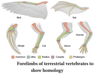
In vertebrates, comparative anatomical studies reveal a basic plan in various structures such as fore limbs and hind limbs. Fore limbs of vertebrates exhibit anatomical similarity with each other and is made of similar bones such as humerus, radius, ulna, carpals, metacarpals and phalanges.

Structures which are similar in origin but perform different functions are called homologous structures that brings about divergent evolution (Fig 6.2).
Evidences For Biological Evolution img 1

Similarly the thorn of Bougainvillea and the tendrils of Curcurbita and Pisum sativum represent homology. The thorn in former is used as a defence mechanism from grazing animals and the tendrils of latter is used as a support for climbing.

Analogous structures

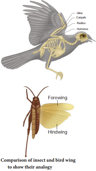
Organs having different structural patterns but similar function are termed as analogous structures. For example, the wings of birds and insects are different structurally but perform the same function of flight that brings about convergent evolution (Fig. 6.3).
Evidences For Biological Evolution img 2

Other examples of analogous organs include the eyes of the Octopus and of mammals and the flippers of Penguins and Dolphins. Root modification in sweet potato and stem modification in potato are considered as analogous organs. Both of these plants have a common function of storage of food.

Vestigial organs

Structures that are of no use to the possessor, and are not necessary for their existence are called vestigial organs. Vestigial organs may be considered as remnants of structures which were well developed and functional in the ancestors, but disappeared in course of evolution due to their nonutilization. Human appendix is the remnant of caecum which is functional in the digestive tract of herbivorous animals like rabbit.

Cellulose digestion takes place in the caecum of these animals. Due to change in the diet containing less cellulose, caecum in human became functionless and is reduced to a vermiform appendix, which is vestigial. Other examples of vestigial organs in human beings include coccyx, wisdom teeth, ear muscles, body hair, mammae in male, nictitating membrane of the eye, etc.

Connecting link

The organisms which possess the characters of two different groups (transitional stage) are called connecting links. Example Peripatus (connecting link between Annelida and Arthropoda), Archeopteryx (connecting link between Reptiles and Aves).

Atavistic organs

Sudden appearance of vestigial organs in highly evolved organisms is called atavistic organs. Example, presence of tail in a human baby is an atavistic organ.

Embryological evidences

Embryology deals with the study of the development of individual from the egg to the adult stage. A detailed study of the embryonic development of different forms makes us to think that there is a close resemblance during development.

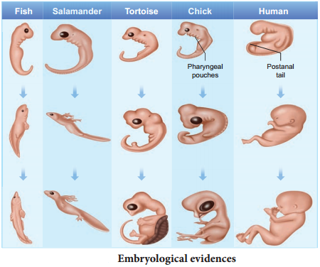
The development of heart in all vertebrates follows the same pattern of development as a pair of tubular structures that later develop into two chambered heart in fishes, three chambered in amphibians and in most reptiles and four chambered in crocodiles, birds and mammals; indicating a common ancestry for all the vertebrates, Hence scientists in the 19th century concluded that higher animals during their embryonic development pass through stages of lower animals (ancestors). Ernst Von Haeckel, propounded the “biogenetic law or theory of recapitulation” which states that the life history of an individual (ontogeny) briefly repeats or recapitulates the evolutionary history of the race (phylogeny).

In other words “Ontogeny recapitulates Phylogeny”. The embryonic stages of a higher animal resemble the adult stage of its ancestors. Appearance of pharyngeal gill slits, yolk sac and the appearance of tail in human embryos are some of the examples (Fig. 6.4). The biogenetic law is not universal and it is now thought that animals do not recapitulate the adult stage of any ancestors. The human embryo recapitulates the embryonic history and not the adult history of the organisms.
Evidences For Biological Evolution img 3

The comparative study of the embryo of different animals shows structural similarities among themselves. The embryos of fish, salamander, tortoise, chick and human start life as a single cell, the zygote, and undergo cleavage to produce the blastula, change to gastrula and are triploblastic. This indicates that all the above said animals have evolved from a common ancestor.

Molecular evidences

Molecular evolution is the process of change in the sequence composition of molecules such as DNA, RNA and proteins across generations. It uses principles of evolutionary biology and population genetics to explain patterns in the changes of molecules.

One of the most useful advancement in the development of molecular biology is proteins and other molecules that control life processes are conserved among species. A slight change that occurs over time in these conserved molecules (DNA, RNA and protein) are often called molecular clocks. Molecules that have been used to study evolution are cytochrome c (respiratory pathway) and rRNA (protein synthesis).

Biological Evolution

Learninsta presents the core concepts of Biology with high-quality research papers and topical review articles.

Biological Evolution

Formation of protobionts

Abiotically produced molecules can spontaneously self assemble into droplets that enclose a watery solution and maintain a chemical environment different from their surroundings. Scientists call these spheres as ‘protobionts’. Liposomes are lipids in a solution that can self assemble into a lipid bilayer. Some of the proteins inside the liposomes acquired the properties of enzymes resulting in fast multiplication of molecules.

The coacervates with nucleoprotein and nutrients had a limiting surface membrane that had the characters of a virus or free living genes. Sub sequently number of genes united to form ‘proto viruses’ somewhat similar to present day viruses. Two major cell types that appeared during this time were significant. One form of the earliest cell contained clumps of nucleoproteins embedded in the cell substance.

Such cells were similar to the Monera. They are considered as ancestral to the modern bacteria and blue green algae. The other form of earliest cells contained nucleoprotein clumps that condensed into a central mass surrounded by a thin membrane.

This membrane separated nucleoproteins from the cell substances. Such cells were referred to as Protista. When the natural sources of food in the ocean declined in course of time the ancestors of Monera and Protista had to evolve different methods for food procurement. These may be summarized as parasitism, saprophytism, predator or animalism and chemosynthesis or photosynthesis. When the number of photosynthetic organisms increased there was an increase in the free O2 in the sea and atmosphere.

CH4 + 2O2 → CO2 + 2H2O
4NH3 + 3O2 → 2N2 + 6H2O

The atmospheric oxygen combined with methane and ammonia to form CO2 and free nitrogen. The presence of the free O2 brought about the evolution of aerobic respiration which could yield large amounts of energy by oxidation of food stuffs. Thus Prokaryotes and Eukaryotes evolved.

Experimental approach to the origin of life

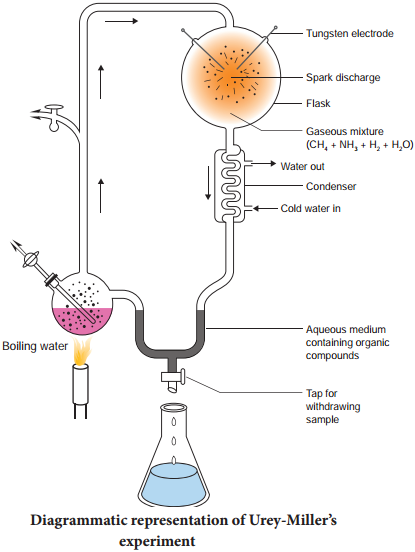
Urey and Miller (1953), paved way for understanding the possible synthesis of organic compounds that led to the appearance of living organisms is depicted in the Fig. 6.1. In their experiment, a mixture of gases was allowed to circulate over electric discharge from an tungsten electrode. A small flask was kept boiling and the steam emanating from it was made to mix with the mixture of gases (ammonia, methane and hydrogen) in the large chamber that was connected to the boiling water.

The steam condensed to form water which ran down the ‘U’ tube. Experiment was conducted continuously for a week and the liquid was analysed. Glycine, alanine, beta alanine and aspartic acid were identified. This Miller’s experiments had an insight as to the possibility of abiogenetic synthesis of large amount of variety of organic compounds in nature from a mixture of sample gases in which the only source of carbon was methane. Later in similar experiments, formation of all types of amino acids, and nitrogen bases were noticed.
Biological Evolution img 1

Geological Time Scale

Learninsta presents the core concepts of Biology with high-quality research papers and topical review articles.

Geological Time Scale

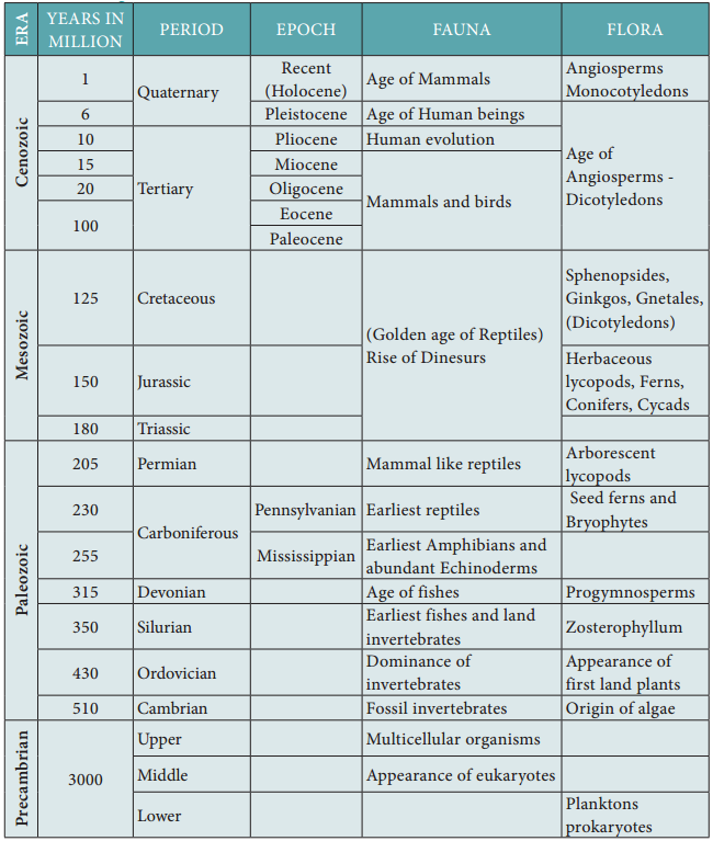
The duration of the earth’s history has been divided into eras that include the Paleozoic, Mesozoic, and Cenozoic. Recent eras are further divided into periods, which are split into epochs. The geological time scale with the duration of the eras and periods with the dominant forms of life is shown in Table 6.1.

The Paleozoic era is characterized by abundance of fossils of marine invertebrates. Towards the later half, other vertebrates (marine and terrestrial) except birds and mammals appeared.

The six periods of Paleozoic era in order from oldest to the youngest are Cambrian (Age of invertebrates), Ordovician (fresh water fihes, Ostracoderms, various types of Molluscs), Silurian (origin of fishes), Devonian (Age of fihes, many types of fishes such as lung fishes, lobe fined fishes and ray fined fihes), Mississippian (earliest amphibians, Echinoderms), Pennsylvanian (earliest reptiles), Permian (mammal like reptiles).

Geological Time Scale img 1

Mesozoic era (dominance of reptiles) called the Golden age of reptiles, is divided into three periods namely, Triassic (origin of egg laying mammals), Jurassic (Dinosaurs were dominant on the earth, fossil bird – Archaeopteryx) and Cretaceous (extinction of toothed birds and dinosaurs, emergence of modern birds).
Cenozoic era (Age of mammals) is sub divided into two periods namely Tertiary and Quaternary.

Tertiary period is characterized by abundant mammalian fauna. This period is subdivided into five epochs namely, Paleocene (placental mammals, Eocene (Monotremes except duck billed Platypus and Echidna, hoofed mammals and carnivores), Oligocene (higher placental mammals appeared), Miocene (origin of first man like apes) and Pliocene (origin of man from man like apes).

Quaternary period witnessed decline of mammals and beginning of human social life. The age of fossils can be determined using two methods namely, relative dating and absolute dating. Relative dating is used to determine a fossil by comparing it to similar rocks and fossils of known age. Absolute dating is used to determine the precise age of a fossil by using radiometric dating to measure the decay of isotopes.

Origin Of Life – Evolution Of Life Forms

Learninsta presents the core concepts of Biology with high-quality research papers and topical review articles.

Origin Of Life – Evolution Of Life Forms

Theory of special creation states that life was created by a supernatural power, respectfully referred to as “God”. According to Hinduism, Lord Brahma created the Earth. Christianity, Islam and most religions believe that God created the universe, the plants and the animals.

According to the theory of spontaneous generation or Abiogenesis, living organisms originated from non-living materials and occurred through stepwise chemical and molecular evolution over millions of years. Thomas Huxley coined the term abiogenesis.

Big bang theory explains the origin of universe as a singular huge explosion in physical terms. The primitive earth had no proper atmosphere, but consisted of ammonia, methane, hydrogen and water vapour. The temperature of the earth was extremely high.

UV rays from the sun split up water molecules into hydrogen and oxygen. Gradually the temperature cooled and the water vapour condensed to form rain. Rain water filled all the depressions to form water bodies. Ammonia and methane in the atmosphere combined with oxygen to form carbon-dioxide and other gases.

According to the theory of biogenesis life arose from pre-existing life. The term biogenesis also refers to the biochemical process of production of living organisms This term was coined by Henry Bastian.

According to the theory of chemical evolution primitive organisms in the primordial environment of the earth evolved spontaneously from inorganic substances and physical forces such, as lightning, UV radiations, volcanic activities, etc.,., Oparin (1924) suggested that the organic compounds could have undergone a series of reactions leading to more complex molecules. He proposed that the molecules formed colloidal aggregates or ‘coacervates’ in an aqueous environment.

The coacervates were able to absorb and assimilate organic compounds from the environment. Haldane (1929) proposed that the primordial sea served as a vast chemical laboratory powered by solar energy. The atmosphere was oxygen free and the combination of CO2, NH3 and UV radiations gave rise to organic compounds. The sea became a ‘hot’ dilute soup containing large populations of organic monomers and polymers.

They envisaged that groups of monomers and polymers acquired lipid membranes and further developed into the first living cell. Haldane coined the term prebiotic soup and this became the powerful symbol of the Oparin-Haldane view on the origin of life (1924-1929). Oparin and Haldane independently suggested that if the primitive atmosphere was reducing and if there was appropriate supply of energy such as lightning or UV light then a wide range of organic compounds can be synthesized.

DNA Finger Printing Technique

Learninsta presents the core concepts of Biology with high-quality research papers and topical review articles.

DNA Finger Printing Technique

The DNA fingerprinting technique was first developed by Alec Jeffreys in 1985 (Recipient of the Royal Society’s Copley Medal in 2014). Each of us have the same chemical structure of DNA. But there are millions of differences in the DNA sequence of base pairs. This makes the uniqueness among us so that each of us except identical twins is different from each other genetically.

The DNA of a person and finger prints are unique. There are 23 pairs of human chromosomes with 1.5 million pairs of genes. It is a well known fact that genes are segments of DNA which differ in the sequence of their nucleotides.

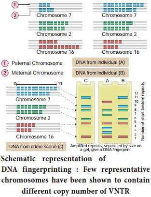
Not all segments of DNA code for proteins, some DNA segments have a regulatory function, while others are intervening sequences (introns) and still others are repeated DNA sequences. In DNA fingerprinting, short repetitive nucleotide sequences are specific for a person. These nucleotide sequences are called as variable number tandem repeats (VNTR).The VNTRs of two persons generally show variations and are useful as genetic markers.

DNA figer printing involves identifying diffrences in some specific regions in DNA sequence called repetitive DNA, because in these sequences, a small stretch of DNA is repeated many times. These repetitive DNA are separated from bulk genomic DNA as different peaks during density gradient centrifugation. The bulk DNA forms a major peak and the other small peaks are referred to as satellite DNA.

Depending on base composition (A : T rich or G : C rich), length of segment and number of repetitive units, the satellite DNA is classified into many sub categories such as micro-satellites, minisatellites, etc., These sequences do not code for any proteins, but they form a large portion of human genome.

These sequences show high degree of polymorphism and form the basis of DNA figerprinting (Fig. 5.15). DNA isolated from blood, hair, skin cells, or other genetic evidences lef at the scene of a crime can be compared through VNTR patterns, with the DNA of a criminal suspect to determine guilt or innocence. VNTR patterns are also useful in establishing the identity of a homicide victim, either from DNA found as evidence or from the body itself.
DNA Finger Printing Technique img 1

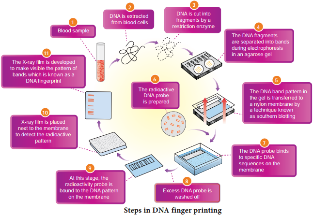
The Steps in DNA Fingerprinting technique is depicted in Fig. 5.16.

1. Extraction of DNA

The process of DNA fingerprinting starts with obtaining a sample of DNA from blood, semen, vaginal fluids, hair roots, teeth, bones, etc.,

2. Polymerase chain reaction (PCR)

In many situations, there is only a small amount of DNA available for DNA figerprinting. If needed many copies of the DNA can be produced by PCR (DNA amplifiation).

3. Fragmenting DNA

DNA is treated with restriction enzymes which cut the DNA into smaller fragments at specific sites.
DNA Finger Printing Technique img 2

4. Separation of DNA by electrophoresis

During electrophoresis in an agarose gel, the DNA fragments are separated into bands of different sizes. The bands of separated DNA are sieved out of the gel using a nylon membrane (treated with chemicals that allow for it to break the hydrogen bonds of DNA so there are single strands).

5. Denaturing DNA

The DNA on gels is denatured by using alkaline chemicals or by heating.

6. Blotting

The DNA band pattern in the gel is transferred to a thin nylon membrane placed over the ‘size fractionated DNA strand’ by Southern blotting.

7. Using probes to identify specific DNA

A radioactive probe (DNA labeled with a radioactive substance) is added to the DNA bands. The probe attaches by base pairing to those restriction fragments that are complementary to its sequence. The probes can also be prepared by using either ‘florescent substance’ or ‘radioactive isotopes’.

8. Hybridization with probe

After the probe hybridizes and the excess probe washed off a photographic film is placed on the membrane containing ‘DNA hybrids’.

9. Exposure on fim to make a genetic/DNA Fingerprint

The radioactive label exposes the film to form an image (image of bands) corresponding to specific DNA bands. The thick and thin dark bands form a pattern of bars which constitutes a genetic fingerprint.

Application of DNA figer printing

Forensic analysis:

1. It can be used in the identification of a person involved in criminal activities, for settling paternity or maternity disputes, and in determining relationships for immigration purposes.

2. Pedigree analysis – inheritance pattern of genes through generations and for detecting inherited diseases.

3. Conservation of wild life – protection of endangered species. By maintaining DNA records for identification of tissues of the dead endangered organisms.

4. Anthropological studies – It is useful in determining the origin and migration of human populations and genetic diversities.

Human Genome Project (HGP)

Learninsta presents the core concepts of Biology with high-quality research papers and topical review articles.

Human Genome Project (HGP)

The international human genome project was launched in the year 1990. It was a mega project and took 13 years to complete. The human genome is about 25 times larger than the genome of any organism sequenced to date and is the first vertebrate genome to be completed. Human genome is said to have approximately 3 × 10⁹ bp. HGP was closely associated with the rapid development of a new area in biology called bioinformatics.

Goals and methodologies of Human Genome Project

The main goals of Human Genome Project are as follows

  • Identify all the genes (approximately 30000) in human DNA.
  • Determine the sequence of the three billion chemical base pairs that makeup the human DNA.
  • To store this information in databases.
  • Improve tools for data analysis.
  • Transfer related technologies to other sectors, such as industries.
  • Address the ethical, legal and social issues (ELSI) that may arise from the project.

The methodologies of the Human Genome Project involved two major approaches. One approach was focused on identifying all the genes that are expressed as RNA (ESTS – Expressed Sequence Tags). The other approach was sequence annotation. Here, sequencing the whole set of genome was taken, that contains all the coding and non-coding sequences and later assigning different regions in the sequences with functions.

For sequencing, the total DNA from a cell is isolated and converted into random fragments of relatively smaller sizes and cloned in suitable hosts using specialized vectors. This cloning results in amplification of pieces of DNA fragments so that it could subsequently be sequenced with ease.

Bacteria and yeast are two commonly used hosts and these vectors are called as BAC (Bacterial Artificial Chromosomes) and YAC (Yeast Artificial Chromosomes). The fragments are sequenced using automated DNA sequencers (developed by Frederick Sanger).

The sequences are then arranged based on few overlapping regions, using specialized computer based programs. These sequences were subsequently annotated and are assigned to each chromosome. The genetic and physical maps on the genome are assigned using information on polymorphism of restriction endonuclease recognition sites and some repetitive DNA sequences, called microsatellites.

The latest method of sequencing even longer fragments is by a method called Shotgun sequencing using super computers, which has replaced the traditional sequencing methods.

Salient features of Human Genome Project:

  • The human genome contains 3 billion nucleotide bases.
  • An average gene consists of 3000 bases, the largest known human gene being dystrophin with 2.4 million bases.
  • Genes are distributed over 24 chromosomes. Chromosome 19 has the highest gene density. Chromosome 13 and Y chromosome have lowest gene densities.
  • The chromosomal organization of human genes shows diversity.
  • There may be 35000-40000 genes in the genome and almost 99.9 nucleotide bases are exactly the same in all people.
  • Functions for over 50 percent of the discovered genes are unknown.
  • Less than 2 percent of the genome codes for proteins.
  • Repeated sequences make up very large portion of the human genome. Repetitive sequences have no direct coding functions but they shed light on chromosome structure, dynamics and evolution (genetic diversity).
  • Chromosome 1 has 2968 genes whereas chromosome ’Y’ has 231 genes.
  • Scientists have identified about 1.4 million locations where single base DNA differences (SNPs – Single nucleotidepolymorphism – pronounce as ‘snips’) occur in humans.
  • Identification of ‘SNIPS’ is helpful in finding chromosomal locations for disease associated sequences and tracing human history.

Applications and future challenges

The mapping of human chromosomes is possible to examine a person’s DNA and to identify genetic abnormalities. This is extremely useful in diagnosing diseases and to provide genetic counselling to those planning to have children.

This kind of information would also create possibilities for new gene therapies. Besides providing clues to understand human biology, learning about non-human organisms, DNA sequences can lead to an understanding of their natural capabilities that can be applied towards solving challenges in healthcare, agriculture, energy production and environmental remediation.

A new era of molecular medicine, characterized by looking into the most fundamental causes of disease than treating the symptoms will be an important advantage.

  • Once genetic sequence becomes easier to determine, some people may attempt to use this information for profit or for political power.
  • Insurance companies may refuse to insure people at ‘genetic risk’ and this would save the companies the expense of future medical bills incurred by ‘less than perfect’ people.
  • Another fear is that attempts are being made to “breed out” certain genes of people from the human population in order to create a ‘perfect race’.