Electron Microscope – Definition, Principle, Parts, Uses

Learninsta presents the core concepts of Microbiology with high-quality research papers and topical review articles.

Electron Microscope – Definition, Principle, Parts, Uses

Examining the ultra structure of cellular components such as nucleus, plasma membrane, mitochondria and others requires 10,000X plus magnification which was just not possible using Light Microscopes. This is achieved by Electron microscopes which have greater resolving power than light microscopes and can obtain higher magnifications.

In an electron microscope, a focused electron beam is used instead of light to examine objects. Electrons are considered as radiation with wavelength in the range 0.001 – 0.01 nm compared to 400 – 700 nm wavelength of visible light used in an optical microscope.

Optical microscopes have a maximum magnification power of 1000X, and resolution of 0.2 μm compared to resolving power of the electron microscope that can reach 1,000,000 times and resolution of 0.2 nm. Hence, electron microscopes deliver a more detailed and clear image compared to optical microscopes. Table 2.1 differentiate electron microscope from light microscope.

Light Microscope Electron Microscope
1. Light is the illuminating source 1. The beam of electrons is the electron source
2. Specimen preparation takes usually few minutes to hours. Live or dead specimen may be seen. 2. Specimen preparation takes usally takes a few days. Only dead or dried specimen are seen.
3. Condenser, objective and eye piece lenses are made up of glasses. 3. All lenses are electromagnetic.
4. Specimen is stained by coloured dyes. 4. Specimen is coated with heavy metals in order to reflect electrons.
5. It has low resolving power (0.25 pm to 0.3 pm). It has a magnification of 500X to 1500X. 5. It has high resolving power (0.001pm), about 250 times higher than light microscope. It has a magnification more than 100,000X
6. Vaccum is not required 6. Vaccum is essential for its operation
7. Image is seen by eyes through ocular lens 7. Image is produced on fluorescent screen or photographic plate.

Types of Electron Microscopes

  • Transmission electron microscopes (TEM)
  • Scanning electron microscopes (SEM)
  • Scanning transmission electron microscopes (STEM)

The electron microscope was invented in 1931 by two German scientists, Ernst Ruska and Max Knoll. Ernst Ruska later received Nobel Prize for his work in 1986. The Transmission Electron Microscope (TEM) was the first type of Electron Microscope to be developed.

Principle

The fundamental principle of electron microscope is similar to light microscope. In electron microscope, a high velocity beam of electrons is used instead of photons. In the electron gun, electrons are emitted from the surface of the cathode and accelerated towards the anode by high voltage to form a high energy electron beam.

All lenses in the electron microscope are electromagnetic. Charged electrons interact with the magnetic fields and magnetic force focuses an electron beam. The condenser lens system controls the beam diameter and convergence angles of the beam incident on a specimen.

The image is formed either by using the transmitted beam or by using the diffracted beam. The image is magnified and focused onto an imaging device, such as a fluorescent screen, on a layer of photographic film, or to be detected by a sensor.

Sample Preparation:-

Preparation of specimens is the most complicated and skillful step in EM. The material to be studied under electron microscopy must be well preserved, fixed, completely dehydrated, ultrathin and impregnated with heavy metals that sharpen the difference among various organelles.

The material is preserved by fixation with glutaraldehyde and then with osmium tetroxide. The fixed material is dehydrated and then embedded in plastic (epoxy resin) and sectioned with the help of diamond or glass razor of ultra-microtome.

In TEM, sample sections are ultrathin about 50 – 100 nm thick. These sections are placed on a copper grid and exposed to electron dense materials like lead acetate, uranylacetate, phosphotungstate. In SEM, samples can be directly imaged by mounting them on an aluminum stub.

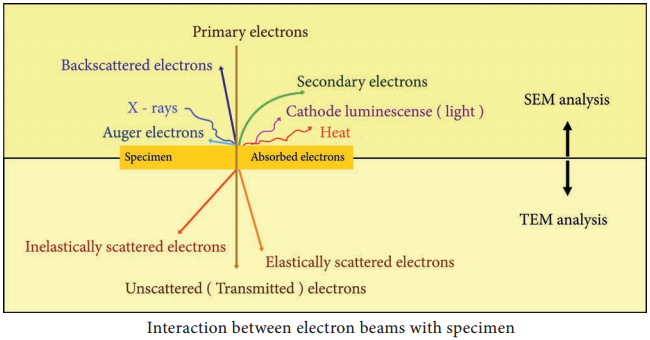
Electron – Sample Interactions:-

Interaction of electron beam with the sample results in different types of electrons: Elastic scattered electrons, Inelastic scattered electrons, secondary electrons and backscattered electrons. Almost all types of electron interactions can be used to retrieve information about the specimen.

Depending on the kind of radiation or emitted electrons which are used for detection, different properties of the specimen such as topography, elemental composition can be concluded. Figure 2.8 shows the interaction of the electron beam with the specimen.

In Transmission electron microscope (TEM), a beam of electrons is transmitted through an ultrathin specimen, interacting with the specimen as it passes through. An image is formed from the interaction of the transmitted unscattered electrons through the specimen.

Secondary electrons are mainly used in scanning electron microscope (SEM) for imaging the surface topography of biological specimens. The interaction of electron beam with samples results in secondary electrons and backscattered electrons that are detected by standard SEM equipment.
Electron Microscope - Definition, Principle, Parts, Uses img 1

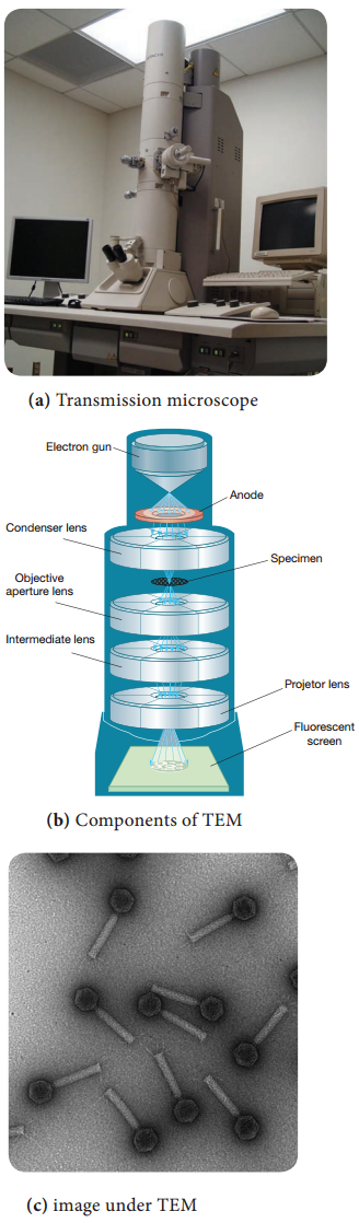
Working Principle and Instrumentation of TEM

The optics of the TEM is similar to conventional transmission light microscope. A transmission electron microscope has the following components,

  1. Electron gun
  2. Condenser lens
  3. Specimen stage
  4. Objective lens and projector lens
  5. Screen/photographic film/Charged Coupled Device (CCD) camera

Electron Gun consists of a tungsten filament or cathode that emits electrons on receiving high voltage electric current (50,000 – 100,000 volts). A high voltage between the electron source (cathode) and an anode plate is applied leading to an electrostatic field through which the electrons are accelerated.

The emitted electrons travel through vacuum in the microscope column. Vacuum is essential to prevent strong scattering of electrons by gases. Electromagnetic condenser lenses focus the electrons into a very thin beam. Electron beam then travels through the specimen and then through the electromagnetic objective
lenses.

In a TEM microscope, the sample is located in the middle of the column. At the bottom of the microscope, unscattered electrons hit the fluorescent screen giving image of specimen with its different parts displayed in varied darkness, according to their density. The image can be studied directly, photographed or digitally
recorded. Figure 2.9 show the arrangement of components for transmission electron microscope.

Information that can be obtained using TEM include,

  • Topography: surface features, texture
  • Morphology: shape and size of the particles
  • Crystallographic arrangement of atoms
  • Composition: elements and the their relative amounts.

Electron Microscope - Definition, Principle, Parts, Uses img 2

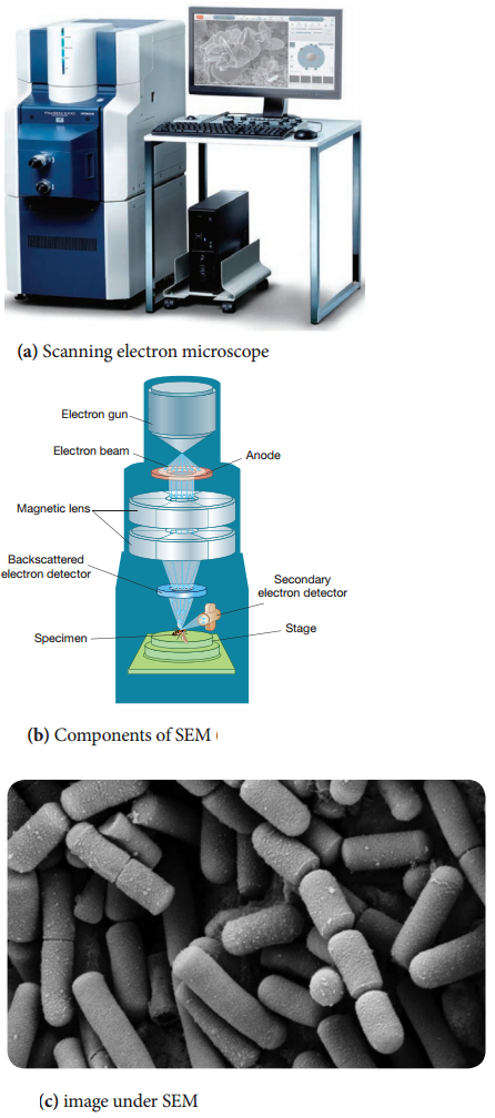
Working Principle and Instrumentation of SEM

It is first built by Knoll in 1935. It is used to study the three dimensional images of the surfaces of cells, tissues or particles. The SEM allows viewing the surfaces of specimens without sectioning. The specimen is first fixed in liquid propane at-180°C and then dehydrated in alcohol at-70°C.

The dried specimen is then coated with a thin film of heavy metal, such as platinum or gold, by evaporation in a vacuum provides a reflecting surface of electrons. In SEMs, samples are positioned at the bottom of the electron column and the scattered electrons (backscattered or secondary) are captured by electron detectors.

In SEM, there are several electromagnetic lenses, including condenser lenses and one objective lens. Electromagnetic lenses are for electron probe formation, not for image formation directly, as in TEM. Two condenser lenses reduce the crossover diameter of the electron beam. The objective lens further reduces the cross-section of the electron beam and focuses the electron beam as probe on the specimen surface (Figure 2.10).

Objective lens thus functions like a condenser. This is in contrast to TEM where objective lens does the magnification. Major difference between SEM and TEM are given in Table 2.2. SEMs are equipped with an energy dispersive spectrometer (EDS) detection system which is able to detect and display most of the X-ray spectrum. The detector normally consists of semiconducting silicon or germanium. Difference between SEM and TEM.

Properties

SEM

TEM

1. Types of electrons It is based on scattered electrons that are emitted from the surface of a specimen It is based on transmitted electrons
2. Sample preparation Sample can be of any thickness and is coated with a thin layer of a heavy metal such as gold or palladium and mounted on an aluminium slab Laborious sample preparation is required. The sample has to be cut into thin sections so as to allow electrons to pass through it and are supported on TEM grids.
3. Resolution The resolution is up to 20nm TEM has much higher resolution than SEM. It can resolve objects as close as 1nm
4. Magnification The magnifying power of SEM is up to 100,000X The magnifying power of TEM is up to 5,000,000 X
5. Image formation SEM provides a 3 dimensional image. Secondary or back scattered electrons are captured, detected and displayed on computer screen TEM provides a 2 dimensional image. Transmitted electrons hit a fluroscent screen giving rise to a shadow image. The image can be studied directly by the operator or photographed with a camera.
6. Application SEM is used to study the topography and atomic composition of specimens TEM is used to study the interior of cells, the structure of protein molecule, the organization of molecules in viruses and cytoskeletal filaments and the arrangement of protein molecules in cell membranes.

Scanning transmission electron microscopy (STEM) combines the principles of transmission electron microscopy and scanning electron microscopy and can be performed on either type of instrument. Like TEM, STEM requires very thin samples and the primary electron beam is transmitted by the sample.

One of its principal advantages over TEM is in enabling the use of other of signals that cannot be spatially correlated in TEM, including secondary electrons, scattered beam electrons, characteristic X-rays, and electron energy loss.
Electron Microscope - Definition, Principle, Parts, Uses img 3

Fluorescence Microscope – Definition, Principle, Parts, Uses

Learninsta presents the core concepts of Microbiology with high-quality research papers and topical review articles.

Fluorescence Microscope – Definition, Principle, Parts, Uses

Fluorescence microscope is a very powerful analytical tool that combines the magnifying properties of light microscope with visualization of fluorescence.

Fluorescence microscope is a type of light microscope which instead of utilizing visible light to illuminate specimens, uses a higher intensity (lower wavelength) light source that excites a fluorescent molecule called a fluorophore (also known as fluorochrome).

Fluorescence is a phenomenon that takes place when the substances (fluorophore) absorbs light at a given wavelength and emits light at a higher wavelength. Thus, fluorescence microscopy combines the magnifying properties of the light microscope with fluorescence technology.
Fluorescence Microscope - Definition, Principle, Parts, Uses img 1

The fluorophore absorbs photons leading to electrons moving to a higher energy state (excited state). When the electrons return to the ground state by losing energy, the fluorophore emits light of a longer wavelength (Figure 2.5). Three of the most common fluorophores used are Diamidino – phenylindole (DAPI) (emits blue), Fluorescein isothiocyanate (FITC) (emits green), and Texas Red (emits red).

Principle

Light source such as Xenon or Mercury Arc Lamp which provides light in a wide range of wavelength, from ultraviolet to the infrared is directed through an exciter filter (selects the excitation wavelength). This light is reflected toward the sample by a special mirror called a dichroic mirror, which is designed to reflect light only at the excitation wavelength.

The reflected light passes through the objective where it is focused onto the fluorescent specimen. The emissions from the specimen are in turn, passed back up through the objective where magnification of the image occurs and through the dichroic mirror.

This light is filtered by the barrier filter, which selects for the emission wavelength and filters out contaminating light from the arc lamp or other sources that are reflected off from the microscope components. Finally, the filtered fluorescent emission is sent to a detector where the image can be digitized.

Components of Fluorescence

Microscope:

The main components of the fluorescent microscope resemble the traditional light microscope. However, the two main difference are the type of light source used and the use of the specialized filter
elements (Figure).

Light source:-
Fluorescence microscopy requires a very powerful light source such as a Xenon or Mercury Arc Lamp. The light emitted from the Mercury Arc Lamp 10 – 100 times brighter than most incandescent lamps and provides light in a wide range of wavelengths from ultra-violet to the infrared. Lasers or high-power LEDs were mostly used for complex fluorescence microscopy techniques.

Filter elements:-
A typical fluorescence microscope consists of three filters: excitation, emission and the dichroic beam splitter.

Excitation filters:
It is placed within the illumination path of a fluorescence microscope. Its purpose is to filter out all
Fluorescence Microscope - Definition, Principle, Parts, Uses img 2

wavelength of the light source, except for the excitation range of the fluorophore in the sample or specimen of interest.

Emission filters:-
The emission filter is placed within the imaging path of a fluorescence microscope. Its purpose is to filter out the entire excitation range and to transmit the emission range of the fluorophore in the specimen.

Dichroic filter or beam splitter:-
The dichroic filter or beam splitter is placed in between the excitation filter and emission filter, at 45° angle. Its purpose is to reflect the excitation wavelength towards the fluorophore in the specimen, and to transmit the emission wavelength towards the detector.

Working Mechanism

The specimen to be observed are stained or labeled with a fluorescent dye and then illuminated with high intensity ultra violet light from mercury arc lamp. The light passes through the exciter filter that allows only blue light to pass through. Then the blue light reaches dichroic mirror and reflected downward to the specimen.

The specimen labeled with fluorescent dye absorbs blue light (shorter wavelength) and emits green light. The emitted green light goes upward and passes through dichroic mirror, reflects back blue light and allows only green light to pass the objective lens then it reaches barrier filter which allows only green light. The filtered fluorescent emission is sent to a detector where the image can be digitized Figure.
Fluorescence Microscope - Definition, Principle, Parts, Uses img 3

Application

  • Fluorescence microscope has become one of the most powerful techniques in biomedical research and clinical pathology.
  • Fluorescence microscope allows the use of multicolour staining, labeling of structures within cells, and the measurement of the physiological state of a cell.
  • Fluorescence microscope helps in observing texture and structure of coal.
  • To study porosity in ceramics, using a fluorescent dye.
  • To identify the Mycobacterium tuberculosis.

Phase Contrast Microscope – Definition, Principle, Parts, Uses

Learninsta presents the core concepts of Microbiology with high-quality research papers and topical review articles.

Phase Contrast Microscope – Definition, Principle, Parts, Uses

Frits Zernike a Dutch Physicist invented the Phase Contrast Microscope and was awarded Nobel Prize in 1953. It is the microscope which allows the observation of living cell. This microscopy uses special optical components to exploit fine differences in the refractive indices of water and cytoplasmic components of living cells to produce contrast.
Phase Contrast Microscope img 1

Principle

The phase contrast microscopy is based on the principle that small phase changes in the light rays, induced by differences in the thickness and refractive index of the different parts of an object, can be transformed into differences in brightness or light intensity. The phase changes are not detectable to human eye whereas the brightness or light intensity can be easily detected.

Optical Components of Phase Contrast Microscope (PCM)

The phase contrast microscope is similar to an ordinary compound microscope in its optical components. It possesses a light source, condenser system, objective lens system and ocular lens system (Figure 2.1).

A phase contrast microscope differs from bright field microscope in having,

(i) Sub-stage annular diaphragm (phase condenser):-

An annular aperture in the diaphragm is placed in the focal plane of the sub-stage which controls the illumination of the object. This is located below the condenser of the
microscope. This annular diaphragm helps to create a narrow, hollow cone of light to illuminate the object.

(ii) Phase – plate (diffraction plate or phase retardation plate):-

This plate is located at the back focal plane of the objective lenses. The phase plate has two portions, in which one is coated with light retarding material (Magnesium fluoride) and the other portion devoid of light retarding material but can absorb light. This plate helps to reduce the phase of the incident light (Figure 2.2).
Phase Contrast Microscope img 2

Working Mechanism of Phase Contrast Microscopy

The unstained cells cannot create contrast under the normal microscope. However, when the light passes through an unstained cell, it encounters regions in the cell with different refractive indexes and thickness. When light rays pass through an area of high refractive index, it deviates from its normal path and such light
rays experience phase change or phase retardation (deviation). Light rays pass through the area of less refractive index remain non-deviated (no phase change). Figure 2.3 shows the light path in phase contrast microscope.
Phase Contrast Microscope img 3

The difference in the phases between the retarded (deviated) and un-retarded (non-deviated) light rays is about ¼ of original wave length (i.e., λ/4). Human eyes cannot detect these minute changes in the phase of light. The phase contrast microscope has special devices such as annular diaphragm and phase plate, which convert these minute phase changes into brightness (amplitude) changes, so that a contrast difference can be created in the final image. This contrast difference can be easily detected by human eyes.

In phase contrast microscope, to get contrast, the diffracted waves have to be separated from the direct waves. This separation is achieved by the sub-stage annular diaphragm.

The annular diaphragm illuminates the specimen with a hollow cone of light. Some rays (direct rays) pass through the thinner region of the specimen and do not undergo any deviation and they directly enter into the objective lens. The light rays passing through the denser region of the specimen get regarded and they
run with a delayed phase than the nondeviated rays.

Both the deviated and non deviated light has to pass through the phase plate kept on the back focal plane
of the objective to form the final image. The difference in phase (Wavelength) gives the contrast for clear visibility of the object. Figure 2.4 Microscopic image comparing phase and bright field microscopy.
Phase Contrast Microscope img 4

Applications (Uses)

  • Phase contrast microscope enables the visualization of unstained living cells.
  • It makes highly transparent objects more visible.
  • It is used to examine various intracellular components of living cells at relatively high resolution.
  • It helps in studying cellular events such as cell division.
  • It is used to visualize all types of cellular movements such as chromosomal and flagellar movements.

Basic Equipments and Microbiological Techniques

Learninsta presents the core concepts of Microbiology with high-quality research papers and topical review articles.

Developments in Microbiology – Equipments

Confocal Microscopy

Confocal microscopy offers several advantages over conventional optical microscopy, including shallow depth of field, elimination of out-of-focus glare, and the ability to collect serial optical sections from thick specimens. In the biomedical sciences, a major application of confocal microscopy involves imaging either fixed or living cells and tissues that have usually been labeled with one or more fluorescent probes.

Confocal microscopy, most frequently confocal laser scanning microscopy (CLSM) or laser confocal scanning microscopy (LCSM), is an optical imaging technique for increasing optical resolution and contrast of a micrograph by means of using a spatial pinhole to block out of-focus light in image formation.

Capturing multiple two-dimensional images at different depths in a sample enables the reconstruction of three-dimensional structures (a process known as optical sectioning) within an object.

This technique is used extensively in the scientific and industrial communities and typical applications are in life sciences, semiconductor inspection and materials science. Light travels through the sample under a conventional microscope as far into the specimen as it can penetrate, while a confocal microscope only focuses a smaller beam of light at one narrow depth level at a time. The CLSM achieves a controlled and highly limited depth of focus.

DNA Sequencing System

Sequencing means finding the order of nucleotides on a piece of DNA. Nucleotide order determines amino acid order, and by extension, protein structure and function (proteomics). An alteration in a DNA sequence can lead to an altered or non functional protein, and hence to a genetic disorder. DNA sequence is important to detect the type of mutations in genetic diseases and offer hope for the eventual development of treatment DNA.

Methods of sequencing

1. Sanger dideoxy (primer extension/chain-termination) method:-
Most popular protocol for sequencing, very adaptable, scalable to large sequencing projects.

2. Maxam-Gilbert chemical cleavage method:-

DNA is labelled and then chemically cleaved in a sequence dependent manner. This method is not easily scaled and is rather tedious.

It provides an important tool for determining the thousands of nucleotide variations associated with specific genetic diseases, like Huntington’s, which may help to better understand these diseases and advance treatment.

Nanoparticles Production Using Microbes

Learninsta presents the core concepts of Microbiology with high-quality research papers and topical review articles.

Nanoparticles Production Using Microbes

Particles with one or more dimensions of the order of 100 nm or less. There are a large number of physical, chemical, biological, and hybrid methods available to synthesize different types of nanoparticles. Although physical and chemical methods are more popular in the synthesis of nanoparticles, the use of toxic chemicals greatly limits their biomedical applications, in particular in clinical fields.

Therefore, development of reliable, nontoxic, and eco-friendly methods for synthesis of nanoparticles is of utmost importance to expand their biomedical applications. One of the options to achieve this goal is to use microorganisms to synthesize nanoparticles.

Nanoparticles are biosynthesized when the microorganisms grab target ions from their environment and then turn the metal ions into the element metal through enzymes generated by the cell activities. It can be classified into intra-cellular and extracellular synthesis according to the location where nanoparticles are formed.

The intracellular method consists of transporting ions into the microbial cell to form nanoparticles in the presence of enzymes. The extracellular synthesis of nanoparticles involves trapping the metal ions on the surface of the cells and reducing ions in the presence of enzymes.

The biosynthesized nanoparticles have been used in a variety of applications including drug carriers for targeted delivery, cancer treatment, gene therapy and DNA analysis, antibacterial agents, biosensors, enhancing reaction rates, separation science, and magnetic resonance imaging (MRI).

Many microorganisms can produce inorganic nanoparticles through either intracellular or extracellular routes. This section describes the production of various nanoparticles via biological methods following the categories of metallic nanoparticles including gold, silver, alloy and other metal nanoparticles, oxide nanoparticles consisting of magnetic and nonmagnetic oxide nanoparticles, sulfide nanoparticles, and other miscellaneous nanoparticles (Figure 1.4).
Nanoparticles Production Using Microbes img 1

Molecular Biology and Genetic Engineering

Learninsta presents the core concepts of Microbiology with high-quality research papers and topical review articles.

Molecular Biology and Genetic Engineering

Molecular biology – is the study of the structure, function & makeup of the molecular building blocks of life. It focuses on the interactions between the various system of a cell, including the interrelationship of DNA, RNA & Protein synthesis &how these interaction are regulated. Bioscience, Molecular biology closely interrelate with the fields of Biochemistry, Genetics & Cell biology.

Molecular biology is a specialised branch of biochemistry, the study of the chemistry of molecules which are specifically connected to living processes. Importance to molecular biology are the nucleicacids (DNA and RNA) and the proteins which are constructed using the genetic instructions encoded in those molecules.

Other biomolecules, such as carbohydrates and lipids may also be studied for the interactions they have with nucleic acids and proteins. Molecular biology is often separated from the field of cell biology, which concentrates on cellular structures (organelles and the like), molecular pathways within cells and cell life cycles.

Genetic Engineering

Genetic Engineering is the act of modifying the genetic makeup of an organism. Modification can be generated by methods such as gene therapy, nuclear transplantation, transfection of synthetic chromosome or viral insertion.

The manipulation of genetic make up of living cells by inserting desired genes through a DNA vector, is the genetic engineering. The gene is a small piece of DNA that encodes for a specific protein. The gene is inserted into a vector DNA so that a new combination of vector DNA is formed.

The DNA formed by joining DNA segments of two different organisms is called recombinant DNA. The organism whose genetic make up is manipulated using recombinant DNA technique, is called genetically manipulated organism (GMO). Genetic engineering has many application in agriculture, animal science, industry and medicines (Figure 1.3).

Biology Lesson Class 12 MCQ Questions with Answers Chapter 1.3 Molecular Biology and Genetic Engineering img 1

Genetically Modified Organism (GMO)

Organism genome has been engineered in the laboratory in order to favour the expression of desired physiological traits or the production of desired biological products. In conventional livestock production, crop farming, and even pet breeding, it has long been the practice to breed select individuals of a species in order to produce offspring that have desirable traits.

In genetic modification, however, recombinant genetic technologies are employed to produce organisms whose genomes have been precisely altered at the molecular level, usually by the inclusion of genes from unrelated species of organisms that code for traits that would not be obtained easily through conventional selective breeding.

GMOs are produced through using scientific methods that include recombinant DNA technology and reproductive cloning. In reproductive cloning, a nucleus is extracted from a cell of the individual to be cloned and is inserted into the enucleated cytoplasm of a host egg.

The process results in the generation of an offspring that is genetically identical to the donor individual. The first animal produced by means of this cloning technique with a nucleus from an adult donor cell (as opposed to a donor embryo) was a sheep named Dolly, born in 1996.

Since then a number of other animals, including pigs, horses, and dogs, have been generated by reproductive cloning technology. Recombinant DNA technology, on the other hand, involves the insertion of one or more individual genes from an organism of one species into the DNA (deoxyribonucleic acid) of another.

Whole-genome replacement, involving the transplantation of one bacterial genome into the “cell body,” or cytoplasm, of another microorganism, has been reported, although this technology is still limited to basic scientific applications.

Developments in Microbiology – Immunology

Learninsta presents the core concepts of Microbiology with high-quality research papers and topical review articles.

Developments in Microbiology – Immunology

Immunology is the study of the immune system and is a very important branch of the medical and biological sciences. The immune system protects us from infection through various lines of defence.

Important initial barriers to infection are physical (Example: the skin), enhanced by substances secreted by the body, such as saliva and tears, that contain molecules that can neutralise bacteria. The internal mucosal tissues (Example: lungs & airways, and the gut) are coated with mucus that is able to trap potential infectants.

In the airways, mobile ciliate hairs work together to transport contaminants away from vulnerable areas. Tissues such as the skin, mucosal surfaces and airways also contain populations of immune cells that can respond to infectants that breach these physical defences.

In its most complex forms, the immune system consists of two branches: the innate immune system that utilises certain ‘hard-wired’ strategies to provide a rapid, general, response when alerted by certain typical signals of infection (essentially forming a first-line of defence); and the adaptive immune system that is able to develop highly specific responses (and a persistent ‘immune memory’) to target infection with extraordinary accuracy.

Both systems work in close cooperation and, to an important extent, the adaptive immune system relies upon the innate immune system to alert it to potential targets, and shape its response to them.

Vaccines currently in development include:-

  • A genetically-modified vaccine for the treatment of pancreatic cancer.
  • A therapeutic vaccine that increases the immune response against the HIV virus.
  • A vaccine that protects infants against meningococcal disease, a leading cause of meningitis.
  • An immunotherapeutic vaccine for the treatment of Alzheimer’s disease.
  • A recombinant vaccine to prevent malaria.

Evolving science has increasingly enabled researchers to explore both promising therapeutic vaccines and new preventative agents for infectious diseases. Although the development process is extremely complex, advances in other scientific fields, such as genomics, are being leveraged in the development of new vaccines.

“Vaccines have been a major contributor in saving countless lives around the world,” said Castellani. “Vaccinations contribute to the public health at large, and they make good economic sense. The many exciting candidates in the pipeline offer great hope for a healthier, more productive future.”

Monoclonal Antibodies 

mAb or moAb are identical immunoglobulins, generated from a single B-cell clone. These antibodies recognize unique epitopes, or binding sites, on a single antigen. Derivation from a single B-cell clones and subsequent targeting of a single epitope is what differentiates monoclonal antibodies from polyclonal antibodies.

The traditional monoclonal antibody (mAb) production process usually starts with generation of mAb-producing cells (i.e. hybridomas) by fusing myeloma cells with desired antibody-producing splenocytes (Example: B cells). These B cells are typically sourced from animals, usually mice. After cell fusion, large numbers of clones are screened and selected on the basis of antigen specificity and immunoglobulin class (Figure 1.1).
Biology Lesson Class 12 MCQ Questions with Answers Chapter 1.2 Immunology img 1

Stem Cell & Therapy

Stem cells are biological cells that can differentiate into other types of cells & they are found in multicellular organism. Stem cells are a class of undifferentiated cells that are able to differentiate into specialized cell types. Commonly, stem cells come from two main sources:

  • Embryos formed during the blastocyst phase of embryological development (embryonic stem cells) and
  • Adult tissue (adult stem cells).

Both types are generally characterized by their potency, or potential to differentiate into different cell types such as skin, muscle, bone, etc., (Figure 1.2).

Stem-cell therapy is the use of stem cells to treat or prevent a disease or condition. Stem Cell Therapy (SCT) is the treatment of various disorders, non-serious to life threatening, by using stem cells. These stem cells can be procured from a lot of different sources and used to potentially treat more than 80 disorders, including neuromuscular and degenerative disorders.

Hematopoietic disorders (Example: leukaemia, thallassemia, aplastic anemia, MDS, sickle cell anemia, storage disorders etc.) affect the bone marrow and manifest with various systemic complications. Stem cells from a donor (either from cord blood or bone marrow) are known to reconstitute the defective bone marrow and permanently overcome the disorder.
Biology Lesson Class 12 MCQ Questions with Answers Chapter 1.2 Immunology img 2

Microbes in Space

Learninsta presents the core concepts of Microbiology with high-quality research papers and topical review articles.

Microbes in Space

The majority of experiments on microorganisms in space were performed using Earth-orbiting robotic spacecraft, Example: the Russian Foton satellites and the European Retrievable Carrier (EURECA) (121), or human-tended spacecraft, such as space shuttles (106, 107) and space stations, Example: MIR and the International Space Station (ISS).

Only twice, during translunar trips of Apollo 16 and 17 in the early 1970s, were microorganisms exposed to space conditions beyond Earth’s magnetic shield, in the MEED (microbial ecology equipment device) facility and in the Biostack experiments.

Arriving in space without any protection, microorganisms are confronted with an extremely hostile environment, characterized by an intense radiation field of galactic and solar origin, high vacuum, extreme temperatures, and microgravity.

Some bacteria were found in International Space Station and on the Mars Rover. Some bacteria and tiny microbes called tardigrades are able to survive for longer periods in space. This ability of surviving in extreme environmental condition leads to forward contamination. Sea planktons and other microorganisms have been identified in cosmonauts’ spacewalk samples.

In July 2016, Kate Rubins was the first to sequence DNA in space. NASA astronaut Peggy Whitson amplified and sequenced the DNA of bacteria that grew as colonies in the petri plate on the surface on the space station.

In June 2018, Professor George Fox and his team have isolated genus Bacillus from spacecraft assembly rooms at the Jet Propulsion Laboratory. They have sequenced the complete genomes of two strains, B. safensis FO-36bT and B. pumilus SAFR-032 and found that they are resistant to radiation.

Los Angeles in great news for India, scientists at NASA have named a new organism discovered by them after the much loved APJ Abdul Kalam. Till date, the new organism – a form of bacteria – has been found only on the International Space Station (ISS) and has been found on earth.

Researchers at the Jet Propulsion Laboratory (JPL) the foremost lab of NASA for work on inter-planetary travel discovered the new bacteria on the filters of the international space station (ISS) and named it Solibacillus kalam to honour the late president, who was a renowned aerospace scientist.

Microbiology – Introduction, Concepts, Free Resources

Microbiology – Introduction, Concepts, Free Resources

Developments in Microbiology

Microscopy

Control of Microorganisms by Chemical Methods

Microbial Metabolism

Food Microbiology

Industrial Microbiology

Medical Bacteriology

Medical Parasitology

Medical Mycology

Medical Virology

Immunology

Microbial Genetics