Carbohydrate Catabolism

Learninsta presents the core concepts of Microbiology with high-quality research papers and topical review articles.

Carbohydrate Catabolism

Most microorganisms oxidize carbohydrates as their primary source of cellular energy. Carbohydrate catabolism is the breakdown of carbohydrate molecule to produce energy and is therefore of great importance in cell metabolism. Glucose is the most common carbohydrate energy source used by cells.

To produce energy from glucose, microorganism use two general processes namely Respiration and Fermentation.

Cellular Respiration

Respiration is defined as an ATP generating process in which organic molecules are oxidized and the final electron acceptor is an inorganic compound.

In aerobic respiration, the final electron acceptor is Oxygen and in anaerobic respiration the final electron acceptor is an inorganic molecule like NO3, SO42- other than Oxygen.

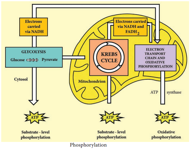
The aerobic respiration of glucose typically occurs in three principal stages. They are Glycolysis Krebs cycle
Electron transport chain.

Glycolysis

Glycolysis is the process of splitting of sugar molecule, where the glucose is enzymatically degraded to produce ATP. Glycolysis is the oxidation of glucose to pyruvic acid with simultaneous production of some ATP and energy containing NADH. It takes place in the cytoplasm of both prokaryotic and eukaryotic cells.
Glycolysis occurs in the extra mitochondrial part of the cell cytoplasm.

Glycolysis was discovered by Emden, Meyerhof and Parnas. So, this cycle is shortly termed as EMP pathway, in honour of these pioneer workers. This cycle occurs in animals, plants and large number of microorganisms. Glycolysis does not require oxygen, it can occur under aerobic or anaerobic condition. Glycolysis is a sequence of ten enzyme catalyzed reactions.

Aerobic condition

Carbohydrate Catabolism img 1

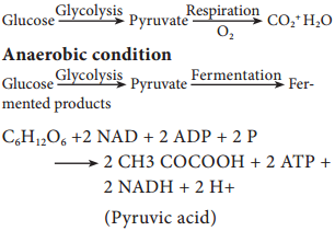
Since glucose is a six carbon molecule and pyruvate is a three carbon molecule, two molecules of pyruvate are produced for each molecule of glucose that enters Glycolysis. Net energy production from each glucose molecule is two ATP molecules The Glycolysis pathway consists of two phases. They are

  1. The preparatory/Investment phase, where ATP is consumed
  2. The pay off phase where ATP is produced (Figure 4.4).

Carbohydrate Catabolism img 2

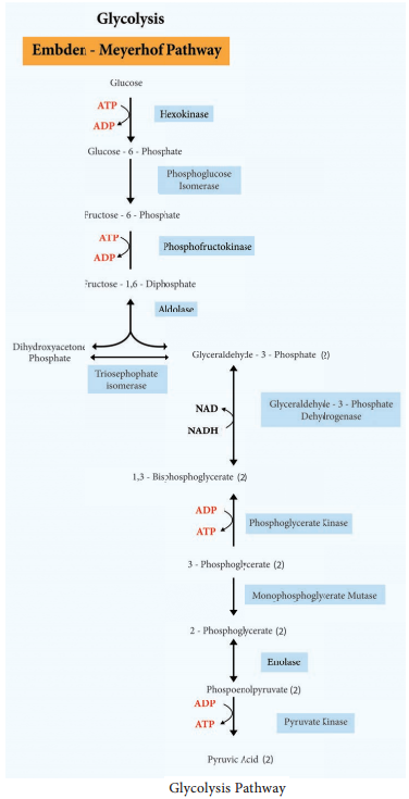
1. In the preparatory stage, two molecules of ATP are utilized and then glucose is phosphorylated, restructured, and split into two 3 carbon compounds namely Glyceraldehyde-3-phosphate and Dihydroxyacetone phosphate.

2. In pay off phase or energy conserving stage, the two 3 carbon molecules are oxidized in several steps to 2 molecules of pyruvic acid and two molecules of NAD+ are reduced to NADH, thus four molecules of ATP are formed by substrate level phosphorylation.

Two molecules of ATP are needed to initiate Glycolysis and four molecules of ATP are generated at the end of the process. Therefore, the net gain of Glycolysis is two ATP for each molecule of glucose oxidized.

Alternatives to Glycolysis

Many bacteria have another pathway in addition to Glycolysis for the oxidation of glucose. Some of the common pathways that occur in most of the bacteria are

• Pentose phosphate pathway (PPP) or Hexose Mono Phosphate shunt
• Entner – Doudoroff Pathway

Generation of ATP

Learninsta presents the core concepts of Microbiology with high-quality research papers and topical review articles.

Generation of ATP

Much of energy released during oxidation reduction reaction is trapped within the cell by the formation of ATP. A phosphate group is added ADP with the input of energy to form ATP. The addition of a phosphate to a chemical compound is called phosphorylation. Organism uses three different mechanisms of phosphorylation to generate ATP from ADP.

Substrate Level Phosphorylation

It is a metabolic reaction that results in the formation of ATP or GTP by the direct transfer of a phosphoryl group to ADP or GDP from another phosphorylated compound.

Oxidative Phosphorylation

In this reaction, electrons are transferred from organic compounds to molecules of Oxygen (O2) or other inorganic molecules through a series of different electron carriers (Example: NAD+ and FAD). Then the electrons are passed through a series of different electron carriers to oxygen. The process of oxidative phosphorylation occurs during electron transport chain (Figure 4.3).
Generation of ATP img 1

Photophosphorylation

It occurs only in photosynthetic cells which contain light trapping pigments. Example: In photosynthesis, photosynthetic pigment, Chlorophyll is involved in the synthesis of organic molecules especially sugars, with the energy of light from the energy poor building blocks like Carbon dioxide and water. In phototropic bacteria (purple, green sulphur bacteria, Cyanobacteria), photosynthetic pigments bateriochlorophylls are involved in ATP production.

Energy of Chemical Reaction

Learninsta presents the core concepts of Microbiology with high-quality research papers and topical review articles.

Energy of Chemical Reaction

Light energy is trapped by phototrophs during photosynthesis, in which it is absorbed by bacteriochlorophyll and other pigments and converted to chemical energy for cellular work. The energy is required by the bacterium for synthesis of cell wall or membrane, synthesis of enzymes, cellular components, repair
mechanism, growth and reproduction.

Some change of energy occurs whenever bonds between atoms are formed or broken during chemical reactions. When a chemical bond is formed, energy is required. Such a chemical reaction which requires energy is called an endergonic reaction (energy is directed inward). When a bond is broken, energy is released. A chemical reaction that release energy is an exergonic reaction (energy is directed outward).

During chemical reaction energy is either released or absorbed and the quantum of energy liberated or taken up is useful energy and is referred to Free Energy Change (ΔG) of the reactions.

High Energy Phosphate

Adenosine Tri-Phosphate (ATP) is the principal energy carrying molecule of all cells and is indispensable to the life of the cell. It stores the energy released by some chemical reactions, and it provides the energy for reactions that require energy. ATP consists of an adenosine unit composed of adenine, ribose with three phosphate groups. In ATP and some other phosphorylated compounds, the outer two phosphate groups are joined by an anhydride bond.

Some of the other high energy nucleotides involved in biochemical processes are given in Table 4.1.

Table 4.1: High energy nucleotides involved in biosynthesis

Name of the Nucleotide

Biosynthesis

Uridine triphosphate (UTP) Polysaccharide
Cytidine triphosphate (CTP) Lipid
Guanidine triphosphate (GTP) Protein

Nutrients are broken from highly reduced compounds to highly oxidized compounds within the cells. Much of the energy released during oxidation reduction reactions is trapped within the cell by the formation of ATP. A phosphate group is added to ADP with the input of energy to form ATP.

ATP + H2O → ADP + pi(ΔG° = – 7.3 K cal/mol)
ATP + H2O → AMP + ppi(ΔG° = – 10.9 K cal/mol)

ATP is ideally suited for its role as an energy currency. It is formed in energy trapping and energy generating processes such as photosynthesis, fermentation, and aerobic respiration. In bacterial and archeal cells, most of the ATP is formed on the cell membrane, while in eukaryotes the reactions occur primarily in the
mitochondria (Figure 4.2).
Energy of Chemical Reaction img 1

Oxidation – Reduction Reactions

Oxidation is the removal of electrons (e) from an atom or molecule and is often an energy producing reaction. Reduction of a substrate refers to its gain or addition of one or more electrons to an atom or molecule. Oxidations and reduction are always coupled. In other words, each time one substance is oxidized, another is simultaneously reduced.
F2 + 2e → 2F
H2 + 2e → 2H+ + 2e
NAD+ + 2H+ + 2e ⇄ NADH + H+.

Microbial Metabolism

Learninsta presents the core concepts of Microbiology with high-quality research papers and topical review articles.

Microbial Metabolism

The term Metabolism refers to the sum of all bio chemical reactions that occur within a living cell. Chemical reaction either release energy or require energy. Metabolism can be viewed as an energy balancing act. It can be divided into two classes of chemical reactions namely Catabolism and Anabolism.

Catabolism:
It is called catabolic or degradative reactions because complex organic compounds are broken down into simples ones. Catabolic reactions are generally hydrolytic reactions. It is enzyme regulated chemical reaction that release energy and they are exergonic. Example: Break down of sugar into Carbon dioxide and water in cells.

Anabolism:
It is called anabolic or biosynthetic reactions because complex organic molecules are formed from simples ones. Anabolic process often involves dehydration, are bio-synthetic reactions (Figure 4.1). It is enzyme regulated energy requiring reaction and they are endergonic. Examples: Formation of proteins from amino acids.
Microbial Metabolism img 1

Catabolic reactions furnish the energy needed to drive anabolic reactions. This coupling of energy requiring and energy releasing reactions is made possible through the molecule Adenosine tri-phosphate (ATP).

Drugs Resistance Mechanisms

Learninsta presents the core concepts of Microbiology with high-quality research papers and topical review articles.

Drugs Resistance Mechanisms

Some microbes respond predictably to certain drugs making selection of treatment easy. Other microbes may vary in their responses, and laboratory tests are usually required to ensure that the selected therapy is appropriate.

Chemotherapeutic effectiveness depends upon the sensitivity of the pathogen to the agent. Antibiotic resistance, however, may develop in microbes within the population. In fact, the history of chemotherapy has been closely paralleled by the history of drug resistance.

None of the therapeutic drugs (antibiotic) inhibits all microbial pathogens and some microbial pathogens possess natural ability to resist to certain antibiotics.

Bacteria become drug resistant using several different resistance mechanisms. A particular type of resistance mechanism is not confined to a single class of drugs. Two bacteria may employ different resistance mechanisms to counter the same antibiotic.

However, bacteria acquire drugs resistance using resistance mechanisms such as reduced permeability to antibiotic, efflux (pumping) antibiotic out of the cell, drug inactivation through chemical modification, target
modification and development of a resistant biochemical pathway (Figure 3.5).
Drugs Resistance Mechanisms img 1

Antimicrobial Susceptibility Testing

Learninsta presents the core concepts of Microbiology with high-quality research papers and topical review articles.

Antimicrobial Susceptibility Testing

Antimicrobial susceptibility tests are used to determine the type and quantity of antimicrobial agents used in chemotherapy. One of the most important functions of a clinical laboratory is to determine the antimicrobial susceptibility.

Antimicrobial susceptibility of pathogens refers to the limitation of pathogens to grow in the presence of effective antibiotics. There are two methods that can be used to determine the susceptibility of a potential pathogen to antimicrobial agents. They are:

  • Disk diffusion method
  • Tube dilution method

Disc Diffusion Method (Kirby – Bauer Test)

William Kirby and Alfred Bauer, in 1966 first introduced the principle of measuring zones of inhibition around antibiotic discs to determine antimicrobial agent susceptibilities. It is a rapid, convenient method to determine the susceptibilities of microorganisms to antimicrobial agents and a most common procedure used in susceptibility testing in clinical laboratory.

Filter paper discs containing known concentrations of antimicrobial agents are placed onto the surface of an agar plate (Muller – Hinton agar medium) inoculated with the test bacterium (Figure 3.3). The plate is incubated for 16 to 18 hours, and the zones of inhibition are read around each paper disc. During the incubation periods, the antimicrobial agent diffuses through the agar, and a concentration gradient of agent is established.

At some point in this gradient, growth of the susceptible bacteria is suppressed, and no growth is observed within a circular zone around disc. The size of a zone of inhibition must be compared to a standard Table for that particular drug before accurate comparisons can be made.

Thus, enabling to classify pathogens as susceptible (S), intermediate or resistant (R) to a drug. The procedure is highly regulated and controlled by the clinical and laboratory standards institute (CLSI) and must be accompanied by a rigorous quality assurance program including performance by certified and/or licensed personnel when the results are to be reported in clinical settings.
Antimicrobial Susceptibility Testing img 1

Minimal Inhibitory Concentration (MIC) Test

The potency of an effective antimicrobial agent is expressed in terms of minimal inhibitory concentration (MIC). It is the minimum concentration of drug that will inhibit the growth of pathogen. The MIC is determined by serial dilutions of antimicrobial agents in tubes with standard amount of bacteria. Turbidity (cloudiness) after incubation indicates bacterial growth and lack of turbidity indicates that the growth of bacteria is inhibited.

E – test

This is another test to determine the minimum inhibitory concentration where a plastic strip containing a gradient of the antimicrobial agent is used (Figure 3.4). An elliptical zone of inhibitory concentration can be noted with the help of a scale printed on the strip.
Antimicrobial Susceptibility Testing img 2

The Minimal Bactericidal Concentration (MBC) Test

MBC test is similar to MIC, the minimal bactericidal concentration test is used to determine the amount of antimicrobial agent required to rather kill the pathogen. In MBC test, samples taken from MIC tubes are transferred to drug free plates. Bacterial growth in these subcultures indicates that some bacterial cells have survived antimicrobial drug. The lowest concentration of drug for which no growth occurs is the minimum bactericidal concentration.

The tube dilution method is considered accurate for determining susceptibility of a pathogen to precise quantities of antimicrobial agent. However, the method is time consuming, expensive, and not practical for use in most clinical laboratories for routine susceptibility testing.

Evaluation of Antimicrobial Chemical Agents Antibiotics

Learninsta presents the core concepts of Microbiology with high-quality research papers and topical review articles.

Evaluation of Antimicrobial Chemical Agents Antibiotics

Testing of antimicrobial agents is a complex process regulated by two different federal agencies.

The U.S. Environmental Protection Agency regulates disinfectants, where as agents used on humans and animals are under the control of the Food and Drug Administration.

Testing of antimicrobial agents often begins with an initial screening test to see if they are effective and at what concentrations.

Laboratory techniques for the evaluation of antimicrobial chemical agents are conducted by one of the following three general procedures. In each procedure, the chemical agent is tested against a specific microorganisms referred to as the test organism.

Agar Plate Method

A plate of agar medium is inoculated with the test organism and the chemical agent is placed on the surface of the medium. The chemical solution is first impregnated in absorbent papers or confined by a hollow cylinder placed on the agar surface. Following incubation, the plate is observed for a zone of inhibition around the chemical agent. This is particularly suitable for semisolid preparations.

Tube Dilution Methods

Appropriately diluted water soluble liquid substances are dispensed into sterile test tubes and are inoculated with a measured amount of the test organism. At specified intervals, a transfer is made from this tube into tubes of sterile media that are then incubated and observed for the appearance of growth.

It is necessary in this type of procedure to ascertain whether the inhibitory action is bactericidal and not bacteriostatic. This approach can also be used to determine the number of organisms killed per unit time by performing a plate count on samples taken at appropriate intervals.

Phenol Coefficient Test

Phenol coefficient is a measure of the bactericidal activity of a chemical compound in relation to phenol. Phenol coefficient is calculated by dividing the concentration of test disinfectant at which it kills the organism in 10 minutes and not in 5 minutes under the same conditions. This method is used for evaluating the efficiency of watermiscible disinfectants.

Series of 10 test tubes with 2ml of distilled water is taken (Figure 3.1). Phenol is added to first test tube and dilution is made by transferring 1ml to next tube up to 5 dilutions. Similarly commercial disinfectant is also diluted. Pure culture of test organisms, such as Staphylococcus aureus or Salmonella typhi, is added to test tubes.

Subcultures from these tubes incubated at 37°C for 48 hours are examined for the presence or absence of growth at intervals of 5, 10 and 15 minutes. The highest dilution that kills the bacteria after 10 minutes, but not after 5 minutes is used to calculate the phenol coefficient (Table 3.3),

Evaluation of Antimicrobial Chemical Agents Antibiotics img 2

Illustration of phenol coefficient determination

Phenol dilution of 1:90 showed growth at 5 minutes but no growth at 10 minutes Test Chemical dilution of 1:450 showed growth at 5 minutes but no growth at 10 minutes phenol coefficient of test chemical as 450/90=5.

Antibiotics

The term ‘antibiotic’ was derived from ‘antibiosis’ which refers to the suppression of microorganisms due to secretion of toxic or inhibitory compounds by other microorganisms. Although antibiosis has been observed by many scientific workers fairly frequently towards the end of the nineteenth century, it was not until the discovery and development of Penicillin that a truly wide ranging search for antibiotics was initiated.

Historical Development

The first chemotherapeutic agent, discovered by Paul Ehrlich, was Salvarsan, used to treat syphilis. Alexander Fleming discovered the first antibiotic, penicillin, in 1929; its first clinical trails were done in 1940. Antibiotics are produced by species of Streptomyces, Bacillus, Penicillium and Cephalosporium.

Drugs such as the sulfonamides are sometimes called antibiotics although they are synthetic chemotherapeutic agents which are not synthesized using microbes.

Classification of Antibiotics

The antibiotics are usually classified on the basis of:-

• Target group of microorganisms
• Antimicrobial spectrum and
• Mode of action

Classification based on target group of microorganisms:-

Based on the target group, the antibiotics can be classified as antibacterial, antifungal and antiviral.

Classification based on Antimicrobial spectrum:-

Antimicrobial spectrum or antibiotic spectrum refers to the range of effectiveness of antibiotics on different kind of microorganisms, i.e. the range of different kind of microorganisms that can be inhibited, killed, or lysed by a particular type of antibiotic.

The susceptibility of microorganisms to individual antibiotic varies significantly and on account of this, the antibiotics can be classified in two groups as,

Broad – spectrum antibiotics:-

These attack different kinds of microbial pathogens and therefore find wider medical use. Antibacterial antibiotics of broad – spectrum are effective against both Gram positive and Gram negative bacteria.
They also attack pathogens belonging to Mycobacteria, Rickettsia, and Chlamydia. Similarly, broad – spectrum antifungal antibiotics attack different type of fungal pathogens.

Narrow – spectrum antibiotics:-

Narrow – spectrum antibiotics are categorized as those that are effective only against a limited variety of microbial pathogens. These antibiotics are quite valuable for the control of microbial pathogens that fail to respond to other antibiotics. For example, vancomycin is a narrow spectrum glycopeptide. It is an effective bactericidal agent for gram – positive penicillin resistant bacterial pathogens belonging to genera Staphylococcus, Bacillus, and Clostridium.

Mode of Action of Antibiotics

The mode of action of antibiotics varies as they damage pathogens in several ways (Flowchart 3.1). Some of the important actions of therapeutic drugs in microbial pathogens are as follows. Cell wall synthesis, Protein synthesis, Nucleic acid synthesis, Cell membrane disruption and Metabolic pathways blockage.
Evaluation of Antimicrobial Chemical Agents Antibiotics img 3

1. Inhibition of Cell Wall Synthesis

The most selective therapeutic antibiotics are those that interfere with the synthesis of bacterial cell walls. These drugs posses a high therapeutic index because bacterial cell walls have a unique structure which is not found in eukaryotic cells. The important cell wall attacking drugs are Penicillin, Cephalosporin, Ampicillin,
Methicillin and Vancomycin.

2. Inhibition of Protein Synthesis

Many therapeutic antibiotics discriminate between prokaryotic and eukaryotic ribosomes and inhibit protein synthesis. The therapeutic index of these drugs is fairly high, but not as favourable as that of cell wall synthesis inhibitors. Several of these drugs are medically useful and effective research tools because they block individual steps in protein synthesis. Some therapeutic drugs bind to 30S while others attach to 50S ribosomal subunits. Example Streptomycin, Chloramphenicol, Tetracyclin and Erythromycin.

3. Inhibition of Nucleic Acid Synthesis

Some antimicrobial drugs or antibiotics inhibit nucleic acid synthesis. These are not selectively toxic as other drugs. This is due to the fact that prokaryotic and eukaryotic nucleic acid synthesis mechanisms do not vary greatly. Example Quinolones, Novobiocin, Actinomycin and Rifampin

4. Disruption of Cell Membrane

There are some antimicrobial drugs or antibiotics that act as cell membrane disorganizing agents. Polymyxins are such drugs of clinical importance. E.g. Polymyxin B and Polymyxin E (colistin)

5. Blocking Metabolic Pathways

Some therapeutic drugs act as antimetabolites and block the functioning of metabolic pathways. They competitively inhibit the key enzymes in the metabolic pathway. Example Sulfonamides, Trimethoprim, Dapsone and Isoniazid (Figure 3.2).
Evaluation of Antimicrobial Chemical Agents Antibiotics img 4
Trimethoprim, Daspone and Isoniazid (Figure 3.2).

Major Groups of Antimicrobial Chemical Agents

Learninsta presents the core concepts of Microbiology with high-quality research papers and topical review articles.

Major Groups of Antimicrobial Chemical Agents

A large number of chemical agents are in common use. Some of the more common groups are listed below.

1. Phenol and Phenolics

Phenol was the first widely used chemical antiseptic and disinfectant. In 1867, Joseph Lister employed carbolic spray to reduce the risk of infection in surgical theatres. Phenol derivatives called phenolics contain altered molecules of phenol useful as antiseptics and disinfectants. The phenolics damage cell membranes and inactivate enzymes of microorganisms, while denaturing the proteins.

Phenolics includes cresols, such as Lysol, as well as several bisphenols, such as hexachlorophene. Today phenol and phenolics such as cresol, xylenol, and orthophenyl phenol are used as disinfectants in laboratories and hospitals.

The commercial disinfectant Lysol is made of mixture of phenolics. Phenolics are tuberculocidal, effective in the presence of organic material, and remain active on surfaces long after application. However, they have a disagreeable odour and can cause skin irritation.

Hexachlorophene is one of the most popular antiseptics because it persists on the skin once applied and reduces skin bacteria for a long period. It is mainly used in soaps and creams. It is an ingredient of various dermatological preparation used for skin disorders.

2. Alcohols

Alcohols are among the most widely used disinfectant and antiseptic. They are bactericidal and fungicidal but not sporicidal. Alcohols can destroy the lipid component of enveloped viruses. The two most popular alcoholic germicides are ethanol and isopropanol. They act by denaturing proteins and dissolving membrane lipids. The recommended optimum concentration of ethanol is 70%, but concentration between 60% and 95% are employed to kill germs as well. Thermometers and small instruments are disinfected by immersing in alcohol for 10 to 20 minutes.

3. Halogens

Halogen compounds are broad spectrum compounds that are considered low toxicity, low cost and easy to use. Among the halogens, iodine and chlorine are important antimicrobial agents. Small quantities of drinking water can be disinfected with halazone tablets.

a. Iodine:-

Iodine compound are broad spectrum and considered effective for a variety of bacteria, mycobacteria, fungi and viruses. The alcoholic tincture of iodine is highly active against gram positive organisms and so is used as a skin antiseptic. It stains the skin. Iodine combines with microbial protein and inhibits their function.

b. Chloride:-

Chloride is also used as a gas to maintain a low microbial count in drinking water. Chlorine together with ammonia called chloramines are used to sanitize glasswall and eating utensils. Sodium hypochlorite (NaOCl) is one of the most widely used chlorine containing disinfectants. Low concentrations (2-500ppm) are active against vegetative bacteria, fungi and most viruses.

Rapid sporicidal action can be obtained around 2500ppm, however this concentration is very corrosive so should be limited in its use. High concentrations are also irritating to the mucous membranes, eyes and skin. Chlorine compounds are rapidly inactivated by light and some metals so fresh solutions should always be used. Hypochlorites should never be mixed with acids or ammonia as this will result in the release of toxic chlorine gas.

c. Iodophores:-

The combinations of iodine and organic molecules are called Iodophores. They include wescodine, betadine and previdone. These iodophore contains surface active agents. They cause less irritation to the skin than free Iodine and do not stain. They are used for cleaning wounds and as a general purpose laboratory disinfectant for discarded jars.

4. Heavy Metals

For many years the ions of heavy metals such as mercury, silver, arsenic, zinc, and copper were used as germicides. More recently these have been superseded by other less toxic and more effective germicides. Many heavy metals are more bacteriostatic than bactericidal. There are a few exceptions. 1% solution of Silver nitrate is often applied to the eyes of infants to prevent ophthalmic gonorrhea. Silver sulfadiazine is used on burns. Copper sulfate is an effective algicide used in lake and swimming pools to retard the growth of algae.

Heavy metals combine with sulfhydryl (SH) groups of proteins and inactivate them. High concentration of metallic salts, particularly those of mercury, silver and copper coagulate cellular proteins that results in damage or death of the microbial cell. The most toxic heavy metals are the mercury, silver, and copper.

5. Quaternary Ammonium Compounds (Quats)

The most widely used surface active agents are the cationic detergents, especially the quaternary ammonium compounds (quats).

Quaternary Ammonium compounds are strongly bactericidal against Gram positive bacteria and less active against gram negative bacteria. These include agents such as cetrimide, bromide and benzalkonium chloride. Their antibacterial activity is antagonized by soaps and certain organisms like Pseudomonas.

They are useful for washing cutlery in catering industry and for cleaning wounds in hospitals. Savlon, a popular antiseptic, is a mixture of cetrimide and chlorohexidine and is active against Gram negative bacteria. They are used as skin disinfectants and as a preservative of ophthalmic solution.

The combined properties of germicidal activity and low toxicity, high solubility in water, stability in solution and non-corrosiveness have resulted in many applications of quaterneries as disinfectants and sanitizing agents.

Quats are also fungicidal, amoebicidal, and virucidal against enveloped viruses. They do not kill endospores or mycobacteria.

6. Aldehydes

Aldehydes are highly effective, broad spectrum disinfectant. The most which typically achieve its anitimicrobial action by denaturing proteins and disrupting nucleic acids. Commonly used aldehydes are formaldehyde and glutaraldehyde. Formaldehyde is usually dissolved in water or alcohol before use. Formaldehyde is used as a surface disinfectat and a fumigant and has been used to decontaminate in animate objects.

A concentration of 2% glutaraldehyde is an effective disinfectant. It is less irritating than formaldehyde and is used to disinfect hospital and laboratory equipments. Glutaraldehyde usually disinfects objects about 10 minutes but may require as long as 12 hours to destroy all spores.

These are highly reactive molecules that combine with nucleic acids and proteins and inactivate them. They disrupt the function of cell organelles and kill the cells probably by cross linking and alkylating the molecules. These are sporicidal and can be used as chemical sterilants.

7. Gaseous Sterilization

Gaseous disinfectants (alkylating agents) are used for the sterilization or disinfection of hospital equipment that is bulky or heat labile. The most widely used gases are ethylene oxide, formaldehyde and β Propiolactone.

Ethylene oxide (EtO):-

Ethylene oxide has a boiling point of 10.8°C. It is highly inflammable and explosive in pure form, but is safe to handle when mixed with Carbon dioxide. It is powerful in the killing of all bacteria, including tubercule bacilli and spores. It is an effective sterilizing agent because it rapidly penetrates packing materials, even
plastic wrappers. To be potent, however, the humidity and temperature must be carefully controlled within narrow limits.

It is highly toxic on contact with the skin or mucous membrane. Materials that have been sterilized with ethylene oxide must be set aside in detoxification chambers for a few days to allow the gases to dissipate. It is frequently used to sterilize heart lung machines and plastic items like catheters.

Formaldehyde:-

It is highly bactericidal. Formaldehyde is used as 40% formalin with humidity at around 50%. It causes irritation. It is used occasionally to fumigate rooms and disinfect respirators.

Betapropiolactone (BPL):-

This is occasionally employed as a sterilizing gas in the liquid form. It has been used to sterilize vaccines, tissue grafts, surgical instrument and enzyme as a sterilants of blood plasma, water, milk and as a vapour – phase disinfectant in enclosed spaces, short-term inhalation exposure to betapropiolactone causes
severe irritation of the eyes, nose, throat and respiratory tract in humans.

BPL decomposes to an inactive form after several hours and is therefore not difficult to eliminate. It destroys microorganisms more readily than ethylene oxide but does not penetrate materials well and may be carcinogenic. For these reasons, BPL has not been used as extensively as EtO.

Mode of Action of Chemical Agents

Learninsta presents the core concepts of Microbiology with high-quality research papers and topical review articles.

Mode of Action of Chemical Agents

Chemical agents act on microorganisms by:

  • They may damage the lipids and proteins of the cytoplasmic membrane of microorganisms.
  • They may denature microbial enzymes and other proteins usually by disrupting the hydrogen and disulfide bonds that give the protein its 3-D shape. This blocks metabolism function.

Modes Of Action

Mechanisms Of Resistance:-

  • Most antimicrobials fall into one of four main categories, based on their site of activity.
  • These include inhibition of cell wall synthesis, protein synthesis, nucleic acid synthesis, or disruption of cell membrane integrity.

There are a number of factors which influence the antimicrobial action of disinfectants and antiseptics, including:-

  • The concentration of the chemical agent.
  • The temperature at which the agent is being used.
  • The number of microorganisms present.
  • The nature of the material bearing the microorganisms.
  • The chemical agent that kills bacteria is Bactericide.

Factors Influencing the Antimicrobial

Learninsta presents the core concepts of Microbiology with high-quality research papers and topical review articles.

Factors Influencing the Antimicrobial

The following factors will affects the activity of a disinfectant or antiseptic and these should be borne in mind during use.

a. The Concentration and kind of a chemical agent used:-

The higher the concentration of the germicide the greater will be the rate of killing. This is particularly important with the phenolic group of compounds, whose activity falls off very rapidly with dilution.

b. Time of exposure to the agent:-

In general germicidal activity is increased with time and a sufficient exposure is imperative for efficient disinfection.

c. Temperature at which the agent is used:-

An increase of temperature will also raise the rate of killing.

d. Presence of Organic matter:-

Most germicides are reduced in activity by the presence of organic matter and particularly by the presence of proteins such as those in body fluids.

e. Number of organisms present:-

The larger the number of organisms, the greater will be the time required for disinfection.

f. The kinds of microorganisms present – Presence of spores:-

Spores are exceptionally resistant to the great majority of disinfection.

Control of Microorganisms by Chemical Methods – Disinfectants, Antiseptics and Antibiotics

Learninsta presents the core concepts of Microbiology with high-quality research papers and topical review articles.

Control of Microorganisms by Chemical Methods –  Disinfectants, Antiseptics and Antibiotics

Disinfection is the elimination of microorganisms from inanimate objects or surfaces. The term disinfectant is used for an agent used to disinfect inanimate objects or surfaces but is generally toxic to use on human tissues.

Antiseptic refers to an agent that kills or inhibits growth of microorganisms but is safe to use on human tissues. Antibiotics produced by microorganisms which kill or inhibit the growth of other microbes.

Following Table gives few examples of antimicrobial chemical agents that destroy unwanted microorganisms.

Disinfectants

Antiseptics

Antibiotics

Chlorine, Copper Phenol, Tincture Iodine Pencillin, Streptomycin

Basic terms used in chemical control of microorganism are mentioned in Table 3.1 and Table 3.2 Describes the difference between Bactericidal and Bacteriostatic agents.

Basic terms used in Chemical sterilization.

Term

Meaning

Disinfection The selective elimination of certain undesirable microorganisms to prevent their transmission directed against their metabolism or structure; applies to the use directly on inanimate objects.
Antisepsis Prevention of the growth or activity of microorganisms by inhibition or killing; applies to the use of chemicals on living tissue
– cide Suffix used to denote agents, usually chemical, that kill. Commonly used terms are bactericide, fungicide, virucide, and algicide. The term germicide is used if the agents kill pathogens but not necessarily spores. An agent that kills bacterial spores is a sporicide.
– Static Suffix used to denote agents, usually chemical, that prevents growth but do not necessarily kill the organism or bacterial spores. Commonly used terms include bacteriostatic and fungistatic.

Difference between Bactericidal and Bacteriostatic

Bactericidal

Bacteriostatic

Bactericidal refers to agents that kill bacteria Bacteriostatic refers to agents that prevent the growth of bacteria
Action is irreversible Action is reversible
Inhibit the cell wall formation of bacteria Inhibit DNA replication and protein synthesis of bacteria
Do not work with the immune system of the host Work with the immune system of the host to prevent the growth and reproduction of bacteria
Minimal Bactericidal Concentration (MBC) refers to the concentration of the drug required to kill 99.99% of the bacterial population. Minimal Inhibitory Concentration (MIC) is the minimum drug concentration which inhibits the bacterial growth.
Examples include betalactam antibiot­ics, cephalosporins, and vancomycin. Examples include tetracyclines, spectinomycin, chloramphenicol, sulfonamides, etc.