Vibrio Cholerae

Learninsta presents the core concepts of Microbiology with high-quality research papers and topical review articles.

Vibrio Cholerae

Vibrio is one of the curved rod bacteria, prominent in the Medical Bacteriology. They are present in marine environment and surface waters worldwide.

Vibrio is a member of the family Vibrionaceae. The most important member of this genus is Vibrio cholerae, the causative agent of cholera. The term Vibrio is derived from Vibrare (Latin word) which means “to shake or vibrate” and the word Cholera is derived from Chole (Greek word) which means, “to bile”.
Vibrio cholerae img 1

Morphology

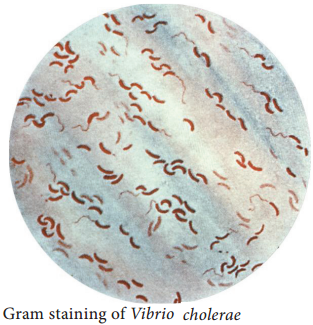
Vibrio cholerae is gram negative, curved or comma shaped, (1.5um × 0.2 – 0.4um in size) non – capsulated. The organism is very actively motile with a single polar flagellum and the characteristic movement is called as darting motility. In stained smears of mucus flakes from acute cholera patients, the Vibriois seen
arranged in parallel rows. This was described by Robert Koch as “fish in stream” appearance.

Culture Characteristics

Vibrio cholera is strongly aerobic. It grows best in alkaline media with the optimum temperature 37°C and pH 8.2. It is nonhalophilic, therefore, cannot grow in media with a concentration of sodium chloride more than 7% (Figure 7.17). Some of the media in which Vibrio cholerae are cultivated are tabulated below
in Table 7.16.

Table 7.16: Colony morphology of Vibrio cholerae on various media

Media

Colony morphology

Nutrient agar The colonies are moist, translucent round disks (1-2mm in diameter) with a bluish tinge in transmitted light.
MacConkey agar The colonies are colorless at first but become reddish on prolonged incubation due to late fermentation of lactose.
Thiosulphate citrate bile sucrose sugar (pH 8.6) It is used as a selective medium for isolation of Vibrios. It produces large yellow convex colonies due to sucrose fermentation.

Enterotoxin

Vibrios multiplying on the intestinal epithelium produce an enterotoxin called Cholera toxin. It is also known as Choleragen (or CT). This toxin molecule is approximately 84,000 Dalton and consists of two major subunits namely A and B There is only one subunit in A (1A) whereas there are five subunits in B (5B) (Figure 7.18).
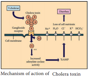
Vibrio cholerae img 2

Mode of Action

  • The B (binding) units of enterotoxin get attached to the GM1 (Ganglioside membrane receptors I) on the surface of jejunal epithelial cells. (target cells).
  • The A (active) subunits then enters the target cell and dissociates into 2 fragments, A1 & A2. The A2  fragment links biologically active A1 fragment to the B – subunit.
  • The A1 fragment causes prolonged activation of cellular adenylate cyclase which in turn accumulates CAMP in the target cell. This leads to outpouring of large quantities of water and electrolytes into small intestinal lumen. Thus, resulting in profuse watery diarrhea.
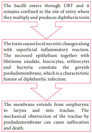
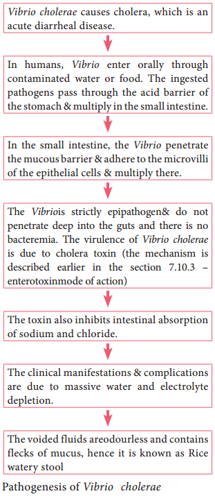
Pathogenesis

The pathogenic mechanism of Vibrio choleraeis discussed below in flowchart 7.7.
Source of Infection – contaminated water or food
Route of entry – fecal – oral route
Site of infection – small intestine
Incubation period – few hours to 5 days (usually 2 – 3 days)
Vibrio cholerae img 3

Clinical Feature

Dehydration, anuria (absence of urine excretion), muscle cramps, hypokalemia (low blood potassium) & metabolic acidosis (low serum concentration of bicarbonates).

Laboratory Diagnosis

Specimen: Stool

Direct microscopy:

It is not a reliable method for rapid diagnosis, the characteristic darting motility of the vibrio can be observed under dark – field microscope.

Culture:

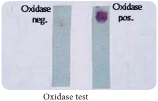
Stool sample is directly inoculated on MacConkey agar and TCBS agar. The plates are examined after overnight incubation at 37°C for typical colonies of Vibrio cholera, and the colonies are identified by gram staining and oxidasetest.

Prophylaxis

1. General Measures:

  • Purification of water supplies
  • Improvement of environment sanitation
  • Infected patients should be isolated, and their excreta must be disinfected

2. Vaccines:

Two types of oral vaccines have been tried recently:

  • Killed oral whole cell vaccines
  • Live oral vaccines

Treatment

1. Oral Rehydrationtherapy:

The severe dehydration & salt depletion can be treated by oral rehydration therapy (as recommended by WHO).

2. Antibiotics:

It is of secondary importance, oral tetracycline was recommended for reducing the period of Vibrio excretion.

Salmonella typhi of Medical Bacteriology

Learninsta presents the core concepts of Microbiology with high-quality research papers and topical review articles.

Salmonella typhi of Bacteriology

The genus Salmonella consists of bacilli that parasites the intestines of vertebrates and human beings. It causes Enteric fever, which includes Typhoid and Paratyphoid fever. The most important species of the genus is Salmonella typhi which causes typhoid fever.

Morphology

Salmonellae are Gram – negative rods (1 – 3µm × 0.5 µm in size). They are motile with peritrichous flagella, non – capsulated and non – sporulated (Figure 7.14).
Salmonella typhi of Bacteriology img 1

Cultural Characteristics

They are aerobic and facultative anaerobe, optimum temperature – 37°C and pH is 7 – 7.5. They grow on the following media and show the following characteristic colony morphology (Table 7.12).

Table 7.12: Colony morphology of Salmonella typhi

Media

Colony Morphology

Nutrient Agar Colonies are large, circular, smooth, translucent
MacConkey Agar Colourless colonies (non – lactose fermenters)
SS – Agar Colourless colonies with black centered.

Pathogenicity

Salmonella typhi causes typhoid fever and its pathogenesis is discussed in flowchart 7.6.

Source of infection – food, feces, fingers, flies
Route of entry – faecal oralroute (ingestion)
Incubation period – 7 – 14 days
Salmonella typhi of Bacteriology img 2

Clinical Manifestations

  • The illness is usually gradual, with headache, malaise (feeling of discomfort), an2orexia (loss of appetite), coated tongue, abdominal discomfort with either constipation or diarrhea.
  • Hepatosplenomegaly (enlargement of liver and spleen), step ladder pyrexia (continuous fever) and rose – spots (during 22nd or 3rd week).

Laboratory Diagnosis

Specimens:

Blood, stool and urine are the clinical samples collected from typhoid patients. The selection of relevant specimen depends upon duration of illness, which is very important for diagnosis (Table 7.13 & Figure 7.15).

Table 7.13: Specimen collection for typhoid

Duration of disease

Specimen examination

% Positivity

1st Week Blood culture 90
22nd Week Blood culture
Faeces culture
Widal test
75
50
Low titer
3rd Week Widal test
Blood culture
Faeces culture
80 – 100
60
80

Salmonella typhi of Bacteriology img 3

The bacteriological diagnosis of enteric fever consists of the following methods, which are:

  • Isolation of the bacilli
  • Demonstration of antibodies

Isolation of the bacilli

The typhoid bacilli are isolatedfrom the following clinical specimens which are tabulated (Table 7.14).

Table 7.14: Isolation method of typhoid bacilli from various clinical speciments.

Demonstration of Antibodies:

Slide – agglutination: The isolate is identified by slide agglutination with ‘O’ and ‘H’ antisera.

Widal Test:

It is an agglutination test for detection of agglutinins ‘H’ and ‘O’ in patients with enteric fever. Salmonella antibodies start appearing in the serum at the end of 1st week and rise sharply during the 3rd week of enteric fever.

Prophylaxis

Various types of vaccine and their doses are given in Table 7.15.

Table 7.15: Various types of vaccine and their doses.

Vaccine

Doses

TAB – Vaccine 2 doses of 0.5 ml at an interval of 4-6 weeks
Typhoral 3 doses on alternate days. It gives 65-96% protection for 3-5 years and is safe
Typhin – Vi A single dose of 25µg

Treatment and Control Measures

  • Antibacterial therapy has been very effective in the treatment of patients.
  • Ampicillin, amoxicillin and cotrimoxazole are useful in the treatment of typhoid fever.
  • At present, ciprofloxacin is the drug of choice.
  • Typhoid fever can be effectively controlled by sanitary measures for disposal of sewage, clean water supply and supervision of food processing and handling.

Medical Shigella dysenteriae

Learninsta presents the core concepts of Microbiology with high-quality research papers and topical review articles.

Medical Shigella dysenteriae

The genus Shigellaare exclusively parasites of human intestine and other primates. Shigella dysenteriae is the causative agent of bacillary dysentery or shigellosis in humans. It is a diarrheal illness which is characterized by frequent passage of bloodstained mucopurulent stools.

The four important species of the genus Shigella are: Shigella dysenteriae, Shigella flexneri, Shigella sonnei and Shigella boydii.

Morphology

Shigella are short, Gram negative rods (0.5µm × 1-3 µm in size). They are non – motile, non – sporing and non – capsulated (Figure 7.12).
Medical Shigella dysenteriae img 1

Cultural Characteristics

  • They are aerobes and facultative anaerobes. Optimum temperature is 37°C and optimum pH – 7.4.
  • They can be grown on the following media and show the characteristic colony morphology (Table 7.10 & Figure 7.13)

Table 7.10: Colony morphology of Shigella

Media

Colony Morphology

Nutrient Agar Colonies are circular, convex smooth and translucent
MacConkey Agar Colourless colonies
SS – Agar Colourless colonies

Medical Shigella dysenteriae img 2

Toxins

Shigella dysenteriae produces toxins, which is of 3 types, namely, endotoxin, exotoxin and verocytotoxin. The mode of action of these toxins is illustrated in the Table 7.11.

Table 7.11: Various toxins of Shigelladysenteriae

Toxins

Mode of Action

Endotoxin It is released ater autolysis, it has irritating effect on intestinal wall which causes diarrhea and subsequently intestinal ulcers.
Exotoxin It is a powerful toxin and acts as Enterotoxin as well as neurotoxin
As Enteroxin – It induces fluid accumulation
As Neurotoxin – It damages the endothelial cells of small blood vessels of CNS which results in polyneuritis and coma
Vero cytotoxin It acts on Vero cells

Pathogenesis

The pathogenic mechanism of Shigella dysenteriaeis discussed below in flowchart 7.5.

Source of Infection – Patient or carriers
Route of entry – faecal – oral route
Site of infection – Large intestine
Incubation Period – Less than 48 hours (1-7 days)
Mode of transmission – Food, finger, faeces and flies

Clinical Manifestations

  • Frequent passage of loose, scanty faeces containing blood and mucus.
  • Abdominal cramps and tenesmus (straining to defecate).
  • Fever and vomiting.
  • Hemolytic uremic syndrome (It is a condition caused by the abnormal destruction of red blood cells)

Laboratory Diagnosis

Specimens:
Fresh stool is collected.

Direct Microscopy:
Saline and Lugol’s iodine preparation of faeces show large number of pus cells, and erythrocytes.

Culture:
For inoculation, it is best to use mucus flakes (if present in the specimen) on MacConkey agar and SS agar. After overnight incubation at 37°C, the plates are observed for characteristic colonies, which is confirmed by Grams staining and biochemical reactions.

Treatment and Prevention

  • Uncomplicated shigellosis is a self – limiting condition that usually recovers spontaneously.
  • In acute cases, oral rehydration therapy (ORT) is done.
  • In all severe cases, the choice of antibiotic should be based on the sensitivity of prevailing strain.
  • Many strains are sensitive to Nalidixic acid and Norfloxacin.
  • Improving personal and environmental sanitation.
  • The detection and treatment of patients and carriers.

Clostridium tetani of Medical Bacteriology

Learninsta presents the core concepts of Microbiology with high-quality research papers and topical review articles.

Clostridium tetani of Medical Bacteriology

The genus Clostridium consists of anaerobic, spore forming Gram positive bacilli. The spores are wider than the bacterial bodies, giving the bacillus a swollen appearance resembling a spindle. The name Clostridium is derived from the word ‘kluster’ (a spindle).

Most species are saprophytes found in soil, water and decomposing plant and animal matter. Some of the pathogens are normal flora of intestinal tract of human and animals.

The genus Clostridium includes bacteria that causes 3 major diseases of human – Tetanus, gas gangrene and food poisoning. Clostridium pathogenicity is mainly due to production of a powerful exotoxin.

Clostridium of medical importance may be classified based on diseases they produce, which is given the Table (7.7).

Table 7.7: Clostridium sp, causing pathogenic diseases.

Organisms

Diseases

Clostridium tetani Tetanus
Clostridium perfringens Gas gangrene
Clostridium botulinum Food poisoning

Morphology

They are Gram positive spore forming rods. The spores are spherical and terminal in position giving a drumstick appearance. They are motile and non – capsulated.

Culture Characteristics

  • They are obligate anaerobes, optimum temperature is 37°C and pH is 7.4.
  • It grows on ordinary media, but growth is enhanced by addition of blood and serum. Clostridia tetani grows on the following media and show the characteristic colony morphology (Table 7.8).

Table 7.8: Colony characteristics of Clostridium tetani

Media

Colony Morphology

Blood agar They produce α – hemolysis which subsequently develop into β – hemolysis (due to tetanolysis) it produces swarming growth.
Cooked meat broth (CMB) Growth occurs as turbidity with gas formation. The meat is not digested but becomes black on prolonged incubation.

Toxins

Clostridium tetani produces two distinct toxins namely,

  • Tetanolysis (haemolysin)
  • Tetanospasmin (neurotoxin)

Tetanolysis

  • Heat labile and oxygen labile toxin.
  • It lysis erythrocytes and also acts as neurotoxin.

Tetanospasmin

  • It is heat labile and oxygen stable powerful neurotoxin.
  • It is protein in nature. consisting of a large polypeptide chain (93,000 Dalton) and a smaller polypeptide chain (52,000 Dalton) joined by a disulphide bond.
  • Mode of Action: Tetanospasminis a neurotoxin, which blocks the release of inhibitory neurotransmitters (glycine and gamma – amino butyric acid) across the synaptic junction.
  • The toxin acts presynaptically, the abolition of spinal inhibition causes uncontrolled spread of impulses in CNS (Central Nerves System).

This results in muscle rigidity and spasms (due to the simultaneous contraction of agonists and antagonists, in the absence of reciprocal inhibition (Figure 7.11).
Clostridium tetani of Medical Bacteriology img 1

Pathogenesis

Clostridium tetani is the causative organism of tetanus or lock jaw disease. pathogenesis of Clostridium tetani was discussed in detail in flowchart 7.4.

Source of infection – Soil, dust, faeces.
Route of entry – Through wound
Incubation period – 6 – 12 days

Clinical Feature

It includes, pain and tingling at the site of wound, Lock jaw ortrismus (It is reduced opening of the jaws), Risus sardonicus (mouth kept slightly open), Dysphasia (impairment of the ability to speak or to understand language) and acute asphyxia.

Laboratory Diagnosis

Specimens:
Wound swab, exudates or tissue from wound.

Microscopy:
Gram staining shows Gram positive bacilli with drumstick appearance.

Culture:
The clinical specimen is inoculated on blood agar and incubated at 37°C for 24-48 hours under anaerobic conditions. The colonies are confirmed by gram staining, where it shows gram positive bacilli with drumstick appearance.

Treatment

Tetanus patients are treated in special isolated units, to protect them from noise and light which may provoke convulsions. The spasm can be controlled by diazepam (0.1 – 0.2 mg/kg) injection. Antibiotic therapy with pencilin or metroniadazole should be done for a week or more.

Prophylaxis

It is done by the following methods, which are as follows.

a. Surgical prophylaxis:

It aims at removal of foreign body, blood clots and damaged tissue in order to prevent anaerobic conditions favorable for the
germination of spores.

b. Immunoprophylaxis:

Tetanus is a preventable disease. Immune prophylaxis is of 3 types, which is given in the (Table 7.9).
Table 7.9: Immunization for tetanus.
Clostridium tetani of Medical Bacteriology img 2

Corynebacterium diphtheriae

Learninsta presents the core concepts of Microbiology with high-quality research papers and topical review articles.

Corynebacterium diphtheriae

Several species of the genus Corynebacterium are normal flora of skin, upper respiratory tract (URT), urogenital and intestinal tract. The most important member of the genus is C. diphtheriae the causative agent of diphtheria, a localized inflammation of the throat with greyish white pseudomembrane and a generalized toxemia due to the secretion and dissemination of a highly potent toxin.

The name Corynebacterium diphtheria is derived from Greek word ‘Coryne’ – “Club shaped swellings” or “Knotted rod” ‘Diphthera’ – Leather.

Morphology

They are Gram positive slender rods, pleomorphic club shape or coryneform bacterium Non – motile, non – sporing and non – capsulated (Figure 7.9 a & b).
Corynebacterium diphtheriae img 1

The bacilli are arranged in a characteristic fashion in angular fashion resembling the letters V or L. This has been called Chinese letter or cuneiform arrangement (Figure 7.10).

They are club shaped due to the presence of metachromatic granules at one or both ends. These granules are composed of polymetaphosphates and represent energy storage depots.
Corynebacterium diphtheriae img 2

Cultural Characteristics

  • They are aerobic and facultative anaerobe. Optimum temperature is 37°C and pH 7.2.
  • They grow on the following media and show the characteristic colony morphology (Table 7.5).

Table 7.5: Colony Morphology of Corynebacterium diphtheriae on cultural media

Media

Colony Morphology

Loeffler’s Serum slope They glow on this medium very rapidly. Colonies appear after 6-8 hours of incubation. The colonies are small, circular white or creamy and glistening.
Tellurite Blood Agar Grey or black colonies. Based on colony morphology on tellurite medium, three main biotypes – Gravis, Intermedius and Mitis.

Toxin

• The pathogenicity is due to production of a very powerful exotoxin by virulent strains of diphtheria bacilli.
• The toxigenicity of diphtheria bacillus depends on the presence of a tox<sup>+</sup> gene which can be transferred from one bacterium to another by lysogenic bacteriophages, of which beta phage is the most important.

Properties

The diphtheria toxin is a heat – labile protein and has a molecular weight of about 62,000 Dalton. It consists of two fragments

  • Fragment A (24,000 Dalton) – It has all enzymatic activity.
  • Fragment B (38,000 Dalton) – It is responsible for binding the toxin to the target cells.

Mode of Action

The toxin acts by inhibiting protein synthesis, specifically fragment A inhibits polypeptide chain elongation in the presence of NAD by inactivating the elongation factor (EF – 2) the toxin has special affinity for myocardium, adrenal gland and nerve endings.

Pathogenicity

Source of infection – Airborne droplets
Route of entry – Upper respiratory tract
Incubation period – 3 – 4 days

Site of infection – Faucial (nasal, otitis, conjunctival, laryngeal, genital) diphtheria is most commonly seen in children of 2-10 years.

Faucial diphtheria is the most common type. The infection is confined to humans only. The toxin has both local (flowchart 7.3) as well as systemic effects.
Corynebacterium diphtheriae img 3
Flowchart 7.3: Localized effect of diphtheria toxin

Systemic effects

The toxin diffuses into the blood stream and causes toxemia. It has got affinity for cardiac muscle, adrenal and nerve endings. It acts on the cells of these tissues.

Clinical Manifestations

  1. Laryngeal obstruction, asphyxia (it is a condition of severe deficient supply of oxygen, causing suffocation).
  2. Diphtheritic myocarditis (inflammation of heart muscle), polyneuropathy (damage of multiple peripheral nerves), paralysis of palatine (the top part of the inside of the mouth) and ciliary muscles.
  3. Degenerative changes in adrenal glands, kidney and liver may occur.

Specimen:
Two swabs from the lesions are collected. One swab is used for smear preparationand other swab for inoculation on culturemedia.

Direct microscopy:
Smears are stained with both Gram stain and Albert stain.

  • Gram Staining – Gram positive slender rods were observed.
  • Albert staining – Club shaped with metachromatic granules were observed.

Culture:
The swabis inoculated on Loeffler’s serum slope, after overnight incubation at 37°C, the plates were observed for characteristic colonies, which are identified by gram staining.

Prophylaxis

Diphtheria can be controlled by immunization. Three methods of immunization are available (Table 7.6).
Corynebacterium diphtheriae img 4
Table 7.6: Immunization for diphtheria

Treatment

The specific treatment for diphtheria consists of administration of antitoxin with dose of 20,000-100,000 units of ADS intramuscularly and antibiotic therapy using penicillin.

Neisseria meningitidis (Meningococcus)

Learninsta presents the core concepts of Microbiology with high-quality research papers and topical review articles.

Neisseria meningitidis (Meningococcus)

The genus Neisseria is included in the family Neisseriaceae (Figure 7.6). It contains two important pathogens Neisseria meningitidis and Neisseria gonorrhoeae, both the species are strict human pathogens. N. meningitides causes meningococcal meningitis (formerly known as cerebrospinal fever).

The word Meningitis is derived from Greek word ‘meninx’ means membrane and ‘it is’ means inflammation. It is an inflammation of meanings of brain or spinal cord. Bacterial meningitis is a much more severe disease than viral meningitis.

Morphology

They are Gram negative diplococci (0.6µm-0.8µm in size) arranged typically in pairs, with adjacent sides flattened. They are non – motile, capsulated (Fresh isolates). Cocci are generally intracellular when isolated from lesions (Figure 7.7).
Neisseria meningitidis (Meningococcus) img 1

Cultural Characteristic

They are strict aerobes, but growth is facilitated by 5-10% CO<sub>2</sub> and high humidity. The optimum temperature is 35°C-36°C and optimum pH is 7.4-7.6. They are fastidious pathogens, growth occurs on media enriched with blood or serum. They grow on the following media and show the characteristic colony morphology (Table 7.4).
Neisseria meningitidis (Meningococcus) img 2

Table 7. 4: Colony morphology of Neisseria Meningitides on media

Name of the Media

Colony Morphology

Chocolate agar Colonies are large, colorless to grey opaque colonies.
Mueller Hinton agar Colonies are small, round, convex grey, translucent with entire edges. The colonies are butyrous in consistency and easily emulsified.

Pathogenesis

N. meningitidis is the causative agent of meningococcal meningitis, also known as pyogenic or septic meningitis. Infection is most common in children and young adults. Meningococci are strict human pathogens. Human nasopharynx is the reservoir of N.meningitidis. The pathogenesis is dicussed in the
flowchart 7.2.

Source of infection – Airborne droplets
Route of entry – Nasopharynx
Site of infection – Meninges
Incubation period – 3 days

Flowchart 7.2: Pathogenesis of Neisseria Meningitides
Neisseria meningitidis (Meningococcus) img 3

Laboratory Diagnosis

Specimens:

CSF, blood, nasopharyngeal scrapings from petechiae lesions are the specimens collected from pyogenic meningitis patients.

Direct Microscopy:

CSF is centrifuged, and smear is prepared from the deposit for gram staining. Meningococci are Gram negative diplococci, present mainly inside polymorphs and many pus cells are also seen.

Culture:

The centrifuged deposit of CSF is inoculated on chocolate agar. The plate is incubated at 36°C under 5-10% CO2 for 18-24 hours. After incubation period, meningococcusis identified by gram staining, colony morphology and biochemical reactions. N. meningitides is catalase and oxidase positive (Figure 7.8).
Neisseria meningitidis (Meningococcus) img 4

Treatment and Prophylaxis

Penicillin – G is the drug of choice. In penicillin allergic cases, chloramphenicol is recommended.

  • Monovalent and polyvalent vaccines (capsular polysaccharide) induce good immunity in older children and adults.
  • Conjugate vaccines are used for children below the age of 2 years.

Streptococcus pyogenes (Flesh eating Bacteria)

Learninsta presents the core concepts of Microbiology with high-quality research papers and topical review articles.

Streptococcus pyogenes (Flesh eating Bacteria)

The genus Streptococcus includes a large and varied group of bacteria. They inhabit various sites, notably the upper respiratory tract. However, some species of which Streptococcus pyogenes is the most important and are highly pathogenic. The name Streptococcus is derived from Greek word ‘Streptos’ which means twisted or coiled.

Morphology

  • They are Gram positive, spherical or oval cocci and arranged in chains (0.6µm-1µm)
  • They are non – motile, non – sporing. Some strains are capsulated (Figure 7.4).

Streptococcus pyogenes (Flesh eating Bacteria) img 1

Cultural Characteristics

  • They are aerobe and facultative anaerobe. Optimum temperature is 37°C and pH is 7.4 to 7.6
  • They grow only in media enriched with blood or serum. It is cultivated on blood agar. On blood agar, the colonies are small, circular, semitransparent, low convex, with an area of clear hemolysis around colonies (Figure 7.5).
  • Crystal violet blood agar – a selective medium for Streptococcus pyogenes.

Streptococcus pyogenes (Flesh eating Bacteria) img 2

Antigenic Structure

Capsule:

It inhibits phagocytosis

Cell wall:

The outer layer of cell wall consists of protein and lipoteichoic acid which helps in attachment to the host cell. Middle layer of cell wall consists of Group Specific C – Carbohydrate that is used for Lancefield grouping. Inner layer of cell wall is made up of peptidoglycan which has pyrogenic and thrombolytic activity.

Toxins and Enzymes:

Streptococcus pyogenes produces several exotoxins and enzymes which contribute to its virulence.

Toxins and Hemolysins:

Streptococci produces two types of hemolysins which are Streptolysin O and Streptolysin S.

Erythrogenic toxin: (Pyrogenic exotoxin)

The induction of fever is the primary effect of this toxin and it is responsible for the rash of scarlet fever.

Enzymes:

The various enzyme of Streptococcus pyogenes which exhibits virulence activity are listed in Table 7.3.

Enzymes of Streptococcus pyogens and its virulence nature

Enzymes

Virulence nature

Streptokinase (fibrinolysin) It promotes the lysis of human fibrin clot by catalyzing the conversion of plasminogen into plasmin. It facilitates the spread of infection by breaking down the fibrin barrier around the lesions.
Deoxyribonucleases It liquefy the highly viscous DNA that accumulate in thick pusand responsible for thin serous character of streptococcal exudates
Hyaluronidase It breaks down hyaluronic acid of the tissues and favours spread of streptococcal lesion along intercellular spaces.
Other enzymes NADase, lipase, amylase, esterase, phosphates and other enzymes.

Pathogenesis

Streptococcus pyogenes is intrinsically a much more dangerous pathogen than Staphylococcus aureus and has a much greater tendency to spread in the tissues.

Mode of transmission:

Streptococcal infections are transmitted by the following ways:
Streptococcus pyogenes (Flesh eating Bacteria) img 3

Streptococcal diseases may be broadly classified, and it is shown in flowchart 7.1
Streptococcus pyogenes (Flesh eating Bacteria) img 4

Suppurative Infections

1. Respiratory tract infection

a. Streptococcal sore throat:

Sore throat (acute tonsillitis and pharyngitis) is the most common streptococcal diseases. Tonsillitis is more common in older children and adults. The pathogen may spread from throat to the surrounding tissues leading to suppurative (pus – formation) complication such as cervical adenitis (inflammation of a lymph node in the neck) otitis media (inflammation of middle ear), quinsy (ulcers of tonsils) Ludwig’s angina (purulent inflammation around the sub maxillary glands) mastoiditis (inflammation of mastoid process).

b. Scarlet fever:

The disease consists of combination of sore throat and a generalized erythematous (redness of skin or mucous membranes) rash.

2. Skin infections

a. Erysipeals:

It is an acute spreading lesion. The skin shows massive brawny oedema with erythema it is seen in elderly persons or elders.

b. Impetigo: (Streptococcal pyoderma)

It is a skin infection that occurs most often in young children. It consists of superficial blisters that break down and eroded areas whose surface is covered with pus. It is the main cause leading to acute glomerulonephritis in children.

c. Necrotizing fasciitis:

It is an invasive, infection characterized by inflammation and necrosis of the skin, subcutaneous fat and fascia. It is a life-threatening infection.

3. Streptococcal toxic shock syndrome

Streptococcal pyrogenic exotoxin leads to streptococcal toxic shock syndrome (TSS). It is a condition in which the entire organ system collapses, leading to death.

4. Genital infections

Streptococcus pyogenes is an important cause of puerperal sepsis or child bed fever (infection occur when bacteria infect the uterus following child birth)

5. Deep infection

Streptococcus pyogenes may cause pyaemia (blood poisoning characterized by pus forming pathogens in the blood) septicemia (A condition in which bacteria circulate and actively multiply in the bloodstream) abscess in internal organs such as brain, lung, liver and kidney.

Non – Suppurative Complication

Streptococcus pyogenes infections are sometimes followed by two important non – suppurative complications which are, acute rheumatic fever and acute glomerulonephritis. These complications occur 1-4 weeks after the acute infection and it is believed to be the result of hypersensitivity to some streptococcal
components.

1. Rheumatic fever

It is often preceded by sore throat and most serious complication of haemolytic streptococcal infection. The mechanism by which Streptococci produce rheumatic fever is still not clear. A common cross – reacting antigen exist in some group A streptococci and heart, therefore, antibodies produced in response to the streptococcal infection could cross react with myocardial and heart valve tissue, causing cellular destruction.

2. Acute glomerulonephritis

It is often preceded by the skin infection. It is caused by only a few “nephritogenic types (strains)”. It develops because some components of glomerular basement membrane are antigenically similar to the cell membranes of nephritogenic streptococci.

The antibodies Formed against Streptococci cross react with glomerular basement membrane and damage. Some patients develop chronic glomerulonephritis with ultimate kidney failure.

Laboratory Diagnosis

Specimens:

Clinical specimens are collected according to the site of lesion. Throat swab, pus or blood is obtained for culture and serum for serology.

Direct Microscopy:

Gram stained smears of clinical specimens is done, where Gram positive cocci in chains were observed. It is indicative of streptococcal infection.

Culture:

The clinical specimen is inoculated on blood agar medium and incubated at 37° C for 18-24 hours. After incubation period, blood agar medium was observed for zone of beta – haemolysis around colonies.

Catalase test:

Streptococci are catalase negative which is an important test to differentiate Streptococci from Staphylococci.

Serology:

Serological tests are done for rheumatic fever and glomerulonephritis. It is established by demonstrating high levels of antibody to streptococci toxins. The standard test is antistreptolysin Otitration. ASOtitres higher than 200 units are indicative of prior Streptococcal infection.

Treatment and Prophylaxis

  • Penicillin G is the drug of choice.
  • In patients allergic to penicillin, erythromycin or cephalexin is used.
  • Antibiotics have no effect on established glomerulonephritis and rheumatic fever.
  • Prophylaxis is indicated only in the prevention of rheumatic fever, it prevents streptococcal reinjection and further damage to the heart.
  • Penicillin is given for a long period in children who have developed early signs of rheumatic fever.

Medical Bacteriology of Staphylococcus aureus

Learninsta presents the core concepts of Microbiology with high-quality research papers and topical review articles.

Medical Bacteriology of Staphylococcus aureus

The genus Staphylococcus is included in the family Micrococcaceae. Staphylococcus is a normal flora of skin and mucous membranes, but it accounts for human infections, which is known as staph infection.

The name Staphylococcus was derived from a Greek word, ‘staphyle’ means bunch of grapes and ‘kokkos’ means berry. Staphylococcus aureus is a pathogenic species that causes pyogenic infections in human.

Morphology

  • Staphylococci are gram positive spherical cocci, (0.8µm-1.0µm in diameter) arranged characteristically in grape like clusters (Figure 7.1).
  • They are non-motile and non-sporing and few strains are capsulated.

Medical Bacteriology of Staphylococcus aureus img 1

Cultural Characteristics

  • They are aerobes and facultative anaerobes, optimal temperature is 37°C and optimum pH is 7.4-7.6.
  • They grow on the following media and shows the characteristic colony morphology (Table 7.1 & Figure 7.2).

Staphylococci aureus colony morphology on various media

Media Colony Morphology
Nutrient Agar Colonies are circular, smooth, convex, opaque and produces golden yellow pigment (most strains).
Blood Agar Beta haemolysis
Mannitol salt Agar (MSA) It is a selective medium for S. aureus produces yellow colored colonies due to fermentation of mannitol.

Medical Bacteriology of Staphylococcus aureus img 2

Virulence Factors

  1. Peptidoglycan → It is a polysaccharide polymer. It activates complement and induces the release of inflammatory cytokines.
  2. Teichoic acid → it facilitates adhesion of cocci to the host cell surface.
  3. Protein A → It is chemotactic, antiphagocytic, anticomplementary and induce platelet injury.

4. Toxins:

  • Hemolysins – It is an exotoxin, those lysis red blood cells. They are of four types namely α-lysin, β-lysin, γ-lysin and delta lysin.
  • Leucocidin – It damages PMNL (polymorphonuclear leucocytes) and macrophages.
  • Enterotoxin – It is responsible for manifestations of Staphylococcus food poisoning.
  • Exfoliative toxin – This toxin causes epidermal splitting resulting in blistering diseases.
  • Toxic shock syndrome toxin – TSST is responsible for toxic shock syndrome.

5. Enzymes:

S. aureus produces several enzymes, which are related to virulence of the bacteria.

  • Coagulase – It clots human plasma and converts fibrinogen into fibrin.
  • Staphylokinase – It has fibrinolytic activity.
  • Hyaluronidase – It hydrolyzes hyaluronic acid of connective tissue, thus facilitates the spread of the pathogens to adjacent cells.
  • Other enzymes – S. aureus also produces lipase, nucleases and proteases.

Pathogenicity

S. aureus is an opportunistic pathogen which causes infection most commonly at sites of lowered host resistance. (Example: damaged skin) Mode of Transmission: Staphylococcus infections are transmitted by the following ways.

Medical Bacteriology of Staphylococcus aureus img 3

It includes the following infections, which are as follows:

Cutaneous infections:

Wound (injury), burn infections (tissue injury caused by heat), pustules (A small elevated skin lesions containing pus), furuncles (boil forms around a hair follicle and containspus), styes (a painful swelling of hair follicle at eyelids), carbuncles (painful cluster of boils of the skin), Impetigo (skin infection with vesicles, pustules which ruptures), pemphigus neonatorum (an auto immune diseases that affect skin and
mucous membranes)

Deep infections:

It includes Osteomyelitis (inflammation of bones), tonsillitis (inflammation of tonsils), pharyngitis (inflammation of pharynx) sinusitis (inflammation of sinuses), periostitis (inflammation of membrane covering bones), bronchopneumonia (inflammation of lungs), empyema (collection of pus in the body cavity), septicemia (blood poisoning caused by bacteria and its toxins), meningitis (inflammation of meninge), endocarditis (inflammation of endocardium), breast and renal abscess.

Food Poisoning:

Staphylococcal food poisoning may follow 2-6 hours after the ingestion of contaminated food (preformed enterotoxin). It leads to nausea, vomiting and diarrhea.

Nosocomial infection:

S. aureus is a leading cause of hospital acquired infections. It is the primary cause of lower respiratory tract (LRT) infections and surgical site infections and the second leading cause of nosocomial bacteremia, pneumonia, and Cardiovascular infections.

Exfoliative diseases:

These diseases are produced due to the production of epidermolytic toxin. The toxin separates the outer layer of epidermis from the underlying tissues leading to blistering disease. The most dramatic manifestation of this toxin is scalded skin syndrome. The patient develops painful rash which slough off and skin surface resembles scalding.

Laboratory Diagnosis

Specimens:

The clinical specimens are collected according to the nature of Staphylococcal infections, which is given in the (Table 7.2).

Table 7.2: Clinical specimen collected for Staphylococcal infections

Infections

Clinical Specimens

Supportive lesions Pus
Respiratory infections Sputum
Septicemia Blood
Meningitis CSF
Food poisoining Faeces, food or vomitus

Specimens should be transported immediately to the laboratory and processed.

Direct Microscopy:

Gram stained smears of clinical specimens is done, where gram positive cocci in clusters were observed.

Culture:

The collected specimen is inoculated on selective media-MSA and the media incubated at 37°C for 18-24 hours. Next day culture plates are examined for bacterial colonies, which are identified by gram staining, colony morphology and biochemical tests such as
Medical Bacteriology of Staphylococcus aureus img 4

a. Catalase test:
The genusStaphylococci are catalase positive. This test distinguishes Staphylococcus from Streptococcus (catalase negative).

b. Coagulase test:
This test helps in differentiating a pathogenic strain from non-pathogenic strain. S. aureus is coagulasepositive (Figure 7.3).

Treatment

Benzyl penicillin is the most effective antibiotic. Cloxacillin is used against beta lactamase. Producing strains (β-lactamase is produced by few strains of S. aureus which cleaves β-lactam ring of penicillin). Vancomycin is used against MRSA (Methicillin Resistant Staphylococcus aureus) strains.

Topical applications:

For mild superficial lesions, topical applications of bacitracin or chlorhexidine is recommended.

Control measures:

Proper sterilization of medical instruments must be done. Intake of antibiotics must be taken under proper medical advice. The detection of source & carriers among hospital staff, their isolation and treatment should be practiced.

Routes of Entry

Learninsta presents the core concepts of Microbiology with high-quality research papers and topical review articles.

Routes of Entry

To establish an infection, pathogen must first enter the host. Normal defense mechanisms and barriers (For example Skin, mucus, ciliated epithelium, lysozyme) make it difficult for the pathogen to enter the body.

Sometimes these barriers are break through for example cut in the skin, wound, tumor, ulcer which provides portal of entry for the bacteria. Some bacterial pathogens have the means to overcome the barriers through various virulence factors and invade the body.

Certain bacteria are infective when introduced through optimal route. The various route of entry of pathogens, which are cut or abrasion or wound (skin), Ingestion, Inhalation, arthropod bite, sexual transmission and congenital transmission.

These are already explained in the XI Standard text book. The various bacterial pathogens, its pathogenesis clinical symptoms, laboratory diagnosis, control, prophylaxis and treatment with appropriate antibiotics are discussed below.

The way a substance is able to enter the body such as ingestion (mouth), inhalation (lungs) or absorption (cintact).

There are four major routes by which a chemical may enter the body:

  • Inhalation (breathing).
  • Skin (or eye) contact.
  • Swallowing (ingestion or eating).
  • Injection.

Pathogenic Attributes of Medical Bacteriology

Learninsta presents the core concepts of Microbiology with high-quality research papers and topical review articles.

Pathogenic Attributes of Medical Bacteriology

The host-parasite relationship is determined by the interaction between host factors and the infecting pathogens.Pathogenicity refers to the ability of a pathogen to produce disease. Virulence is the ability of the pathogen to cause disease.

Adhesion, invasiveness (Streptococcal infections), Bacterial toxins (endotoxins and exotoxins), capsule enzymes (proteases, collagenase, coagulase and other enzymes). These are already explained in the XI Standard text book.

Pathogenicity refers to the ability of an organism to cause disease (ie, harm the host). This ability represents a genetic component of the pathogen and the overt damage done to the host is a property of the host-pathogen interactions. Commensals and opportunistic pathogens lack this inherent ability to cause disease.

Pathogen types. There are different types of pathogens, but we’re going to focus on the four most common types: viruses, bacteria, fungi, and parasites.

The definition of a pathogenic organism is an organism capable of causing disease in its host. A human pathogen is capable of causing illness in humans. Common examples of pathogenic organisms include specific strains of bacteria like Salmonella, Listeria and E. coli, and viruses such as Cryptosporidium.

Decrease your risk of infecting yourself or others:

  1. Wash your hands often.
  2. Get vaccinated.
  3. Use antibiotics sensibly.
  4. Stay at home if you have signs and symptoms of an infection.
  5. Be smart about food preparation.
  6. Disinfect the ‘hot zones’ in your residence.
  7. Practice safer sex.
  8. Don’t share personal items.

Immobilization of Industrail Microbiology

Learninsta presents the core concepts of Microbiology with high-quality research papers and topical review articles.

Immobilization of Industrail Microbiology

It is technique used for the physical or chemical fixation of plant, animal cells, organelles, enzymes or other proteins (monoclonal antibodies) onto a solid matrix or retained by a membrane, in order to increase their stability and make possible their repeated or continued use.

The immobilized enzyme is defined as the enzyme physically confined or localized in a certain defined region of space with retention of its catalytic activity which can be used repeatedly and continuously.

The selection of appropriate carrier and immobilization procedure is very essential procedure is very essential for the immobilization technique.

Various types of materials like cellulose, dextran, agarose, gelatin, albumin polystyrene, Calcium alginate polyacrylamide, collagen carrageenan and polyurethane, inorganic materials (brick, rand, glass, and ceramics, magnetic) are used for immobilization.

The linkage is mediated by ionic bonds, physical absorption or bio specific binding.

The immobilization methods can be classified into four categories:-

  1. Carrier–binding
  2. Cross–linking
  3. Entrapping
  4. Combining

Among all these methods entrapping is discussed in brief.

Entrapping

The enzymes, cells are not directly attached to the support surface, but simply trapped inside the polymer matrix. Entrapping is carried out by mixing the biocatalyst into a monomer solution followed by a polymerization. It is done by change in temperature or by chemical reactions.

Advantages of immobilization

  1. Immobilized growing cells serve as self proliferating and self regenerating bio catalyst
  2. They are stable
  3. They are used either repeatedly in a series of batch wise reactions or continuously in flow systems.