Food Microbiology of Cheese

Learninsta presents the core concepts of Microbiology with high-quality research papers and topical review articles.

Food Microbiology of Cheese

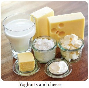
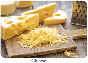
There are about 2000 varieties of cheese made from mammalian milk. Cheese is thought to have originated in south western Asia some 8000 years ago. The Romans encouraged technical improvements and stimulated the development of new varieties during their invasion in Europe between 60 B.C and A.D. 300. The cheese name is derived from Latin name caseus (Figure 5.5).
Food Microbiology of Cheese img 1

They are two groups of cheese, fresh cheese and ripened cheese. The fresh cheese are made up of milk coagulated by acid or high heat. Example: cottage cheese. Ripened cheese are made through lactic acid bacterial fermentation and coagulated by an enzyme preparation. The curd is removed and salted and whey
is separated. The salted curd is held in controlled environment.

During this process, various physical and chemical changes occur to give a characteristic flavour and texture. So the mammalian origin of milk influences the flavour and aroma of a natural ripened cheese.

Microbiology of cheese

A large number of microorganisms plays a role in the ripening process. On the first day of cheese making process, the microbial number in the starting material ranges from one to two billion. Therefore, the production declines because of insufficient oxygen, high acidity and the presence of inhibitory
compounds that are produced as the cheese ripens.

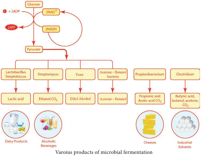
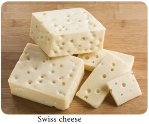
It is mainly the action of their cellular enzymes on lactose, fat and proteins that creates the ripened cheese flavour. The gas forming culture of Propionibacterium shermanii is essential for giving swiss cheese its eye, or holes and flavour (Figure 5.6).
Food Microbiology of Cheese img 2

The specificity of cheese depends upon the varieties of microorganisms used. The process of cheese making, involves nine steps:

a. Preparing the milk
b. Forming a curd.
c. Cutting
d. Cooking
e. Separating the whey
f. Salting the residue
g. Applying microbes
h. Pressing the curd
i. Ripening the young cheese
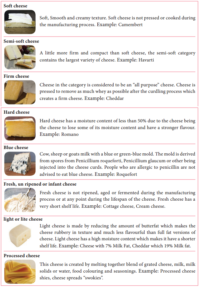
Types of Cheese

Cheese can be divided among different categories or types, according to their firmness. There are various system for classifying cheese and there are variations within each system (Table 5.6).

Types of Cheese
Food Microbiology of Cheese img 3

Dairy Microbiology

Learninsta presents the core concepts of Microbiology with high-quality research papers and topical review articles.

Dairy Microbiology

The area of dairy microbiology is large and diverse. The bacteria in dairy products may cause disease or spoilage. Some bacteria may be specifically added to milk for fermentation to produce products like yoghurts and cheese (Figure 5.3).
Dairy Microbiology img 1

MILK

Milk is the fluid, secreted by mammals for the nourishment of their young ones. It is in liquid form without having any colostrum. The milk contains water, fat, protein and lactose. About 80 – 85% of the protein is casein. Due to moderate pH (6.4 – 6.6), good quantity of nutrients and high water content, milk an excellent nutrient for the microbial growth. (Flowchart 5.2).
Dairy Microbiology img 2

Flowchart 5.2: Various products obtained from raw milk.

pH – Hydrogen ion concentration
T – Elevated temperature
H – Reduced water pressure
aw – water activity diet. It is an extremely complex mixture and usally contains (Table 5.3).

Complex mixture

Composition

Approximate percentage

1. Liquid (Water) 87%
2. Solids 13%
3. Fat 4%
4. Protein 3.3%
5. Lactose (Milk Sugar) 5%
6. Ash content (Vitamins and minerals) 0.7%

Sources of Microorganisms in Milk

Three sources contribute to the microorganism found in milk the udder interior, the teat exterior and its immediate surroundings, and the milking and milk handling equipment.

Bacteria that get on to the outside of the teat may be able to invade the opening and hence the udder interior. The organisms most commonly isolated are Micrococcus, Streptococci and the diptheroid Corynebacterium bovis. Aseptically taken milk from a healthy cow normally contains low number of organisms, typically fewer than 102 – 103 cfu ml-1.

The udder exterior and its immediate environment can be contaminated with organisms from the cow’s general environment.

Heavily contaminated teats have been reported to contribute up to 105 cfu ml-1 in the milk. Contamination
from bedding and manure can be source of human pathogens such as E.Coli, Campylobacter, Salmonella, Bacillus spp. and Clostridia spp.

Milk – handling equipment such as teat cups, pipe work, milk holders and storage tanks is the principal source of the microorganisms found in raw milk. Micrococcus and Enterococcus.

Microbiological Standard and Grading of Milk

In India, raw milk is graded by Bureau of Indian standards (BIS) 1977. The Indian standard institute (ISI) has prescribed microbiological standard for quality of milk.

  1. Coliforms count in raw milk is satisfactory if, coliforms are absent in 1:100 dilution.
  2. Coliforms count in pasteurized milk is satisfactory is coliforms are absent in 1:10 dilution (Table 5.4).

Microbiological Standard and Grading of Milk
Dairy Microbiology img 3

Grading of milk

The quality of milk is judged by certain standards and it is known as grading milk. Grading of milk is based upon regulations pertaining to production, processing and distribution.

This includes sanitation, pasteurization, holding conditions and microbiological standards. The U.S public health secrine publication “Milk ordinance and code” shows the following chemical, bacteriological and temperature standards for grade A milk and milk products.

Methylene Blue dye Reduction Test (MBRT)

Methylene blue dye reduction test commonly known as MBRT test is used as a quick method to access the microbiological quality of raw and pasteurized milk. This test is based on the fact that the blue colour of the dye solution added to the milk get decolorized when the oxygen present in the milk get exhausted due to microbial activity.

The sooner the de colorization, more inferior is the bacteriological quality of milk assumed to be MBRT test may be utilized for grading of milk which may be useful for the milk processor to take a decision on further processing of milk.

Procedure

The test has to be done under sterile conditions. Take 10ml milk sample in sterile MBRT test tube. Add 1 ml Methylene Blue dye solution (dye concentration 0.005%). Stopper the tubes with sterilized rubber stopper and carefully place them in a test tube stand dipped in a serological water bath maintained at 37°C, records this time as the beginning of the incubation period. Decolourization is considered complete when only a faint blue ring (about 5mm) persists at the top (Figure 5.4).
Dairy Microbiology img 4

Recording of Results – During incubation, observe colour changes as follows:

a. If any sample is decolourized on incubation for 30 minutes, record the reduction time as MBRT 30 minutes.

b. Record such readings as, reduction times in whole hours. For example, if the colour disappears between 0.5 and 1.5 hour readings, record the result as MBRT 1 hour, similarly, if between 1.5 and 2.5 hours as MBRT-2 hour and so on.

c. Immediately after each, reading, remove and record all the decolourized samples and then gently invert the remaining tubes if the decolourization has not yet begun (Table 5.5).

Microbilogical Quality of MilkDairy Microbiology img 5

Food Preservation Methods – An Overview

Learninsta presents the core concepts of Microbiology with high-quality research papers and topical review articles.

Food Preservation Methods – An Overview

Foods can be preserved by a variety of methods. It is vital to eliminate or reduce the populations of spoilage and disease – causing microorganisms and to maintain the microbiological quality of a food with proper storage and packaging. Contamination often occurs after a package or can is opened and just before
the food is served.

This can proved an ideal opportunity for growth and transmission of pathogens, if care is not taken. Preservation of food is the process by which food is stored by special methods. Cooked or uncooked food can be preserved in different ways to be used later Table 5.2. Some methods of preservation are:

Basic Approaches to Food Preservation

Food Preservation Methods img 1

1. Freezing

Food kept in a refrigerator remains fresh for some day. Germs do not grow easily in cool places. We preserve food items, like milk, fruit, vegetable and cooked food by keeping them in a refrigerator.

2. Boiling

By this method, we can preserve food for a short period of time. Germs in milk are killed by pasteurization. It is done by boiling milk for sometimes and then cooling it quickly.

3. Salting

Add salt to preserve pickles and fish.

4. Sweetening

Sugar act as a preservative when added in large quantities. For example, food can be stored for a long time in the form of jams, jellies and murabbas (Figure 5.2) by adding sugar.
Food Preservation Methods img 2

5. Drying

In this method, the food items are dried in sun to stop the growth of bacteria in them. Certain foods, like raw mangoes, fishes, potato chips and papads are preserved by this method.

6. Canning

In this method, food is processed and sealed in airtight cans. Food items like vegetables, seafood, and dairy product are preserved through this method.

Advantages of food preservation:-

  • Germs do not grow easily in preserved food and make it safe to eat.
  • Preservation enables us to enjoy seasonal fruits like strawberries and mangoes even during the off-season.

Disadvantages:-

  • Excess salt and sugar are used in the preservation of food which is not good for health.
  • Some methods of food preservation may lead to loss of nutrients.

Principles of Food preservation

In accomplishing the preservation of foods by the various methods, the following principles are involved.

1. Prevention or delay of microbial decomposition.

  • By keeping out microorganism (asepsis)
  • By removal of microorganism. Example: Filtration
  • By hindering the growth and activity of microorganism Example: Low temperature, drying, anaerobic conditions or chemicals.
  • By killing the microorganism Example: Heat or radiation

2. Prevention or delay of self – decomposition of the food.

  • By destruction or inactivation of food enzymes Example: Blanching
  • By prevention or delay of purely chemical reactions Example: Prevention of oxidation by means of antioxidants.

3. Prevention of damage because of insects, animals, mechanical causes, etc.

Food Borne Disease

Learninsta presents the core concepts of Microbiology with high-quality research papers and topical review articles.

Food Borne Disease

Food borne disease has been defined by the world health organization (WHO) as a disease of an infectious or toxic nature caused by or thought to be caused by the consumption of food or water.

The term “food poisoning” as applied to diseases caused by microorganisms is used very loosely to include both illness caused by the ingestion of toxins elaborated by the organisms and those resulting from infection of the host through the intestinal tract. A further classification of food borne disease is shown in flowchart 5.1.

All these food – borne diseases are associated with poor hygienic practices.

Whether by water or food transmission, the fecal – oral route is maintained, with the food providing the vital link between hosts. Fomites, such as sink faucets, drinking cups, and cutting boards, also play a role in the maintenance of fecal – oral route of contamination.

There are two primary types of food related diseases: food – borne infections and food intoxications or food poisoning.

Food Borne Disease img 1

Food Borne Infection

Food borne infection involves the ingestion of the pathogen followed by growth in the host, including tissue invasion and/or the release of toxins. The major diseases of this type are summarized in table (5.1).

Major Food – Borne Infectious Diseases
Food Borne Disease img 2

Food Poisoning

Food borne intoxication (or) food poisonings is caused by ingesting food containing toxins formed by bacteria which resulted from the bacterial growth in the food item. Food poisoning refers to the toxicity introduced into food by microorganisms and their products.

Microbial growth in food products also can results in food intoxication.

Intoxication produces symptoms shortly after the food is consumed because growth of the disease – causing microorganism is not required. Toxins produced in the food can be associated with microbial cells or can be released from the cells.

Food poisoning is caused by various factors as follows.

  1. Microorganism of plant food products.
  2. Microorganism of Animal food products.
  3. Microorganism of processed food.
  4. Standard chemicals added to the food.
  5. Excess use of preservatives in food.
  6. Presence of higher population of Microorganism in food.
  7. Toxin produced by various types of Microorganism.

Food Spoilage and its Causes

Learninsta presents the core concepts of Microbiology with high-quality research papers and topical review articles.

Food Spoilage and its Causes

Spoilage of food can be defined as any visible or invisible change which can make food or product derived from food unfit for human consumption. Spoilage of food not only causes health hazard to the consumer but also causes great economic losses. Spoilage leads to loss of nutrients from food and cause change in
original flavor and texture.

It is estimated that about 25% of total food produced is spoilt due to microbial activities despite a range of preservation methods available. Food spoilage is considered as a complex phenomenon where by a combination of microbial and bio-chemical activities take place. Due to such activities various types
of metabolites are formed which aid in spoilage (Figure 5.1).

Food Spoilage img 1

i. Perishable foods

These foods are readily spoiled; require special preservation and storage condition for use. This includes, foods such as dairy products, eggs, poultry, meat, fish, fruits and vegetable. These foods get spoiled easily by natural enzymes.

ii. Semi – perishable foods

This class of foods if properly stored can be used for a longer duration. These foods include processed cereals, pulses and their products like flour, semolina, parched rice and popcorn. Shelf life of these products depends on the storage temperature and moisture in the air.

Foods like potato, onion, nuts, frozen foods and certain canned foods can be stored for a week to a couple of months at room temperature without any undesirable changes in the products.

iii. Non – perishable foods

These foods remain stable for long period unless handled improperly. Nonperishable foods include sugar, jaggery, hydrogenated fat, vegetable oil, ghee, whole grains, dhals, whole nuts and processed foods like dry salted fish/meat, papads, canned foods, jams and murabbas. These foods do not spoil unless they are
handled carelessly.

Causes of Food Spoilage

Food and water may be infected by germs. Fly carries germs to food. There are various factors which are responsible for food spoilage such as.

  • Microorganism
  • Insects
  • Rough handling
  • Transport
  • improper storage
  • enzyme activity (Chemical reaction)
  • unhygienic conditions
  • physical changes, such as those caused by freezing, burning, drying pressure.

Signs of food spoilage include difference in appearance from the fresh food such as a change in colour, a change in texture and an unpleasant odour or taste.

Scope of Food Microbiology

Learninsta presents the core concepts of Microbiology with high-quality research papers and topical review articles.

Scope of Food Microbiology

The field of food microbiology is very broad, encompassing the study of microorganisms which have both beneficial and deleterious effects on the quality and safety of raw and processed foods. The primary tool of microbiologists is the ability to identify and quantitate foodborne microorganisms. Microorganisms in food include bacteria, molds, yeasts, algae, viruses, parasitic worms and protozoans.

Microorganisms are associated with the food we eat in a variety of ways. They may influence the quality of our food. Naturally occurring foods such as fruits and vegetables normally contain some microorganisms and may be contaminated with additional organisms during handling and processing.

Food can serve as a medium for the growth of microorganism, and microbial growth may cause the food to undergo decomposition and spoilage.

Food may also carry pathogenic microorganisms which when ingested can cause disease. When food with microorganisms that produce toxic substances is ingested, it results in food poisoning. Some microorganisms are used in the preparation and preservation of food products.

Classification of Foods

Foods may be classified as

a. Fresh foods

These are foods which have not been preserved and not spoiled yet. For example; vegetables, fruits and meat spoil immediately after harvesting or slaughtering.

b. Preserved foods

Foods are preserved by adding salt, sugar, acetic acids and ascorbic acids. Example: Jam, Pickles. In this way their shelf life is improved.

c. Canned foods

In canning, food products are processed and sealed in the air tight containers. It provides longer shelf life ranging from one to five years. Example: Baked beans, Olives.

d. Processed foods

During food processing, original nature of food is changed or altered. It is done by Freezing, Canning, Baking and Drying. Example: Breakfast cereals, Cakes, Biscuits and Bread.

e. Fermented food products

These foods are subjected to fermentation by the action of microorganisms. Example: Kefir, Cheese.

Sources of Microorganism in Food

The primary sources of microorganisms in food include,

  1. Soil and water
  2. Plant and plant products
  3. Food utensils
  4. Intestinal tract of human and animals
  5. Food handlers
  6. Animal hides and skins
  7. Air and dust

Factors that Influence Growth of Microorganisms in Food

Many factors influence the growth of the microorganisms in food. Some of the factors are intrinsic and some others are extrinsic.

1. Intrinsic factors

The intrinsic factors include pH, moisture content, oxidation – reduction potential, nutrient status, antimicrobial constituents and biological structures.

a. pH:

Every microorganisms has a minimal or maximal, and an optimal pH for its growth. Microbial cells are significantly affected by the pH of food because they apparently have no mechanism for adjusting their internal pH. In general, yeasts and molds are more acid tolerant than bacteria.

Foods with low pH values (below 4.5) are usually not readily spoiled by bacteria and are more susceptible to spoilage by yeast and molds. Most of the microorganisms grow best at pH value around 7.0.

b. Moisture content:

The preservation of food by drying is a direct consequence of removal of moisture, without which microorganisms do not grow. The water requirement of microorganism is defined in terms of the water activity (aw) in the environment. Water activity is defined as the ratio of the water vapour pressure of food substrate to the vapour pressure of pure water at the same temperature.

The water activity of most fresh food is above 0.99. The minimum value of aw for the growth of the microorganisms in foods should be around 0.86.

c. Oxidation reduction (O/R) potential

The oxygen tension or partial pressure of oxygen around a food and the O-R potential or reducing and oxidizing power of the food itself influence the type of organisms which can grow and the changes produced in the food. The O-R potential of the food is determined by,

  • The O-R potential of the original food.
  • The poisoning capacity (the resistance of the food against change).

d. Nutrient Content

The kinds and proportions of nutrients in the food are all important in determining what organism is most likely to grow. Consideration must be given to (i) foods for energy (ii) foods for growth and (iii) accessory food substances or vitamins which may be necessary for energy or growth.

e. Antimicrobial constituents

The stability of foods against attack by microorganism is due to the presence of certain naturally occurring substances that have been shown to have antimicrobial activity. Some species contain essential oils that possess antimicrobial activity. Among these are allicin in garlic, eugenol in cloves and cinnamon.

2. Extrinsic factors

These include those properties of the storage environment that affect both the foods and microorganisms present in them. Storage temperature, pH, presence and concentration of gases in the environment are some of the extrinsic factors that affect the growth of microorganisms.

Fermentation

Learninsta presents the core concepts of Microbiology with high-quality research papers and topical review articles.

Fermentation

In 1856 fermentation, reaction was first demonstrated by Louis Pasteur in yeast. The study of fermentation and its practical uses is named as Zymology. Any energy releasing metabolic process that takes place only under anaerobic condition is called fermentation. It can also be defined as a metabolic process that release energy from a sugar or other organic molecule.

It does not require oxygen or an electron transport system, and uses an organic molecule as the final electron acceptor. Fermentation reaction yields only a small amount of energy (2 ATP). (Figure 4.9). Organic electron acceptors such as pyruvate or acetaldehyde react with NADH to form NAD+, producing CO2 and organic solvent like ethanol. Fermentation can be classified as Lactic acid fermentation and alcohol fermentation.
Fermentation img 1

Lactic acid fermentation

During Glycolysis, in the first step of lactic acid fermentation, a molecule of glucose is oxidized to 2 molecules of pyruvic acid and it generates the energy. In the next step pyruvic acid is reduced by NADH to form lactic acid. Lactobacillus and Streptococcus are some of the lactic acid producing genera (Figure 4.10).
Fermentation img 2

Anaerobes do not use an electron transport chain to oxidize NADH to NAD+ and therefore use fermentation as alternative method to maintain a supply of NAD+ for the proper function of normal metabolic pathways. Facultative anaerobes can use fermentation under anaerobic condition and carryout aerobic respiration when oxygen is present.

Fermentation reoxidizes NADH to NAD+ by converting pyruvic acid into various organic acids.

During fermentation, NADH is converted back into the coenzyme NAD+ so that it can be used again for Glycolysis.

Milk is converted into fermented products such as curd, yogurt and cheese. The fermentation of lactose in milk by these bacteria produces lactic acid which acts on milk protein to give yogurt its texture and characteristic tart flavour. Here lactase enzyme is produce by the bacteria which convert the lactose into
lactic acid.

Homolactic acid fermentation

In this type of fermentation, organism produces lactic acid alone. So it is referred to as homolactic fermentation.

Glucose + 2ADP + 2P → Lactic acid + 2 ATP

Heterolactic acid fermentation

In this type of fermentation, organism produces Lactic acid as well as other acids or alcohol. So it is known as hetero fermentation or heterolactic and often uses the pentose phosphate pathway.
Fermentation img 3

Alcohol Fermentation

Alcohol fermentation begins with the Glycolysis which yields two molecules of pyruvic acid and two molecules of ATPs. In the next step, the two molecules of pyruvic acid are converted to two molecules of acetaldehyde and two molecules of CO2.

The acetaldehydes are then reduced by NADH to form ethanol. The ethanol and CO2 produced by the yeast Saccharomyces is used in alcoholic beverages and to raise bread dough respectively.

Protein Metabolism

Learninsta presents the core concepts of Microbiology with high-quality research papers and topical review articles.

Protein Metabolism

Many microbes use protein as their source of carbon and energy. Pathogenic microorganisms secrete protease enzyme that hydrolyze proteins and polypeptides to amino acids which are then transported into the cell and catabolized.

Protease (Peptidase or proteinase) helps in proteolysis (Figure 4.8). These proteolytic enzymes break the long chains of proteins into peptides and eventually into amino acids. The enzymes are classified based on the sites at which they catalyse the cleavage of proteins as exopeptidase and endopeptidase.

The protein catabolism involves two reactions namely,

  • Deamination and
  • Transamination

Deamination is the removal of the amino group from an amino acid. Transamination is the transferring of amino group from an amino acid to an amino acid acceptor.

The organic acid resulting from deamination can be converted to pyruvate, acetyl CoA or TCA cycle intermediates to release energy. Excess nitrogen from deamination may be excreted as ammonium ion.
Protein Metabolism img 1

Lipid Catabolism

Learninsta presents the core concepts of Microbiology with high-quality research papers and topical review articles.

Lipid Catabolism

Microorganisms frequently use lipids such as triglyceride or triacylglycerol (esters of glycerol and fatty acids) as common reserve energy sources. These can be hydrolyzed to glycerol and fatty acid by microbial lipases. The glycerol is then phosphorylated and oxidized to Dihydroxyacetone phosphate and then catabolized in the Glycolysis pathway.

Fatty acids from triacylglycerols and other lipids are often oxidized in the β-oxidation pathway. In this pathway fatty acids are degraded to acetyl CoA (2C segment), then it enters into the TCA cycle.

Lipid catabolism comprises two major spatially and temporarily separated steps, namely lipolysis, which releases fatty acids and head groups and is catalyzed by lipases at membranes or lipid droplets, and degradation of fatty acids to acetyl-CoA, which occurs in peroxisomes through the β-oxidation pathway in green.

The released fatty acids are catabolized in a process called β-oxidation, which sequentially removes two-carbon acetyl groups from the ends of fatty acid chains, reducing NAD+ and FAD to produce NADH and FADH2, respectively, whose electrons can be used to make ATP by oxidative phosphorylation.

Lipid metabolism begins in the intestine where ingested triglycerides are broken down into smaller chain fatty acids and subsequently into monoglyceride molecules by pancreatic lipases, enzymes that break down fats after they are emulsified by bile salts.

Lipid metabolism is the process that most of the fat ingested by the body is emulsified into small particles by bile and then the lipase secreted by the pancreas and small intestine hydrolyzes the fatty acids in the fat into free fatty acids and monoglycerides.

Electron Transport Chain

Learninsta presents the core concepts of Microbiology with high-quality research papers and topical review articles.

Electron Transport Chain

An electron transport chain consists of a sequence of carrier molecules that are capable of oxidation and reduction. In and FADH2 to acceptor such as molecular Oxygen. In the process, protons are pumped from the mitochondrial matrix to the inner membrane space, and eventually combine with O2 and H+ to form water (Figure 4.6).

As the electrons flow through the chain, much of their free energy is conserved in the form of ATP. The process by which energy from electron transport is used to make ATP is called as oxidative phosphorylation.

Respiratory chain is an electron transport chain where a pair of electrons or hydrogen atoms containing electron from the substrate oxidized is coupled to reduction of oxygen to water.

The mitochondrial system is arranged Eukaryotic cell, the ETC is contained in the inner membrane of mitochondria or chloroplast membrane, whereas in prokaryotic cells, it is found in plasma membrane or cytoplasmic membrane.

The ETC is carried out through a series of electron transporters embedded in the inner mitochondrial membrane that transfer electrons from electron donors NADH into three complexes of electron carriers.
They are:

1. Flavoproteins:
These proteins contain flavin, a coenzyme derived from riboflavin (Vit B12). One important flavoprotein is flavin mono nucleotide.

2. Ubiquinones (coenzyme Q):
These are small non protein carriers.

3. Cytochromes:
These are proteins with iron containing group, capable of existing alternately as reduced (Fe2+) and oxidized form (Fe3+). Cytochromes involved in ETC include cyt (b),cyt c1, cyt c, cyt a, cyt a3.

The first step in electron transport chain is the transfer of high energy electrons from NADH to FMN. This transfer actually involves the passage of hydrogen atom with 2e to FMN, which then picks up an additional H+ from the surrounding aqueous medium.
Electron Transport Chain img 1

As a result of the first transfer, NADH is oxidized to NAD+, and FMN is reduced to FMNH2.

In the second step, FMNH2 passes 2 H+ to the other side of the mitochondrial membrane and passes 2 e to coenzyme Q. As a result, FMNH2 is oxidized to FMN. Coenzyme Q also picks up additional 2H+ from the surrounding aqueous and releases to other side of the membrane.

In the next step, electrons are passed successively from coenzyme Q to cyt b1, cyt c1, cyt c, cyt a, cyt a3.
Each cytochrome in the chain is reduced, as it picks up electrons and is oxidized as it gives up electrons. The last cytochrome cyt a3 passes its electrons to molecular O2 which picks up protons from the surrounding medium to form H2O.

FADH2 derived from the Krebs cycle is another source of electrons. Thus at the end of ETC, NADH pumps three protons (synthesizes 3ATPs) whereas FADH2 pumps only two protons (synthesizes 2ATPs).

Chemiosmotic Mechanism of ATP

Chemiosmotic mechanism of ATP synthesis was first proposed by the Biochemist, Peter Mitchell in 1961. In ETC, when energetic electrons from NADH pass down the carriers, some of the carriers (proton pumps) in the chain pump [actively transport] protons across the membrane to inner membrane space.

Thus in addition to a concentration gradient, an electrical charge gradient is created. The resulting electro chemical gradient has potential energy called proton motive force.

The proton diffuses across the membrane through protein channels that contain an enzyme called ATP synthase. When this flow occurs, energy is released and is used by the enzyme to synthesize ATP from ADP and phosphate.

At the end of the chain, electrons join with protons and O2 in the matrix fluid to form H2O. Thus O2 is the final electron acceptor. ETC also operates in photophosphorylation and is located in thylakoid membrane of Cyanobacteria (BGA), and of eukaryotic chloroplasts. Overview of Aerobic respiration (Figure 4.7):
Electron Transport Chain img 2

1. Electron transport chain regenerates NAD and FAD which can be used again in Glycolysis and Krebs cycle.

2. Various electrons transfer in the electron transport chain generates about 34 ATP, (10 NADH = 10 × 3 = 30 + 2 FADH2 = 2 × 2 = 4).

3. A total of 38 ATP molecules is generated from one molecule of glucose oxidized in prokaryotes, whereas in eukaryotes, 36 molecules of ATP is generated because in eukaryotes, some energy is lost when electrons are shuttled across the mitochondrial membranes that separate Glycolysis (in the cytoplasm) from the electron transport chain (Table 4.2). There is no such separation exists in prokaryotes.
C6H12O6 + 6CO2 + 38ADP + 38Pi → 6CO2 + 6H2O + 38 ATP

Glycolysis

1. Oxidation of glucose to Pyruvic acid.
2. Production of 2 NADH

Preparatory step

2 ATP (substrate level phosphorylation)
6 ATP (Oxidative phosphorylation in ETC)
Preparatory step

1. Formation of acetyl CoA produces 2NADH

6 ATP (Oxidative phosphorylation in ETC)
Krebs cycle

1. Oxidation of succinyl CoA to succinic acid
2. Production of 6 NADH
3. Production of 2 FADH

2 ATP (Substrate level phosphorylation)
18 ATP (Oxidative phosphorylation in ETC)
4 ATP (Oxidative phosphorylation in ETC)
Total 38 ATP  

1 NADH = 3 ATPs and 1 FADH2 = 2 ATP

Tricarboxylic Acid Cycle

Learninsta presents the core concepts of Microbiology with high-quality research papers and topical review articles.

Tricarboxylic Acid Cycle

TCA cycle was first elucidated by Sir Hans Adolf Krebs, a German Biochemist in 1937. It is also known as Tricarboxylic acid cycle, Citric acid cycle or Amphibolic cycle. In prokaryotic cells, the citric acid cycle occurs in the cytoplasm; in eukaryotic cells it takes place in the matrix of the mitochondria.

The process oxidizes glucose derivatives, fatty acids, and amino acids to carbon dioxide (CO2) through a series of enzyme controlled steps. The purpose of the Krebs cycle is to collect high energy electrons from these fuels by oxidizing them, which are transported by activated electron carriers such as NADH and FADH2 to electron transport chain.

The Krebs cycle is also the source for the precursor of many other molecules and is therefore an amphibolic pathway (both anabolic and catabolic reactions take place in this cycle) and therefore, it can be used for both the synthesis and degradation of bio molecules.
Tricarboxylic Acid Cycle img 1

Pyruvate cannot enter the Krebs cycle directly. In a preparatory step, it must lose one molecule of CO2 and becomes a two-carbon compound. This process is called decarboxylation. The two-carbon compound, called acetyl group, attaches to coenzyme A through a high-energy bond, the resulting is a complex known as acetyl coenzyme (acetyl CoA).

During this reaction, pyruvic acid is also oxidized and NAD+ is reduced to NADH by pyruvate dehydrogenase complex (PDHC). This multi enzyme complex is responsible for the conversion of pyruvate to acetyl-coA. Therefore PDHC contribute to linking the Glycolysis pathway to the citric acid pathway.

The Krebs cycle generates a pool of chemical energy (ATP, NADH, and FADH2) from the oxidation of Pyruvic acid and it loses one carbon atom as CO2 and reduces NAD+ to NADH. The resulting two carbon acetyl molecule is joined to Co enzyme A. Acetyl CoA transfers its acetyl group to a 4C compound (oxaloactate) to make a 6C compound (Citrate) and the Coenzyme A is released which goes back to the link reaction to form another molecule of acetyl CoA. Oxaloacetate is both the first reactant and the product of the metabolic pathway (creating a loop).

After citrate has been formed, the cycle machinery continues through seven distinct enzyme catalyzed reactions that produce in order isocitrate, α – ketoglutarate, succinyl CoA, succinate, fumarate, malate and oxaloacetate.

At the end of Krebs cycle, each pyruvic acid produces 2 CO2, 1 ATP (substrate level phosphorylation), 3 NADH and 1 FADH2. Then NADH and FADH2 can be oxidized by electron transport chain to provide more ATPs.