Dark Reaction or Cycle or Biosynthetic Phase or Photosynthetic Carbon Reduction (PCR) Cycle

Learninsta presents the core concepts of Biology with high-quality research papers and topical review articles.

Dark Reaction or Cycle or Biosynthetic Phase or Photosynthetic Carbon Reduction (PCR) Cycle

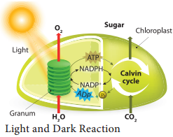
Biosynthetic phase of photosynthesis utilises assimilatory powers (ATP and NADPH + H+) produced during light reaction are used to fix and reduce carbon dioxide into carbohydrates. This reaction does not require light. Therefore, it is named Dark reaction. Ribulose 1, 5 bisphosphate (RUBP) act as acceptor molecule of carbon dioxide and fix the CO2 by RUBISCO enzyme.

The first product of the pathway is a 3 – carbon compound (Phospho Glyceric Acid) and so it is also called as C3 Cycle. It takes place in the stroma of the chloroplast.

M. Melvin Calvin, A.A. Benson and their co-workers in the year 1957 found this path way of carbon fixation. Melvin Calvin was awarded Nobel Prize for this in 1961 and this pathway named after the discoverers as Calvin-Benson Cycle. Dark reaction is temperature dependent and so it is also called thermo-chemical reaction.

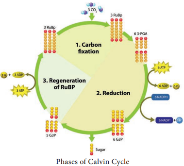
Dark reaction consists of three phases: (Figure 13.16).
Dark Reaction or Cycle or Biosynthetic Phase or Photosynthetic Carbon Reduction (PCR) Cycle img 1

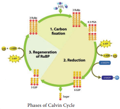
  1. Carboxylation (fixation)
  2. Reduction (Glycolytic Reversal)
  3. Regeneration

Phase 1 – Carboxylation (Fixation)

The acceptor molecule Ribulose 1, 5 Bisphosphate (RUBP) a 5 carbon compound with the help of RUBP carboxylase oxygenase (RUBISCO) enzyme accepts one molecule of carbon dioxide to form an unstable 6 carbon compound. This 6C compound is broken down into two molecules of 3-carbon compound phospho glyceric acid (PGA) (Figure 13.17).
Dark Reaction or Cycle or Biosynthetic Phase or Photosynthetic Carbon Reduction (PCR) Cycle img 2

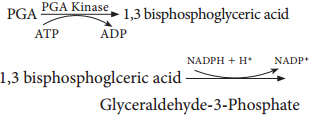
Phase 2 – Glycolytic Reversal / Reduction

Phospho glyceric acid is phosphorylated by ATP and produces 1, 3 bis phospho glyceric acid by PGA kinase. 1, 3 bis phospho glyceric acid is reduced to glyceraldehyde 3 Phosphate (G-3-P) by using the reducing power NADPH + H+. Glyceraldehyde 3 phosphate is converted into its isomeric form di hydroxy acetone phosphate (DHAP).
Dark Reaction or Cycle or Biosynthetic Phase or Photosynthetic Carbon Reduction (PCR) Cycle img 3

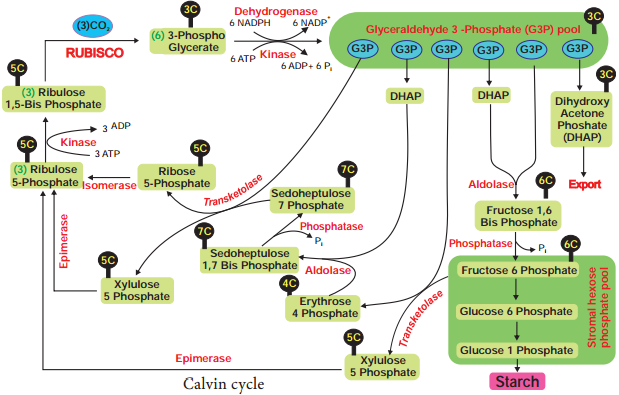
Phase 3 – Regeneration

Regeneration of RUBP involves the formation of several intermediate compounds of 6-carbon, 5-carbon,4-carbon and 7- carbon skeleton. Fixation of one carbon dioxide requires 3 ATPs and 2 NADPH + H+, and for the fixation of 6 CO2 requires 18 ATPs and 12 NADPH + H+ during C3 cycle. One 6 carbon compound is the net gain to form hexose sugar.
Dark Reaction or Cycle or Biosynthetic Phase or Photosynthetic Carbon Reduction (PCR) Cycle img 4

Overall Equation for Dark Reaction:
6CO2 + 18ATP + 12NADPH + H+ → C6H12O6 + 6H2O + 18Pi + 12NADP+

Photophosphorylation

Learninsta presents the core concepts of Biology with high-quality research papers and topical review articles.

Photophosphorylation

Phosphorylation taking place during respiration is called as oxidative phosphorylation and ATP produced by the breakdown of substrate is known as substrate level phosphorylation. In this topic, we are going to learn about phosphorylation taking place in chloroplast with the help of light. During the movement of electrons through carrier molecules ATP and NADPH + H+ are produced.

Phosphorylation is the process of synthesis of ATP by the addition of inorganic phosphate to ADP. The addition of phosphate here takes place with the help of light generated electron and so it is called as photophosphorylation. It takes place in both cyclic and non-cyclic electron transport.

Cyclic Photophosphorylation

Cyclic photophosphorylation refers to the electrons ejected from the pigment system I (Photosystem I) and again cycled back to the PS I. When the photons activate P700 reaction centre photosystem II is activated. Electrons are raised to the high energy level.

The primary electron acceptor is Ferredoxin Reducing Substance (FRS) which transfers electrons to Ferredoxin (Fd), Plastoquinone (PQ), cytochrome b6-f complex, Plastocyanin (PC) and finally back to chlorophyll P700 (PS I).

During this movement of electrons Adenosine Di Phosphate (ADP) is phosphorylated, by the addition of inorganic phosphate and generates Adenosine Tri Phosphate (ATP). Cyclic electron transport produces only ATP and there is no NADPH + H+ formation.

At each step of electron transport, electron loses potential energy and is used by the transport chain to pump H+ ions across the thylakoid membrane. The proton gradient triggers ATP formation in ATP synthase enzyme situated on the thylakoid membrane.

Photosystem I need light of longer wave length (> P700 nm). It operates under low light intensity, less CO2 and under anaerobic conditions which makes it considered as earlier in evolution (Figure 13.13).
Photophosphorylation img 1

Non-Cyclic Photophosphorylation

When photons are activated reaction centre of pigment system II(P680), electrons moved to the high energy level. Electrons from high energy state passes through series of electron carriers like pheophytin, plastoquinone, cytochrome complex, plastocyanin and finally accepted by PS I (P700). During this movement of electrons from PS II to PS I ATP is generated (Figure 13.16).
Photophosphorylation img 2

PS I (P700) is activated by light, electrons are moved to high energy state and accepted by electron acceptor molecule ferredoxin reducing Substance (FRS). During the downhill movement through ferredoxin, electrons are transferred to NADP+ and reduced into NADPH + H+ (H+ formed from splitting of water by light).

Electrons released from the photosystem II are not cycled back. It is used for the reduction of NADP+ into NADPH + H+. During the electron transport it generates ATP and hence this type of photophosphorylation is called non-cyclic photophosphorylation. The electron flow looks like the appearance of letter ‘Z’ and so known as Z scheme.

When there is availability of NADP+ for reduction and when there is splitting of water molecules both PS I and PS II are activated (Table 13.3). Non-cyclic electron transport PS I and PS II both are involved co operatively to transport electrons from water to NADP+ (Figure 13.14).
Photophosphorylation img 3
Photophosphorylation img 4

Bio Energetics of Light Reaction

  • To release one electron from pigment system it requires two quanta of light.
  • One quantum is used for transport of electron from water to PS I.
  • Second quantum is used for transport of electron from PS I to NADP+
  • Two electrons are required to generate one NADPH + H+
  • During Non-Cyclic electron transport two NADPH + H+ are produced and it requires 4 electrons.
  • Transportation of 4 electrons requires 8 quanta of light

Chemiosmotic Theory

Chemiosmosis theory was proposed by P. Mitchell (1966). According to this theory electrons are transported along the membrane through PS I and PS II and connected by Cytochrome b6-f complex. The flow of electrical current is due to difference in electrochemical potential of protons across the membrane.

Splitting of water molecule takes place inside the membrane. Protons or H+ ions accumulate within the lumen of the thylakoid (H+ increase 1000 to 2000 times). As a result, proton concentration is increased inside the thylakoid lumen. These protons move across the membrane because the primary acceptor of electron is located outside the membrane.

Protons in stroma less in number and creates a proton gradient. This gradient is broken down due to the movement of proton across the membrane to the stroma through CFO of the ATP synthase enzyme. The proton motive force created inside the lumen of thylakoid or chemical gradient of H+ ion across the membrane stimulates ATP generation (Figure 13.15).
Photophosphorylation img 5

The evolution of one oxygen molecule (4 electrons required) requires 8 quanta of light. C3 plants utilise 3 ATPs and 2 NADPH + H+ to evolve one Oxygen molecule. To evolve 6 molecules of Oxygen 18 ATPs and 12 NADPH + H+ are utilised. C4 plants utilise 5 ATPs and 2 NADPH + H+ to evolve one oxygen molecule. To evolve 6 molecules of Oxygen 30 ATPs and 12 NADPH + H+ are utilised.

Photo Chemical Phase of Light Reaction

Learninsta presents the core concepts of Biology with high-quality research papers and topical review articles.

Photo Chemical Phase of Light Reaction

In this phase electrons pass through electron carrier molecules and generate assimilatory powers ATP and NADPH + H+. Splitting of water molecule generates electrons replacing electrons produced by the light.

Photolysis of Water

The process of Photolysis is associated with Oxygen Evolving Complex (OEC) or water splitting complex in pigment system II and is catalysed by the presence of Mn++ and Cl. When the pigment system II is active it receives light and the water molecule splits into OH ions and H+ ions. The OH ions unite to form water molecules again and release O2 and electrons (Figure 13.11).
Photo Chemical Phase of Light Reaction img 1

Electron Transport Chain of Chloroplast
Electron Transport Chain in each photosystem involves four complexes:

Core Complex (CC):
CC I in PS I the reaction centre is P700, CC II in PS II the reaction centre is P680

Light Harvesting Complex or Antenna

Complex (LHC):
Two types: LHC I in PS I and LHC II in PS II.

Cytochrome b6 f Complex:

It is the non-pigmented protein complex connecting PS I and PS II. Plastoquinone (PQ) and Plastocyanin (PC) are intermediate complexes acting as mobile or shuttle electron carriers of Electron Transport Chain. PQ acts as shuttle between PS II and Cytochrome b6 – f complex and PC connects.

Cytochrome b6-f and PS I Complex

ATPase complex or Coupling Factor:
It is found in the surface of thylakoid membrane. This complex is made up of CF1 and CF0 factors. This complex utilizes energy from ETC and converts ADP and inorganic phosphate (Pi) into ATP (Figure 13.12).
Photo Chemical Phase of Light Reaction img 2

Photo – Oxidation Phase of Light Reaction

Learninsta presents the core concepts of Biology with high-quality research papers and topical review articles.

Photo – Oxidation Phase of Light Reaction

The action of photon plays a vital role in excitation of pigment molecules to release an electron. When the molecules absorb a photon, it is in excited state. When the light source turned off the high energy electrons return to their normal low energy orbitals as the excited molecule goes back to its original stable condition known as ground state.

When molecules absorb or emit light they change their electronic state. Absorption of blue light excites the chlorophyll to higher energy state than absorption of Red light, because the energy of photon is higher when their wavelength is shorter.

When the pigment molecule is in an excited state, this excitation energy is utilised for the phosphorylation. Phosphorylation takes place with the help of light generated electron and hence it is known as photophosphorylation.

Photosystem and Reaction Centre

  1. Thlakoid membrane contains Photosystem I (PS I) and Photosystem II (PS II).
  2. PS I is in unstacked region of granum facing stroma of chloroplast.
  3. PS II is found in stacked region of thylakoid membrane facing lumen of thylakoid.
  4. Each Photosystem consists of central core complex (CC) and light harvesting Complex (LHC) or Antenna molecules (Figure 13.10).
  5. The core complex consists of respective reaction centre associated with proteins, electron donors and acceptors.
  6. PS I – CC I consists of reaction centre P700 and LHC I.
  7. PS II – CC II consists of reaction centre P680 and LHC II (Table 13.2).
  8. Light Harvesting Complex consists of several chlorophylls, carotenoids and xanthophyll molecules.
  9. The main function of LHC is to harvest light energy and transfer it to their respective reaction centre.

Photo - Oxidation Phase of Light Reaction img 1
Photo - Oxidation Phase of Light Reaction img 2

The action of photon plays a vital role in excitation of pigment molecules to release an electron. The action of photon plays a vital role in excitation of pigment molecules to release an electron. When the molecules absorb a photon, it is in excited state.

Photo-oxidation is a chain process incorporating a large number of chemical reactions which are subsequent to the outcome of the primary event absorption of a photon, which induces breakdown to free-radical products.

In photosynthesis, the light-dependent reactions take place on the thylakoid membranes. The four photosystems absorb light energy through pigments-primarily the chlorophylls, which are responsible for the green color of leaves. The light-dependent reactions begin in photosystem II.

The light-dependent generation of active oxygen. species is termed photooxidative stress. This can occur in two ways:

  1. The donation of energy or electrons directly to oxygen as a result of photosynthetic activity
  2. Exposure of tissues to ultraviolet irradiation.

Oxidation is defined as a process in which an electron is removed from a molecule during a chemical reaction. What happens in oxidation? During oxidation, there is a transfer of electrons.

The Light Reactions of Photosynthesis. Light is absorbed and the energy is used to drive electrons from water to generate NADPH and to drive protons across a membrane. These protons return through ATP synthase to make ATP.

This is an intrinsic feature of the regulation of photosynthetic electron transport. Photoinhibition and photooxidation only usually occur when plants are exposed to stress. Active oxygen species are part of the alarm signalling processes in plants.

Light reactions harness energy from the sun to produce chemical bonds, ATP, and NADPH. These energy-carrying molecules are made in the stroma where carbon fixation takes place. The light-independent reactions of the Calvin cycle can be organized into three basic stages: fixation, reduction, and regeneration.

Photo-oxidation means the release of electrons after absorption of a photon occurs during non-cyclic photophosphorylation. It involves an outsized number of chemical reactions.

Photolysis is an oxidative process that means the splitting of water to form oxygen, protons and electrons in the presence of light. As a result, oxygen evolution happens and the electron travels to PS-IIvia the Mn-protein.

The light-dependent reactions use light energy to make two molecules needed for the next stage of photosynthesis: the energy storage molecule ATP and the reduced electron carrier NADPH. In plants, the light reactions take place in the thylakoid membranes of organelles called chloroplasts.

Modern Concept of Photosynthesis

Learninsta presents the core concepts of Biology with high-quality research papers and topical review articles.

Modern Concept of Photosynthesis

Photosynthesis is an Oxidation and Reduction process. Water is oxidised to release O2 and CO2 is reduced to form sugars. The first phase requires light and is called light reaction or Hill’s reaction.

1. Light Reaction:

It is a photochemical reaction whereas dark reaction is a thermochemical reaction. Solar energy is trapped by chlorophyll and stored in the form of chemical energy (assimilatory power) as ATP and reducing power NADPH + H+, NADPH + H+ alone are known as reducing powers. This reaction takes place in thylakoid membrane of the chloroplast. Oxygen is evolved as a result of splitting of water molecules by light.

Light reaction is discussed in two phases:

(i) Photo-Oxidation Phase:

  • Absorption of light energy.
  • Transfer of energy from accessory pigments to reaction centre.
  • Activation of Chlorophyll ‘a’ molecule.

(ii) Photo Chemical Phase:

  • Photolysis of water and oxygen evolution
  • Electron transport and synthesis of assimilatory power.

2. Dark reaction (Biosynthetic Phase):

Fixation and reduction of CO2 into carbohydrates with the help of assimilatory power produced during light reaction. This reaction does not require light and is not directly light driven. Hence, it is called as Dark reaction or CalvinBenson cycle (Figure 13.9).
Modern Concept of Photosynthesis img 1

photosynthesis, the process by which green plants and certain other organisms transform light energy into chemical energy. During photosynthesis in green plants, light energy is captured and used to convert water, carbon dioxide, and minerals into oxygen and energy-rich organic compounds.

During photosynthesis, plants take in carbon dioxide (CO2) and water (H2O) from the air and soil. This transforms the water into oxygen and the carbon dioxide into glucose. The plant then releases the oxygen back into the air, and stores energy within the glucose molecules.

Recent advances include combining photosystem complexes with hydrogenases for hydrogen production, using isolated thylakoids, photosystems on nanostructured electrodes such as gold nanoparticles, carbon nanotubes, ZnO nanoparticles for electricity generation.

The process by which green plants make their own food (like glucose) from carbon dioxide and water by using sunlight energy (in the presence of chlorophyll) is called photosynthesis.

Green plants are the main producers of food in the ecosystem. All other organisms directly or indirectly depend on green plants for food. The process of photosynthesis also helps in maintaining the balance of carbon dioxide and oxygen in the air.

The three events that occur during the process of photosynthesis are:

  1. Absorption of light energy by chlorophyll
  2. Conversion of light energy to chemical energy and splitting of water molecules into hydrogen and oxygen
  3. Reduction of carbon dioxide to carbohydrates.

Photosynthesis and respiration, both using electron flow coupled with phosphorylation, have a common origin (‘conversion hypothesis’), but photosynthesis came first. Anaerobic (nitrate or sulphate) respiration cannot have preceded photosynthesis as neither nitrate nor sulphate existed on the early earth.

It fixes and balances the amount of carbon dioxide and oxygen in the atmosphere by plants exhaling oxygen and animals and humans exhaling carbon dioxide. It helps in the synthesis of organic compounds from inorganic compounds. It provides nutrition to the plants and thus helps in their growth and development.

The first stage of photosynthesis is called the light reactions. During this stage, light is absorbed and transformed to chemical energy in the bonds of NADPH and ATP.

The Light Reactions of Photosynthesis. Light is absorbed and the energy is used to drive electrons from water to generate NADPH and to drive protons across a membrane. These protons return through ATP synthase to make ATP.

Scientists think that glycolysis evolved before the other stages of cellular respiration. This is because the other stages need oxygen, whereas glycolysis does not, and there was no oxygen in Earth’s atmosphere when life first evolved about 3.5 to 4 billion years ago.

Emerson’s Experiments and Hill’s Reaction

Learninsta presents the core concepts of Biology with high-quality research papers and topical review articles.

Emerson’s Experiments and Hill’s Reaction

Red Drop or Emerson’s First Effect

Emerson conducted experiment in Chlorella using only one wavelength of light (monochromatic light) at a time and he measured quantum yield. He plotted a graph of the quantum yield in terms of O2 evolution at various wavelengths of light. His focus was to determine at which wavelength the photochemical yield of oxygen was maximum.

He found that in the wavelength of 600 to 680 the yield was constant but suddenly dropped in the region above 680 nm (red region). The fall in the photosynthetic yield beyond red region of the spectrum is referred as Red drop or Emerson’s first effect.

Emerson’s Enhancement Effect

Emerson modified his first experiment by supplying shorter wavelength of light (red light) along with longer wavelength of light (far red light). He found that the monochromatic light of longer wavelength (far red light) when supplemented with shorter wavelength of light (red light) enhanced photosynthetic yield and recovered red drop. This enhancement of photosynthetic yield is referred to as Emerson’s Enhancement Effect (Figure 13.8).
Emerson's Experiments and Hill's Reaction img 1

  • Photosynthetic rate at far red light (710 nm) = 10
  • Photosynthetic rate at red light (650 nm) = 43.5
  • Photosynthetic rate at red + far red (650 + 1710 nm) = 72.5 (Enhancement effect).

Hill’s Reaction

R. Hill (1937) isolated chloroplasts and when they were illuminated in the presence of suitable electron acceptors such as ferricyanide, they were reduced to ferrocyanide and oxygen is evolved. Hill’s Reaction is now considered to be equivalent to Light Reaction.

Conclusions of Hill’s Reaction:

  1. During photosynthesis oxygen is evolved from water.
  2. Electrons for the reduction of CO2 are obtained from water.
  3. Reduced substance produced, later helps to reduce CO2
    2H2O + 2A → 2 AH2 + O2

A is the Hydrogen acceptor, the common in vitro hydrogen acceptors are ferricyanide, benzoquinone and Di Chloro Phenol Indole Phenol (DCPIP).

Absorption Spectrum and Action Spectrum

Learninsta presents the core concepts of Biology with high-quality research papers and topical review articles.

Absorption Spectrum and Action Spectrum

The term absorption refers to complete retention of light, without reflection or transmission. Pigments absorb different wavelengths of light. A curve obtained by plotting the amount of absorption of different wavelengths of light by a pigment is called its absorption spectrum.

  1. Chlorophyll ‘a’ and chlorophyll ‘b’ absorb quanta from blue and red region.
  2. Maximum absorption peak for different forms of chlorophyll ‘a’ is 670 to 673, 680 to 683 and 695 to 705nm.
  3. Chlorophyll ‘a’ 680 (P680) and Chlorophyll ‘a’ 700 (P700) function as trap centre for PS II and PS I respectively.

Action Spectrum

The effectiveness of different wavelength of light on photosynthesis is measured by plotting against quantum yield. The curve showing the rate of photosynthesis at different wavelengths of light is called action spectrum.

From the graph showing action spectrum, it can be concluded that maximum photosynthesis takes place in blue and red region of the spectrum. This wavelength of the spectrum is the absorption maxima for Chlorophyll (a) and Chlorophyll (b). The Action Spectrum is instrumental in the discovery of the existence of two photosystems in O2 evolving photosynthesis (Figure 13.7).
Absorption Spectrum and Action Spectrum img 1

Absorption spectrum deals with wavelengths of light absorbed by each pigment whereas the action spectrum indicates the overall rate of photosynthesis at each wavelength of light. It shows the overall rate of photosynthesis at each wavelength of light.

An absorption spectrum defines the spectrum of electromagnetic radiation, or light, plants absorb. An action spectrum defines the spectrum of electromagnetic radiation most effective for photosynthesis. In other words, it is the part of the light spectrum that does the work.

The action spectrum for photosynthesis shows which wavelengths are used by plants to create energy, while the absorption spectrum shows which wavelengths are most absorbed by a specific molecule. But other molecules play a part as well, which is why there is some difference in the absorption and action spectra.

An action spectrum is a graph of the rate of biological effectiveness plotted against wavelength of light. It shows which wavelength of light is most effectively used in a specific chemical reaction. Some reactants are able to use specific wavelengths of light more effectively to complete their reactions.

Absorption spectroscopy is employed as an analytical chemistry tool to determine the presence of a particular substance in a sample and, in many cases, to quantify the amount of the substance present. Infrared and ultraviolet-visible spectroscopy are particularly common in analytical applications.

The similarity of the action spectrum of photosynthesis and the absorption spectrum of chlorophyll tells us that chlorophylls are the most important pigments in the process. The spectra are not identical, though, because carotenoids, which absorb strongly in the blue, play a role as well.

Absorption Spectrum. Definition. The range of a pigment’s ability to absorb various wavelengths of light. A graph plotting light absorption of a pigment versus wavelength.

An action spectrum is measured by plotting a response to light such as oxygen evolution, as a function of wavelength. If the pigments used to obtain the absorption spectrum are the same as those that cause the response, the absorption and action spectra will match.

Because more absorption leads to more action, peaks in an absorption spectrum will have corresponding peaks in an action spectrum. Thus, if a pigment can be found that has an absorption spectrum that matches a process’s action spectrum, it is likely that pigment is the photoreceptor for that process.

The pattern of absorption lines in a spectrum is diagnostic of the types of atoms and molecules present, for example, in the surface layers of a star or the atmosphere of a planet. Absorption lines are seen in the spectra of the Sun and other stars.

To get an absorption spectrum, just shine white light on a sample of the material that you are interested in white light is made up of all the different wavelengths of visible light put together. In the absorption spectrum there will be gaps.

An absorption spectrum occurs when light passes through a cold, dilute gas and atoms in the gas absorb at characteristic frequencies; since the re-emitted light is unlikely to be emitted in the same direction as the absorbed photon, this gives rise to dark lines (absence of light) in the spectrum.

The action spectrum for photosynthesis is much broader than the absorption spectrum of chlorophyll a. This is because accessory pigments with different absorption spectra also present in chloroplasts broaden the spectrum of colors that can be used for photosynthesis.

Photosynthetic Unit (Quantasome)

Learninsta presents the core concepts of Biology with high-quality research papers and topical review articles.

Photosynthetic Unit (Quantasome)

Quantasomes are the morphological expression of physiological photosynthetic units, located on the inner membrane of thylakoid lamellae. Each quantasome measures about 180 A° × 160 A° and 100 A° thickness. In 1952, Steinman observed granular structures in chloroplast lamellae under electron microscope. Later, Park and Biggins (1964) confirmed these granular structures as physiological units of photosynthesis and coined the term Quantasome.

According to them one quantasome contains about 230 chlorophyll molecules. A minimum number of chlorophyll and other accessory pigments act together in a photochemical reaction to release one oxygen or to reduce one molecule of CO2. It constitutes a photosynthetic unit. (Figure 13.6) Emerson and Arnold (1932) based on flashing light experiment found 2500 chlorophyll molecules are required to fix one molecule of CO2.
Photosynthetic Unit (Quantasome) img 1

However, the reduction or fixation of one CO2 requires 10 quanta of light and so each unit would contain 1/10 of 2500 i.e. 250 molecules. Usually 200 to 300 chlorophyll molecules are considered as a physiological unit of photosynthesis. According to Emerson 8 quanta of light are required for the release of one oxygen molecule or reduction of one Carbon dioxide molecule. The quantum yield is 1/8 or 12%.

Quantasomes are particles found in the thylakoid membrane of chloroplasts in which photosynthesis takes place. They are composed of lipids and proteins that include various photosynthetic pigments and redox carriers. For this reason they are considered to be photosynthetic units.

The photosynthetic unit is experimentally defined as the number of oxygen. molecules evolved or carbon dioxide molecules reduced per molecule of chlorophyll when the chloroplast pigments are excited by one flash of light so.

Quantasomes are particles found in a paracrystalline array of the thylakoid membrane of chloroplasts in which photosynthesis takes place. They are believed to be the structural and functional units of the chloroplast.

The cell is the smallest structural and functional unit of living organisms, which can exist on its own. Therefore, it is sometimes called the building block of life. Some organisms, such as bacteria or yeast, are unicellular consisting only of a single cell while others, for instance, mammalians, are multicellular.

Quantasomes are particles found in the thylakoid membrane of chloroplasts in which photosynthesis takes place. They occur in 2 sizes: the smaller quantasome is thought to represent the site of photosystem I, the larger to represent the site of photosystem II.

The two stages of photosynthesis: Photosynthesis takes place in two stages: light-dependent reactions and the Calvin cycle (light-independent reactions). Light-dependent reactions, which take place in the thylakoid membrane, use light energy to make ATP and NADPH.

The average minimal quantum number remains 10 (efficiency 0.1) as in many algae and typical higher plants. Most of the time the optimal efficiency depends on the availability of some far-red radiation, particularly in the blue region of the spectrum where blue light alone is rather inefficient.

Cyclic photophosphorylation can be defined as the synthesis of ATP coupled to electron transport activated by Photosystem I solely, and can therefore proceed in long-wave-length light (03BB 2265 700 nm). This proces is unaffected by the specific inhibitors of Photosystem II, such as CMU, DCMU and orthophenanthroline.

The reaction centre contains pigments such as chlorophyll and phaeophytin. These absorb light, promoting an electron to a higher energy level within the pigment. The free energy created is used to reduce an electron acceptor and is critical for the production of chemical energy during photosynthesis.

The chlorophyll molecules are grouped into antenna complexes, clusters of several hundred molecules that are anchored onto the thylakoid membrane by special proteins. Within each antenna complex is a specialized set of proteins and chlorophyll molecules that form a reaction.

As you rise from low light intensity to higher light intensity, the rate of photosynthesis will increase because there is more light available to drive the reactions of photosynthesis. At a very high intensity of light, the rate of photosynthesis would drop quickly as the light starts to damage the plant.

Chlorophyll a is the most important photosynthetic pigment because it is directly involved in the conversion of light energy (photons) to chemical energy. All other photosynthetic pigments found in the chloroplasts of higher plants are called “accessory pigments”.

Spectrum of Electromagnetic Radiation

Learninsta presents the core concepts of Biology with high-quality research papers and topical review articles.

Spectrum of Electromagnetic Radiation

In the total electromagnetic spectrum, visible light is the smallest part. The entire life on earth depends on light and is the driving force for all organisms. Plants have natural potential to utilize solar energy directly. In the given picture electromagnetic radiation spectrum and components of visible spectrum are mentioned. The wavelength of solar radiation which reaches the earth is between 300 to 2600 nm.

The visible spectrum ranges between 390 to 763 nm (3900 å to 7630 å). The colour of the light is determined by the wavelength. Energy of the quantum is inversely proportional to wavelength. Shorter wavelength has more energy than longer wavelength. Electromagnetic spectrum consists of 7 types of radiations such as gamma rays, X rays, U-V rays, Visible light spectrum, infrared rays, electric rays and radio rays (Figure 13. 4).
Spectrum of Electromagnetic Radiation img 1

Properties of Light

  1. Light is a transverse electromagnetic wave.
  2. It consists of oscillating electric and magnetic fields that are perpendicular to each other and perpendicular to the direction of propagation of the light.
  3. Light moves at a speed of 3 × 10⁸ ms-1
  4. Wavelength is the distance between successive crests of the wave.
  5. Light as a particle is called photon. Each photon contains an amount of energy known as quantum.
  6. The energy of a photon depends on the frequency of the light (Figure 13.5).Spectrum of Electromagnetic Radiation img 2

The entire electromagnetic spectrum, from the lowest to the highest frequency (longest to shortest wavelength), includes all radio waves (e.g., commercial radio and television, microwaves, radar), infrared radiation, visible light, ultraviolet radiation, X-rays, and gamma rays.

The EM spectrum is generally divided into seven regions, in order of decreasing wavelength and increasing energy and frequency. The common designations are: radio waves, microwaves, infrared (IR), visible light, ultraviolet (UV), X-rays and gamma rays.

The electromagnetic spectrum is a continuum of all electromagnetic waves arranged according to frequency and wavelength. The sun, earth, and other bodies radiate electromagnetic energy of varying wavelengths. Electromagnetic energy passes through space at the speed of light in the form of sinusoidal waves.

The characteristics of the electromagnetic spectrum are the propagation features and the amount of information, which signals can carry. In general, signals sent using the higher frequencies have shorter propagation distances but a higher data-carrying capacity.

Radio waves, microwaves, visible light, and x rays are all examples of electromagnetic waves that differ from each other in wavelength.

  • Longer Wavelength
  • Shorter Wavelength

Electromagnetic waves are produced by the motion of electrically charged particles. The different types of waves have different uses and functions in our everyday lives. The most important of these is visible light, which enables us to see. Radio waves have the longest wavelengths of all the electromagnetic waves. They range from around a foot long to several miles long.

“Electromagnetic spectrum” refers to the spectrum of electromagnetic radiation, and electromagnetic radiation is so named because it consists of electric and magnetic fields. In fact, light does affect charges and currents.

These observations enable astronomers to determine certain physical characteristics of objects, such as their temperature, composition and velocity. The electromagnetic spectrum consists of much more than visible light. It includes wavelengths of energy that human eyes can’t perceive.

Cell phones use antennae to transmit and receive radio waves that carry binary information. Every cell tower presides over an area of land, where it receives and transmits radio waves. When a text message is written, it is transmitted as binary code using a particular frequency of radio waves specific to that user.

Almost all of the energy available at Earth’s surface comes from the sun. The sun gets its energy from the process of nuclear fusion. This energy eventually makes its way to the outer regions of the sun and is radiated or emitted away in the form of energy, known as electromagnetic radiation.

Yes, all objects, including human bodies, emit electromagnetic radiation. The wavelength of radiation emitted depends on the temperature of the objects. Such radiation is sometimes called thermal radiation. Most of the radiation emitted by human body is in the infrared region, mainly at the wavelength of 12 micron.
Spectrum of Electromagnetic Radiation img 3

Photosynthetic Pigments

Learninsta presents the core concepts of Biology with high-quality research papers and topical review articles.

Photosynthetic Pigments

A photosynthetic pigment is a pigment that is present in chloroplasts or photosynthetic bacteria which captures the light energy necessary for photosynthesis (Table 13.1).
Photosynthetic Pigments img 1

Chlorophyll

Chlorophyll ‘a’ is the primary pigment which acts as a reaction centre and all other pigments act as accessory pigments and trap solar energy and then transfer it to chlorophyll ‘a’. Chlorophyll molecules have a tadpole like structure. It consists of Mg-Porphyrin head (Hydrophilic Head) and (Lipophilic tail) Phytol tail. The Porphyrin head consists of four pyrrol rings linked together by C-H bridges.

Each pyrrole ring comprises of four carbons and one nitrogen atom. Porphyrin ring has several side groups which alter the properties of the pigment. Different side groups are indicative of various types of chlorophyll. The Phytol tail made up of 20 carbon alcohol is attached to carbon 7 of the Pyrrole ring IV. It has a long propionic acid ester bond. Long lipophilic tail helps in anchoring chlorophyll to the lamellae.

Carotenoids

Carotenoids are yellow to orange pigments, mostly tetraterpens and these pigments absorb light strongly in the blue to violet region of visible spectrum. These pigments protect chlorophyll from photo-oxidative damage. Hence, they are called as shield pigments.

These pigments absorb light and transfer these to chlorophyll. Almost all carotenoid pigments have 40 carbon atoms. Ripening of fruits, floral colours and leaf colour change during autumn is due to Carotenoids (Carotene and Xanthophyll) (Figure 13.2).
Photosynthetic Pigments img 2

(i) Carotenes:

Orange, Red, Yellow and Brownish pigments, hydrocarbons (Lipids) and most of them are tetraterpenes (C40H56). Carotene is the most abundant Carotene in plants and it is a precursor of Vitamin A. Lycopene is the red pigment found in the fruits of tomato, red peppers and roses.

(ii) Xanthophylls:

Yellow (C40H56O2) pigments are like carotenes but contain oxygen. Lutein is responsible for yellow colour change of leaves during autumn season. Examples: Lutein, Violaxanthin and Fucoxanthin.

Phycobilins

They are proteinaceous pigments, soluble in water, and do not contain Mg and Phytol tail. They exist in two forms such as:-

  1. Phycocyanin found in cyanobacteria
  2. Phycoerythrin found in rhodophycean algae (Red algae).

Definition – Significance and Site of Photosynthesis

Learninsta presents the core concepts of Biology with high-quality research papers and topical review articles.

Definition – Significance and Site of Photosynthesis

Photosynthesis is referred as photochemical oxidation and reduction reactions carried out with the help of light, converting solar energy into chemical energy. It is the most important anabolic process. Plants and photosynthetic bacteria use simple raw materials like carbon dioxide water and with the help of light energy synthesize carbohydrates and evolve oxygen. The overall chemical equation for photosynthesis is:
Definition - Significance and Site of Photosynthesis img 1

Ruben and Kamen (1941) demonstrated six molecules of water as insufficient for the evolution of 6 molecules of O2 and modified the equation as:
Definition - Significance and Site of Photosynthesis img 2

Photosynthesis is a collection of oxidation and reduction reactions (Redox reaction).

Oxidation:
Water is oxidised into oxygen (loss of electrons).

Reduction:
CO2 is reduced into Carbohydrates (gain of electrons).

In some bacteria, oxygen is not evolved and is called as non-oxygenic and anaerobic photosynthesis. Examples: Green sulphur, Purple sulphur and green fiamentous bacteria.

Significance of Photosynthesis

  1. Photosynthetic organisms provide food for all living organisms on earth either directly or indirectly.
  2. It is the only natural process that liberates oxygen in the atmosphere and balances the oxygen level.
  3. Photosynthesis balances the oxygen and carbon cycle in nature.
  4. Fuels such as coal, petroleum and other fossil fuels are from preserved photosynthetic plants.
  5. Photosynthetic organisms are the primary producers on which all consumers depend for energy.
  6. Plants provide fodder, fire, fie wood, timber, useful medicinal products and these sources come by the act of photosynthesis.

Site of Photosynthesis

Chloroplasts are the main site of photosynthesis and both energy yielding process (Light reaction) and fixation of carbon di oxide (Dark reaction) that takes place in chloroplast. It is a double wall membrane bounded organelle, discoid or lens shaped, 4-10 µm in diameter and 1-33 µm in thickness. The membrane is a unit membrane and space between them is 100 to 200 A °. A colloidal and proteinaceous matrix called stroma is present inside.

A sac like membranous system called thylakoid or lamellae is present in stroma and they are arranged one above the other forming a stack of coin like structure called granum (plural grana). Each chloroplast contains 40 to 80 grana and each granum consists of 5 to 30 thylakoids.

Thlakoids found in granum are called grana lamellae and in stroma are called stroma lamellae. Thlakoid disc size is 0.25 to 0.8 micron in diameter. A thinner lamella called Fret membrane connects grana. Pigment system I is located on outer thylakoid membrane facing stroma and Pigment system II is located on inner membrane facing lumen of thylakoid.

Grana lamellae have both PS I and PS II whereas stroma lamellae have only PS I. Chloroplast contains 30-35 Proteins, 20-30% phospholipids, 5-10% chlorophyll, 4-5% Carotenoids, 70S ribosomes, circular DNA and starch grains.

Inner surface of lamellar membrane consists of small spherical structure called as Quantasomes. Presence of 70S ribosome and DNA gives them status of semi-autonomy and proves endosymbiotic hypothesis which says chloroplast evolved from bacteria. Thlakoid contains pigment systems which produces ATP and NADPH + H+ using solar energy. Stroma contains enzyme which reduces carbondioxide into carbohydrates. In Cyanobacteria thylakoid lies freely in cytoplasm without envelope (Figure 13.1).
Definition - Significance and Site of Photosynthesis img 3