Life Cycle Patterns in Plants

Learninsta presents the core concepts of Biology with high-quality research papers and topical review articles.

Life Cycle Patterns in Plants

Alternation of Generation

Alternation of generation is common in all plants. Alternation of the haploid gametophytic phase (n) with diploid sporophytic phase (2n) during the life cycle is called alternation of generation. Following type of life cycles are found in plants (Figure 2.2).
Life Cycle Patterns in Plants img 1

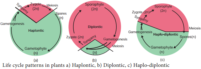
Haplontic Life Cycle

Gametophytic phase is dominant, photosynthetic and independent, whereas sporophytic phase is represented by the zygote. Zygote undergoes meiosis to restore haploid condition. Example: Volvox, Spirogyra.

Diplontic Life Cycle

Sporophytic phase (2n) is dominant, photosynthetic and independent. The gametophytic phase is represented by the single to few celled gametophyte. The gametes fuse to form zygote which develops into sporophyte. Example: Fucus, gymnosperms and angiosperms.

Haplodiplontic Life Cycle

This type of life cycle is found in bryophytes and pteridophytes which is intermediate between haplontic and diplontic type. Both the phases are multicellular but they differ in their dominant phase.

In bryophytes dominant independent phase is gametophyte and it alternates with short-lived multicellular sporophyte totally or partially dependent on the gametophyte. In pteridophytes sporophyte is the independent phase. It alternates with multicellular saprophytic or autotrophic, independent, short lived gametophyte (n).

There are three different plant life cycles: haploid (1n), diploid (2n), and the more common haploid-diploid (1n-2n). A haploid organism consists of a multicellular structure of cells that contain only one set of chromosomes, whereas, a diploid organism’s multicellular stage contains two sets of chromosomes.

Plants have two distinct stages in their lifecycle: the gametophyte stage and the sporophyte stage. The haploid gametophyte produces the male and female gametes by mitosis in distinct multicellular structures. Fusion of the male and females gametes forms the diploid zygote, which develops into the sporophyte.

Alternation of generations is a type of life cycle found in terrestrial plants and some algae in which subsequent generations of individuals alternate between haploid and diploid organisms. This can be contrasted to sexual reproduction in animals, in which both haploid and diploid cells are found in every generation.

Plants alternate between the diploid sporophyte and haploid gametophyte, and between asexual and sexual reproduction. Therefore, the life cycle of plants is known as alternation of generations. In tracheophytes, the dominant generation is diploid and the sporophyte comprises the main plant.

Flowering plants all go through the same stages of a life cycle, but the length of time they take varies a lot between species. Some plants go though their complete cycle in a few weeks – others take many years. Annuals are plants that grow from a seed, then flower and make new seeds, then die, all in less than a year.

Some animals lay eggs with shells as the first stage of their life cycle. Birds and reptiles lay eggs that are covered by protective shells. The eggs hatch when the baby animal breaks through the protective shell. The young animal that emerges has many of the same features as the adult.

Fungi Definition – Types, Characteristics, Classification and its Types

Learninsta presents the core concepts of Biology with high-quality research papers and topical review articles.

Fungi Definition – Types, Characteristics, Classification and its Types

World War II and Penicillin
History Speaks on Fungi

Sir Alexander Fleming

Discovery of Penicillin in the year 1928 is a serendipity in the world of medicine. The History of World War II recorded the use of Penicillin in the form of yellow powder to save lives of soldiers. For this discovery – The wonderful antibiotic he shared Nobel Prize in Medicine in the year 1945 along with Ernest Boris chain and Sir Howard Walter Florey.

Milestones in Mycology

1729 – P.A. Micheli conducted spore culture experiments
1767 – Fontana proved that Fungi could cause disease in plants
1873 – C.H. Blackley proved fungi could cause allergy in Human beings
1904 – A.F.Blakeslee reported heterothallism in fungi
1952 – Pontecorvo and Roper reported Parasexual cycle

The word ‘fungus’ is derived from Latin meaning ‘mushroom’. Fungi are ubiquitous, eukaryotic, achlorophyllous heterotrophic organisms. They exist in unicellular or multicellular forms. The study of fungi is called mycology. (Gr. mykes – mushroom: logos – study). P.A. Micheli is considered as founder of Mycology. Few renowned mycologists include Arthur H.R. Buller, John Webster, D.L.Hawksworth, G.C.Ainsworth, B.B.Mundkur, K.C.Mehta, C.V. Subramanian and T.S. Sadasivan.

General Characteristic Features

Majority of fungi are made up of thin, filamentous branched structures called hyphae. A number of hyphae get interwoven to form mycelium. The cell wall of fungi is made up of a polysaccharide called chitin (polymer of N-acetyl glucosamine) and fungal cellulose.

The fungal mycelium is categorised into two types based on the presence or absence of septa (Figure 1.18). In lower fungi the hypha is aseptate, multinucleate and is known as coenocytic mycelium (Example: Albugo). In higher fungi a septum is present between the cells of the hyphae. Example: Fusarium.
Bacteria img 17

The mycelium is organised into loosely or compactly interwoven fungal tissues called plectenchyma. It is further divided into two types prosenchyma and pseudoparenchyma. In the former type the hyphae are arranged loosely but parallel to one another. In the latter hyphae are compactly arranged and loose their identity.

In holocarpic forms the entire thallus is converted into reproductive structure whereas in Eucarpic some regions of the thallus are involved in the reproduction other regions remain vegetative. Fungi reproduce both by asexual and sexual methods. The asexual phase is called Anamorph and the sexual phase is called Teleomorph. Fungi having both phases are called Holomorph.

General sexual reproduction in fungi includes three steps:-

  • Fusion of two protoplasts (plasmogamy)
  • Fusion of nuclei (karyogamy) and
  • Production of haploid spores through meiosis. Methods of reproduction in fungi is given in Figure 1.19.

Bacteria img 18
Bacteria img 18a

Methods of Reproduction in Fungi

Asexual Reproduction

1. Zoospores:
They are flagellate structures produced in zoosporangia (Example: Chytrids).

2. Conidia:
The spores produced on condiophores (Example: Aspergillus, Penicillium).

3. Oidia/Thallospores/Arthrospores:
The hypha divided and developed in to spores called oidia (Example: Erysiphe).

4. Fission:
The vegetative cell divides into 2 daughter cells. (Example: Schizosaccharomyces-yeast).

5. Budding:
A small outgrowth is developed on parent cell, which gets detached and becomes independent. (Example: Saccharomycesyeast)

6. Chlamydospore:
Thick walled resting spores are called chlamydospores (Example: Fusarium).

Sexual Reproduction

1. Planogametic Copulation:

Fusion of motile gamete is called planogametic copulation.

a. Isogamy:
Fusion of morphologically and physiologicall similar gametes. (Example: Synchytrium).

b. Anisogamy:
Fusion of morphologically or physiologically dissimilar gametes (Example: Allomyces).

c. Oogamy:
Fusion of both morphologically and physiologically dissimilar gametes. (Example: Monoblepharis).

2. Gametangial Contact:

During sexual reproduction a contact is established between antheridium and Oogonium (Example: Albugo).

3. Gametangial Copulation:

Fusion of gametangia to form zygospore (Example: Mucor, Rhizopus).

4. Spermatization:

In this method a uninucleate pycniospore/microconidium is transferred to receptive hyphal cell (Example: Puccinia, Neurospora)

5. Somatogamy:

Fusion of two somatic cells of the hyphae (Example: Agaricus)

Classification of Fungi

Many mycologists have attempted to classify fungi based on vegetative and reproductive characters. Traditional classifications categorise fungi into 4 classes – Phycomycetes, Ascomycetes, Basidiomycetes and Deuteromycetes. Among these ‘Phycomycetes’ include fungal species of Oomycetes, Chytridiomycetes and Zygomycetes which are considered as lower fungi indicating algal origin of fungi.

Constantine J. Alexopoulos and Charles W. Mims in the year 1979 proposed the classification of fungi in the book entitled ‘Introductory Mycology’. They classified fungi into three divisions namely Gymnomycota, Mastigomycota and Amastigomycota. There are 8 subdivisions, 11 classes, 1 form class and 3 form subclasses in the classification proposed by them.

The salient features of some of the classes – Oomycetes, Zygomycetes, Ascomycetes, Basidiomycetes and Form class Deuteromycetes are discussed below.

Oomycetes

Coenocytic mycelium is present. The cell wall is made up of Glucan and Cellulose. Zoospore with one whiplash and one tinsel flagellum is present. Sexual reproduction is oogamous. Example: Albugo.

Zygomycetes

  • Most of the species are saprophytic and live on decaying plant and animal matter in the soil. Some lead parasitic life (Example: Entomophthora on housefly).
  • Bread mold fungi (Example: Mucor, Rhizopus) and coprophilous fungi (Fungi growing on dung Example: Pilobolus) belong to this group (Figure 1.20).
    Bacteria img 27
  • The mycelium is branched and coenocytic.
  • Asexual reproduction by means of spores produced in sporangia.
  • Sexual reproduction is by the fusion of the gametangia which results in thick walled zygospore. It remains dormant for long periods.
  • The zygospore undergoes meiosis and produce spores.

Ascomycetes

  1. Ascomycetes include a wide range of fungi such as yeasts, powdery mildews, cup fungi, morels and so on (Figure 1.21).
  2. Although majority of the species live in terrestrial environment, some live in aquatic environments both fresh water and marine.
  3. The mycelium is well developed, branched with simple septum.
  4. Majority of them are saprophytes but few parasites are also known (Powdery mildew – Erysiphe).
  5. Asexual reproduction takes place by fission, budding, oidia, conidia, and chlamydospore.
  6. Sexual reproduction takes place by the fusion of two compatible nuclei.
  7. Plasmogamy is not immediately followed by karyogamy, instead a dikaryotic condition is prolonged for several generations.
  8. A special hyphae called ascogenous hyphae is formed.
  9. A crozier is formed when the tip of the ascogenous hyphae recurves forming a hooked cell. The two nuclei in the penultimate cell of the hypha fuse to form a diploid nucleus. This cell forms young ascus.
  10. The diploid nucleus undergo meiotic division to produce four haploid nuclei, which further divide mitotically to form eight nuclei. The nucleus gets organised into 8 ascospores.
  11. The ascospores are found inside a bag like structure called ascus. Due to the presence of ascus, this group is popularly called “Sac fungi”.
  12. Asci gets surrounded by sterile hyphae forming fruit body called ascocarp.
  13. There are 4 types of ascocarps namely Cleistothecium (Completely closed), Perithecium (Flask shaped with ostiole), Apothecium (Cup shaped, open type) and Pseudothecium.

Bacteria img 19

Basidiomycetes

Basidiomycetes include puff balls, toad stools, Bird’s nest fungi, Bracket fungi, stink horns, rusts and smuts (Figure 1.22).

Bacteria img 20

  • The members are terrestrial and lead a saprophytic and parasitic mode of life.
  • The mycelium is well developed, septate with dolipore septum (bracket like). Three types of mycelium namely primary (Monokaryotic), secondary (Dikaryotic) and tertiary are found.
  • Clamp connections are formed to maintain dikaryotic condition.
  • Asexual reproduction is by means of conidia, oidia or budding.
  • Sexual reproduction is present but sex organs are absent. Somatogamy or spermatisation results in plasmogamy.
  • Karyogamy is delayed and dikaryotic phase is prolonged.
  • Karyogamy takes place in basidium and it is immediately followed by meiotic division.
  • The four nuclei thus formed are transformed into basidiospores which are borne on sterigmata outside the basidium (Exogenouz).
  • The basidium is club shaped with four basidiospores, thus this group of fungi is popularly called “Club fungi”. The fruit body formed is called Basidiocarp.

Deuteromycetes or Fungi Imperfecti

The fungi belonging to this group lack sexual reproduction and are called imperfect fungi. A large number of species live as saprophytes in soil and many are plant and animal parasites. Asexual reproduction takes place by the production of conidia, chlamydospores, budding, oidia etc., Conidia are also produced in special structures called pycnidium, acervulus, sporodochium and synnemata (Figure 1.23). Parasexual cycle operates in this group of fungi. This brings genetic variation among the species.
Bacteria img 21

Economic Importance

Fungi provide delicious and nutritious food called mushrooms. They recycle the minerals by decomposing the litter thus adding fertility to the soil. Dairy industry is based on a single celled fungus called yeast. They deteriorate the timber. Fungi cause food poisoning due the production of toxins. The Beneficial and harmful activities of fungi are discussed below:

Beneficial Activities

Food

Mushrooms like Lentinus edodes, Agaricus bisporus, Volvariella volvaceae are consumed for their high nutritive value. Yeasts provide vitamin B and Eremothecium ashbyii is a rich source of Vitamin B12.

Medicine

Fungi produce antibiotics which arrest the growth or destroy the bacteria. Some of the antibiotics produced by fungi include Penicillin (Penicillium notatum) Cephalosporins (Acremonium chrysogenum) Griseofulvin (Penicillium griseofulvum). Ergot alkaloids (Ergotamine) produced by Claviceps purpurea is used as vasoconstrictors.

Industries

Production of Organic Acid:

For the commercial production of organic acids fungi are employed in the Industries. Some of the organic acids and fungi which help in the production of organic acids are: citric acid and gluconic acid – Aspergillus niger, Itaconic acid – Aspergillus terreus, Kojic acid – Aspergillus oryzae.

Bakery and Brewery

Yeast(Saccharomyces cerevisiae) is used for fermentation of sugars to yield alcohol. Bakeries utilize yeast for the production of Bakery products like Bread, buns, rolls etc., Penicillium roquefortii and Penicillium camemberti were employed in cheese production.

Production of Enzymes

Aspergillus oryzae, Aspergillus niger were employed in the production of enzymes like amylase, protease, lactase etc. Rennet which helps in the coagulation of milk in cheese manufacturing is derived from Mucor spp.

Agriculture

Mycorrhiza forming fungi like Rhizoctonia, Phallus, Scleroderma helps in absorption of water and minerals. Fungi like Beauveria bassiana, Metarhizium anisopliae are used as Biopesticides to eradicate the pests of crops. Gibberellin, produced by a fungus Gibberella fujikuroi induce the plant growth and is used as growth promoter.

Harmful Activities

Fungi like Amanita phalloides, Amanita verna, Boletus satanus are highly poisonous due to the production of Toxins. These fungi are commonly referred as “Toad stools”.

Aspergillus, Rhizopus, Mucor and Penicilium are involved in spoilage of food materials. Aspergillus flavus infest dried foods and produce carcinogenic toxin called aflatoxin.

Patulin, ochratoxin A are some of the toxins produced by fungi.Fungi cause diseases in Human beings and plants (Table 1.11).
Bacteria img 22

Activity 1.4

Get a button mushroom. Draw diagram of the fruit body. Take a thin longitudinal section passing through the gill and observe the section under a microscope. Record your observations.

Activity 1.5

Keep a slice of bread in a clean plastic tray or plate. Wet the surface with little water. Leave the setup for 3 or 4 days. Observe the mouldy growth on the surface of the bread. Using a needle remove some mycelium and place it on a slide and stain the mycelium using lactophenol cotton blue. Observe the mycelium and sporangium under the microscope and record your observation and identify the fungi and its group based on characteristic features.

Mycorrhizae

The symbiotic association between fungal mycelium and roots of plants is called as mycorrhizae. In this relationship fungi absorb nutrition from the root and in turn the hyphal network of mycorrhizae forming fungi helps the plant to absorb water and mineral nutrients from the soil (Figure 1.24). Mycorrhizae is classified into three types (Table 1.12)
Bacteria img 23
Bacteria img 24

Importance of Mycorrhizae

  • Helps to derive nutrition in Monotropa, a saprophytic angiosperm,
  • Improves the availability of minerals and water to the plants.
  • Provides drought resistance to the plants
  • Protects roots of higher plants from the attack of plant pathogens

Lichens

The symbiotic association between algae and fungi is called lichens. The algal partner is called Phycobiont or Photobiont., and the fungal partner is called Mycobiont. Algae provide nutrition for fungal partner in turn fungi provide protection and also help to fix the thallus to the substratum through rhizinae.

Asexual reproduction takes place through fragmentation, Soredia and Isidia. Phycobionts reproduce by akinetes, hormogonia, aplanospore etc., Mycobionts undergo sexual reproduction and produce ascocarps.

Classification

  • Based on the habitat lichens are classified into following types: Corticolous (on Bark) Lignicolous (on Wood) Saxicolous (on rocks) Terricolous (on ground) Marine (on siliceous rocks of sea) Fresh water (on siliceous rock of fresh water).
  • On the basis of morphology of the thallus they are divided into Leprose (a distinct fungal layer is absent) Crustose-crust like; Foliose-leaf like; Fruticose- branched pendulous shrub like (Figure 1.25).
    Bacteria img 25
  • The distribution of algal cells distinguishes lichens into two forms namely Homoiomerous (Algal cells evenly distributed in the thallus) and Heteromerous (a distinct layer of algae and fungi present).
  • If the fungal partner of lichen belongs to ascomycetes, it is called Ascolichen and if it is basidiomycetes it is called Basidiolichen.

Lichens secrete organic acids like Oxalic acids which corrodes the rock surface and helps in weathering of rocks, thus acting as pioneers in Xerosere. Usnic acid produced from lichens show antibiotic properties. Lichens are sensitive to air pollutants especially to sulphur-di-oxide. Therefore, they are considered as pollution indicators.

The dye present in litmus paper used as acid base indicator in the laboratories is obtained from Roccella montagnei. Cladonia rangiferina (Reindeer mose) is used as food for animals living in Tundra regions.

Bacteria – Types, Characteristics and its Silent Features

Learninsta presents the core concepts of Biology with high-quality research papers and topical review articles.

Bacteria – Types, Characteristics and its Silent Features

Bacteria Friends or Foes?

Have you noticed the preparation of curd in our home? A little drop of curd turns the milk into curd after some time. What is responsible for this change? Why it Sours? The change is brought by Lactobacillus lactis, a bacterium present in the curd. The sourness is due to the formation of Lactic acid. Have you been a victim of Typhoid? It is a bacterial disease caused by Salmonella typhi, a bacterium. So we can consider this prokaryotic organism as friend and foe, due to their beneficial and harmful activities.

Milestones in Bacteriology

1829 – C.G. Ehrenberg coined the term Bacterium
1884 – Christian Gram introduced Gram staining method
1923 – David H. Bergy published First edition of Bergey’s Manual
1928 – Fredrick Griffith discovered Bacterial transformation
1952 – Joshua Lederberg discovered of Plasmid

Bacteria are prokaryotic, unicellular, ubiquitous, microscopic organisms. The study of Bacteria is called Bacteriology. Bacteria were first discovered by a Dutch scientist, Anton van Leeuwenhoek in 1676 and were called “animalcules”.

General Characteristic Features of Bacteria

  • They are Prokaryotic organisms and lack nuclear membrane and membrane bound organelles
  • The Genetic material is called nucleoid or genophore or incipient nucleus
  • The cell wall is made up of Polysaccharides and proteins
  • Most of them lack chlorophyll, hence they are heterotrophic (Vibrio cholerae) but some are autotrophic and possess Bacteriochlorophyll (Chromatium)
  • They reproduce vegetatively by Binary fission and endospore formation.
  • They exhibit variations which are due to genetic recombination and is achieved through conjugation, transformation and transduction.

The shape and flagellation of the bacteria varies and is given in Figure 1.8.
Bacteria img 1

Ultrastructure of a Bacterial Cell

The bacterial cell reveals three layers

  • Capsule/Glycocalyx
  • Cell wall and
  • Cytoplasm (Figure 1.9).

Capsule/Glycocalyx

Some bacteria are surrounded by a gelatinous substance which is composed of polysaccharides or polypeptide or both. A thick layer of glycocalyx bound tightly to the cell wall is called capsule. It protects cell from desiccation and antibiotics. The sticky nature helps them to attach to substrates like plant root surfaces, Human teeth and tissues. It helps to retain the nutrients in bacterial cell.
Bacteria img 2

Cell Wall

The bacterial cell wall is granular and is rigid. It provides protection and gives shape to the cell. The chemical composition of cell wall is rather complex and is made up of peptidoglycan or mucopeptide (N-acetyl glucosamine, N-acetyl muramic acid and peptide chain of 4 or 5 aminoacids). One of the most abundant polypeptide called porin is present and it helps in the diffusion of solutes.

Plasma Membrane

The plasma membrane is made up of lipoprotein. It controls the entry and exit of small molecules and ions. The enzymes involved in the oxidation of metabolites (i.e., the respiratory chain) as well as the photosystems used in photosynthesis are present in the plasma membrane.

Cytoplasm

Cytoplasm is thick and semitransparent. It contains ribosomes and other cell inclusions. Cytoplasmic inclusions like glycogen, poly-β-hydroxybutyrate granules, sulphur granules and gas vesicles are present.

Bacterial Chromosome

The bacterial chromosome is a single circular DNA molecule, tightly coiled and is not enclosed in a membrane as in Eukaryotes. This genetic material is called Nucleoid or Genophore. It is amazing to note that the DNA of E.coli which measures about 1mm long when uncoiled, contains all the genetic information of the organism. The DNA is not bound to histone proteins.

The single chromosome or the DNA molecule is circular and at one point it is attached to the plasma membrane and it is believed that this attachment may help in the separation of two chromosomes after DNA replication.

Plasmid

Plasmids are extra chromosomal double stranded, circular, self-replicating, autonomous elements. The size of a plasmid varies from 1 to 500 kb usually plasmids contribute to about 0.5 to 5.0% of the total DNA of bacteria.

They contain genes for fertility, antibiotic resistant and heavy metals. It also help in the production of bacteriocins and toxins which are not found in bacterial chromosome. The number of plasmids per cell varies. Plasmids are classified into different types based on the function. Some of them are F (Fertility) factor, R (Resistance) plasmids, Col (Colicin) plasmids, Ri (Root inducing) plasmids and Ti (Tumour inducing) plasmids.

Mesosomes

These are localized infoldings of plasma membrane produced into the cell in the form of vesicles, tubules and lamellae. They are clumped and folded together to maximize their surface area and helps in respiration and in binary fission.

Polysomes / Polyribosomes

The ribosomes are the site of protein synthesis. The number of ribosome per cell varies from 10,000 to 15,000. The ribosomes are 70S type and consists of two subunits (50S and 30S). The ribosomes are held together by mRNA and form polyribosomes or polysomes.

Flagella

Certain motile bacteria have numerous thin hair like projections of variable length emerge from the cell wall called flagella. It is 20-30 μm in diameter and 15 μm in length.

The flagella of Eukaryotic cells contain 9+2 microtubles but each flagellum in bacteria is made up of a single fibril. Flagella are used for locomotion. Based on the number and position of flagella there are different types of bacteria (Figure 1.8)
Bacteria img 3

Fimbriae or Pili

Pili or fimbriae are hair like appendages found on surface of cell wall of gram-negative bacteria (Example: Enterobacterium). The pili are 0.2 to 20 µm long with a diameter of about 0.025µm. In addition to normal pili there are special type of pili which help in conjugation called sex pili are also found.

Gram Staining Procedure

The Gram staining method to differentiate bacteria was developed by Danish Physician Christian Gram in the year 1884. It is a differential staining procedure and it classifies bacteria into two classes – Gram positive and Gram negative.

The steps involved in Gram staining procedure is given in Figure 1.10. The Gram positive bacteria retain crystal violet and appear dark violet whereas Gram negative type loose the crystal violet and when counterstained by safranin appear red under a microscope.
Bacteria img 4

Most of the gram positive cell wall contain considerable amount of teichoic acid and teichuronic acid. In addition, they may contain polysaccharide molecules. The gram negative cell wall contains three components that lie outside the peptidoglycan layer.

  • Lipoprotein
  • Outer membrane
  • Lipopolysaccharide.

Thus the different results in the gram stain are due to differences in the structure and composition of the cell wall. The difference between Gram Positive and Gram negative bacteria is given in Table 1.6.
Bacteria img 5

Life Processes in Bacteria

Respiration

Two types of respiration are found in Bacteria. They are:-

  1. Aerobic Respiration
  2. Anaerobic Respiration.

1. Aerobic Respiration

These bacteria require oxygen as terminal acceptor and will not grow under anaerobic conditions. (i.e. in the absence of (O2) Example: Streptococcus.

Obligate Aerobes:
Some Micrococcus species are obligate aerobes (i.e. they must have oxygen to survive).

2. Anaerobic Respiration

These bacteria do not use oxygen for growth and metabolism but obtain their energy from fermentation reactions. Example: Clostridium.

Facultative Anaerobes

There are bacteria that can grow either using oxygen as a terminal electron acceptor or anaerobically using fermentation reaction to obtain energy. When a facultative anaerobe such as E. coli is present at a site of infection like an abdominal abscess, it can rapidly consume all available O2 and change to anaerobic metabolism producing an anaerobic environment and thus allow the anaerobic bacteria that are present to grow and cause disease. Example: Escherichia coli and Salmonella.

Capnophilic Bacteria

Bacteria which require CO2 for their growth are called as capnophilic bacteria. Example: Campylobacter

Nutrition

On the basis of their mode of nutrition bacteria are classified into two types namely autotrophs and heterotrophs.

I. Autotrophic Bacteria

Bacteria which can synthesise their own food are called autotrophic bacteria. They may be further subdivided as:-

A. Photoautotrophic Bacteria

Bacteria use sunlight as their source of energy to synthesize food. They may be

1. Photolithotrophs

In photolithotrophs the hydrogen donor is an inorganic substance.

a. Green Sulphur Bacteria:

In this type of bacteria the hydrogen donor is H2S and possess pigment called Bacterioviridin. Example: Chlorobium.

b. Purple Sulphur Bacteria:

For bacteria belong to this group the hydrogen donor is thiosulphate, Bacteriochlorophyll is present. Chlorophyll containing chlorosomes are present Example: Chromatium.

2. Photoorganotrophs

They utilize organic acid or alcohol as hydrogen donor. Example: Purple non sulphur bacteria – Rhodospirillum.

B. Chemoautotrophic Bacteria

They do not have photosynthetic pigment hence they cannot use sunlight energy. This type of bacteria obtain energy from organic or inorganic substance.

1. Chemolithotrophs

This type of bacteria oxidize inorganic compound to release energy.

Examples:

  • Sulphur bacteria – Thiobacillus thiooxidans
  • Iron bacteria – Ferrobacillus ferrooxidans
  • Hydrogen bacteria – Hydrogenomonas
  • Nitrifying bacteria – Nitrosomonas and Nitrobacter

2. Chemoorganotrophs

This type of bacteria oxidize organic compounds to release energy.

Examples:

  • Methane bacteria – Methanococcus
  • Acetic acid bacteria – Acetobacter
  • Lactic acid bacteria – Lactobacillus

II. Heterotrophic Bacteria

They are Parasites (Mycobacterium) Saprophytes (Bacillus mycoides) or Symbiotic (Rhizobium in root nodules of leguminous crops).

Reproduction in Bacteria

Bacteria reproduces asexually by binary fission, conidia and endospore formation (Figure 1.11). Among these, binary fission is the most common one.

Binary Fission

Under favourable conditions the cell divides into two daughter cells. The nuclear material divides first and it is followed by the formation of a simple median constriction which finally results in the separation of two cells.
Bacteria img 6

Endospores

During unfavourable condition bacteria produce endospores. Endospores are produced in Bacillus megaterium, Bacillus sphaericus and Clostridium tetani. Endospores are thick walled resting spores. During favourable condition, they germinate and form bacteria.

Sexual Reproduction

Typical sexual reproduction involving the formation and fusion of gametes is absent in bacteria. However gene recombination can occur in bacteria by three different methods they are:-

  • Conjugation
  • Transformation
  • Transduction

1. Conjugation

J. Lederberg and Edward L. Tatum demonstrated conjugation in E. coli. in the year 1946. In this method of gene transfer the donor cell gets attached to the recipient cell with the help of pili. The pilus grows in size and forms the conjugation tube.

The plasmid of donor cell which has the F+ (fertility factor) undergoes replication. Only one strand of DNA is transferred to the recipient cell through conjugation tube. The recipient completes the structure of double stranded DNA by synthesizing the strand that complements the strand acquired from the donor (Figure 1.12).
Bacteria img 7

2. Transformation

Transfer of DNA from one bacterium to another is called transformation (Figure 1.13). In 1928 the bacteriologist Frederick Griffith demonstrated transformation in Mice using Diplococcus pneumoniae. Two strains of this bacterium are present. One strain produces smooth colonies and are virulent in nature (S-type).

In addition another strain produce rough colonies and are avirulent (R-type). When S-type of cells were injected into the mouse, the mouse died. When R-type of cells were injected, the mouse survived. He injected heat killed S-type cells into the mouse.

The mouse did not die. When the mixture of heat killed S-type cells and R-type cells were injected into the mouse, the mouse died. The avirulent rough strain of Diplococcus had been transformed into S-type cells. The hereditary material of heat killed S-type cells had transformed R-type cell into virulent smooth strains. Thus the phenomenon of changing the character of one strain by transferring the DNA of another strain into the former is called Transformation.
Bacteria img 8

3. Transduction

Zinder and Lederberg (1952) discovered Transduction in Salmonella typhimurum. Phage mediated DNA transfer is called Transduction (Figure 1.14).
Bacteria img 9

Transduction is of Two Types

  1. Generalized transduction
  2. Specialized or Restricted transduction

(i) Generalized Transduction

The ability of a bacteriophage to carry genetic material of any region of bacterial DNA is called generalised transduction.

(ii) Specialized or Restricted

Transduction

The ability of the bacteriophage to carry only a specific region of the bacterial DNA is called specialized or restricted transduction.

Economic Importance of Bacteria
Bacteria are both beneficial and harmful. The beneficial activities of bacteria are given in table 1.7.
Bacteria img 10

Bacteria are known to cause disease in plants, animals and Human beings. The List is given in Table 1.8, 1.9, 1.10 and Figure 1.15.
Bacteria img 11
Bacteria img 12
Bacteria img 26
Bacteria img 14

Activity 1.3

Collect some root nodules of leguminous crops. Draw diagram. Wash it in tap water and prepare a smear by squeezing the content into a clean slide. Follow Gram staining method and identify the bacteria.

Archaebacteria

Archaebacteria are primitive prokaryotes and are adapted to thrive in extreme environments like hot springs, high salinity, low pH and so on. They are mostly chemoautotrophs.

The unique feature of this group is the presence of lipids like glycerol & isopropyl ethers in their cell membrane. Due to the unique chemical composition the cell membrane show resistance against cell wall antibiotics and lytic agents. Example: Methanobacterium, Halobacterium, Thermoplasma.

Cyanobacteria (Blue Green Algae)

How old are Cyanobacteria? Stromatolites reveals the truth.

Stromatolites are deposits formed when colonies of cyanobacteria bind with calcium carbonate. They have a geological age of 2.7 billion years. Their abundance in the fossil record indicates that cyanobacteria helped in raising the level of free oxygen in the atmosphere.

Cyanobacteria are popularly called as ‘Blue green algae’ or ‘Cyanophyceae’. They are photosynthetic, prokaryotic organisms. According to evolutionary record Cyanobacteria are primitive forms and are found in different habitats. Most of them are fresh water and few are marine (Trichodesmium and Dermacarpa) Trichodesmium erythraeum a cyanobacterium imparts red colour to Red sea.

Species of Nostoc, Anabaena lead an endophytic life in the coralloid root of Cycas, leaves of aquatic fern Azolla by establishing a symbiotic association and fix atmospheric nitrogen. Members like Gloeocapsa, Nostoc, Scytonema are found as phycobionts in lichen thalli.

Salient Features

  1. The members of this group are prokaryotes and lack motile reproductive structures.
  2. The thallus is unicellular in Chroococcus, Colonial in Gloeocapsa and filamentous trichome in Nostoc.
  3. Gliding movement is noticed in some species (Oscillatoria).
  4. The protoplasm is differentiated into central region called centroplasm and peripheral region bearing chromatophore called chromoplasm.
  5. The photosynthetic pigments include c-phyocyanin and c-phycoerythrin along with myxoxanthin and myxoxanthophyll.
  6. The reserve food material is Cyanophycean starch.
  7. In some forms a large colourless cell is found in the terminal or intercalary position called Heterocysts. They are involved in nitrogen fixation.
  8. They reproduce only through vegetative methods and produce Akinetes (thick wall dormant cell formed from vegetative cell), Hormogonia (a portion of filament get detached and reproduce by cell division), fission and endospores.
  9. The presence of mucilage around the thallus is characteristic feature of this group. Therefore, this group is also called Myxophyceae.
  10. Sexual reproduction is absent.
  11. Microcystis aeruginosa, Anabaena flos-aquae cause water blooms and release toxins and affect the aquatic organism.

Most of them fix atmospheric nitrogen and are used as biofertilizers (Example: Nostoc, Anabaena). Spirulina is rich in protein hence it is used as single cell protein. The thallus organisation and methods of reproduction is given in Figure 1.16.
Bacteria img 15

Mycoplasma or Mollicutes

The Mycoplasma are very small (0.1-0.5µm), pleomorphic gram negative microorganisms. They are first isolated by Nocard and coworkers in the year 1898 from pleural fluid of cattle affected with bovine pleuropneumonia.

They lack cell wall and appear like “Fried Egg” in culture. The DNA contains low Guanine and Cytosine content than true bacteria. They cause disease in animals and plants. Little leaf of brinjal, witches broom of legumes phyllody of cloves, sandal spike are some plant diseases caused by mycoplasma. Pleuropneumonia is caused by Mycoplasma mycoides. The structure of Mycoplasma is given in Figure 1.17.
Bacteria img 16

Actinomycetes (Actinobacteria)

Actinomycetes are also called ‘Ray fungi’ due to their mycelia like growth. They are anaerobic or facultative anaerobic microorganisms and are Gram positive. They do not produce an aerial mycelium. Their DNA contains high guanine and cytosine content (Example: Streptomyces).

Frankia is a symbiotic actinobacterium which produces root nodules and fixes nitrogen in non – leguminous plants such as Alnus and Casuarina. They produce multicellular sporangium. Actinomyces bovis grows in oral cavities and cause lumpy jaw.

Streptomyces is a mycelial forming Actinobacteria which lives in soil, they impart “earthy odour” to soil after rain which is due to the presence of Geosmin (volatile organic compound). Some important antibiotics namely, Streptomycin, Chloramphenicol, and Tetracycline are produced from this genus.

Classification of Living World

Learninsta presents the core concepts of Biology with high-quality research papers and topical review articles.

Classification of Living World

From the previous chapter we know that the planet earth is endowed with living and non-living things. In our daily life we see several things in and around us. Imagine, you are on a trip to Hill station. You are enjoying the beauty of mountains, dazzling colour of the flowers, and melodious sound of the birds. You may be capturing most of the things you come across in the form of photography.

Now, from this experience can you mention the objects you have come across? Can you record your observations and tabulate them? How will you organize the things? Will you place mountain and flowers together or tall trees and trailing herbs in one category or place it in different category?

If you place it in different category, what made you to place them in different category? So classification is essential and could be done only by understanding and comparing the things based on some characters. In this chapter we shall learn about classification of living world.

Many attempts have made in the past to classify the organisms on earth. Theophrastus, “Father of Botany” used the morphological characters to classify plants into trees, shrubs and herbs. Aristotle classified animals into two groups. i.e., Enaima (with red blood) and Anaima (without red blood).

Carl Linnaeus classified living world into two groups namely Plants and Animals based on morphological characters. His classification faced major setback because Prokaryotes and Eukaryotes were grouped together. Similarly fungi, heterotrophic organisms were placed along with the photosynthetic plants.

In course of time, the development of tools compelled taxonomists to look for different areas like cytology, anatomy, embryology, molecular biology, phylogeny etc., for classifying organisms on earth. Thus, new dimensions to classifications were put forth from time to time.

Need of Classification

Classification is essential to achieve following needs:

  • To relate things based on common characteristic features.
  • To define organisms based on the salient features.
  • Helps in knowing the relationship amongst different groups of organisms.
  • It helps in understanding the evolutionary relationship between organisms.

Classification of Living World

A comparison of classification proposed for classification of living world is given in Table 1.4.
Classification of Living World img 1

Five Kingdom Classification

R.H.Whittaker, an American taxonomist proposed five Kingdom classification in the year 1969. The Kingdoms include Monera, Protista, Fungi, Plantae and Animalia (Figure 1.7). The criteria adopted for the classification include cell structure, thallus organization, mode of nutrition, reproduction and phylogenetic relationship. A comparative account of the salient features of each Kingdom is given in Table 1.5
Classification of Living World img 2

Merits

  • The classification is based on the complexity of cell structure and organization of thallus.
  • It is based on the mode of nutrition
  • Separation of fungi from plants
  • It shows the phylogeny of the organisms

Demerits

  • The Kingdom Monera and protista accommodate both autotrophic and heterotrophic organisms, cell wall lacking and cell wall bearing organisms thus making these two groups more heterogeneous.
  • Viruses were not included in the system.

Carl Woese and co-workers in the year 1990 introduced three domains of life viz., Bacteria, Archaea and Eukarya based on the difference in rRNA nucleotide sequence, lipid structure of the cell membrane. A revised six Kingdom classification for living world was proposed by Thomas Cavalier-Smith in the year 1998 and the Kingdom Monera is divided in to Archaebacteria and Eubacteria.

Recently Ruggierio et al., 2015 published a seven Kingdom classification which is a practical extension of Thomas Cavalier’s six Kingdom scheme. According to this classification there are two Super Kingdoms. (Prokaryota and Eukaryota) Prokaryota includes two Kingdoms namely Archaebacteria and Eubacteria. Eukaryota includes the Protozoa, Chromista, Fungi, Plantae and Animalia.

A new Kingdom, the Chromista was erected and it included all algae whose chloroplasts contain chlorophyll a and c, as well as various colourless forms that are closely related to them. Diatoms, Brown algae, Cryptomonads and Oomycetes were placed under this Kingdom.

Activity 1.2

Visit to a pond and record the names of the biotic components of it with the help of your teacher. Tabulate the data and segregate them according to Five Kingdom Classification.

Viruses – Definition | Structure | Various Types of Viruses and its Function

Learninsta presents the core concepts of Biology with high-quality research papers and topical review articles.

Viruses – Definition | Structure | Various Types of Viruses and its Function

Did you go through the headlines of newspapers in recent times? Have you heard of the terms EBOLA, ZIKA, AIDS, SARS, H1N1 etc.? There are serious entities which are considered as “Biological Puzzle” and cause disease in man. They are called viruses. We have learnt about the attributes of living world in the previous chapter. Now we shall discuss about viruses which connect the living and nonliving world.

The word virus is derived from Latin meaning ‘Poison’. Viruses are submicroscopic, obligate intracellular parasites. They have nucleic acid core surrounded by protein coat. Viruses in their native state contain only a single type of nucleic acid which may be either DNA or RNA. The study of viruses is called Virology.

Milestones in Virology

  • 1796 – Edward Jenner used vaccination for small pox
  • 1886 – Adolf Mayer demonstrated the infectious nature of Tobacco mosaic virus using sap of mosaic leaves
  • 1892 – Dimitry Ivanowsky proved that viruses are smaller than bacteria
  • 1898 – M.W. Beijierink defined the infectious agent in tobacco leaves as ‘Contagium vivum fluidum’
  • 1915 – F.W.T wort identified Viral infection in Bacteria
  • 1917 – d’Herelle coined the term ‘Bacteriophage’
  • 1984 – Luc Montagnier and Robert Gallo discovered HIV (Human Immuno Deficiency Virus).

Size and Shape

Viruses are ultramicroscopic particles. They are smaller than bacteria and their diameter range from 20 to 300 nm. (1nm = 10-9 metres). Bacteriophage measures about 10-100 nm in size. The size of TMV is 300×20 nm.

Generally viruses are of three types based on shape and symmetry (Figure 1.4).

  • Cuboid Symmetry – Example: Adenovirus, Herpes virus.
  • Helical Symmetry – Example: Influenza virus, TMV.
  • Complex or Atypical – Example: Bacteriophage, Vaccinia virus.

Characteristic Features of Viruses

Living Characters

  • Presence of nucleic acid and protein
  • Capable of mutation
  • Ability to multiply within living cells
  • Able to infect and cause diseases in living beings
  • Show irritability
  • Host – specific

Non-living Characters

  • Can be crystallized
  • Absence of metabolism
  • Inactive outside the host
  • Do not show functional autonomy
  • Energy producing enzyme system is absent

Classification of Viruses

Among various classifications proposed for viruses the classification given by David Baltimore in the year 1971 is given below. The classification is based on mechanism of RNA production, the nature of the genome (single stranded – ss or double stranded – ds), RNA or DNA, the use of reverse transcriptase (RT), ss RNA may be (+) sense or (–) antisense. Viruses are classified into seven classes (Table 1.2).
Viruses img 1

Viral Genome

Each virus possesses only one type of nucleic acid either DNA or RNA. The nucleic acid may be in a linear or circular form. Generally nucleic acid is present as a single unit but in wound tumour virus and in influenza virus it is found in segments. The viruses possessing DNA are called ‘Deoxyviruses’ whereas those possessing RNA are called ‘Riboviruses’.

Majority of animal and bacterial viruses are DNA viruses (HIV is the animal virus which possess RNA). Plant viruses generally contain RNA (Cauliflower Mosaic virus possess DNA). The nucleic acids may be single stranded or double stranded. On the basis of nature of nucleic acid viruses are classified into four Categories. They are Viruses with ssDNA (Parvo viruses), dsDNA (Bacteriophages), ssRNA (TMV) and dsRNA(Wound Tumour Virus).

Tobacco Mosaic Virus (TMV)

Tobacco mosaic virus was discovered in 1892 by Dimitry Ivanowsky from the Tobacco plant. Viruses infect healthy plants through vectors like aphids, locusts etc. The first visible symptom of TMV is discoloration of leaf colour along the veins and show typical yellow and green mottling which is the mosaic symptom. The downward curling and distortion of young apical leaves occurs, plant becomes stunted and yield is affected.

Structure

Electron microscopic studies have revealed that TMV is a rod shaped (Figure 1.4b) helical virus measuring about 300x20nm with a molecular weight of 39×106 Daltons. The virion is made up of two constituents, a protein coat called capsid and a core called nucleic acid.

The protein coat is made up of approximately 2130 identical protein subunits called capsomeres which are present around a central single stranded RNA molecule. The genetic information necessary for the formation of a complete TMV particle is contained in its RNA. The RNA consists of 6,500 nucleotides.
Viruses img 2

Bacteriophage

Viruses infecting bacteria are called Bacteriophages. It literally means ‘eaters of bacteria’ (Gr: Phagein = to eat). Phages are abundant in soil, sewage water, fruits, vegetables, and milk.

Structure of T4 Bacteriophage

The T4 phage is tadpole shaped and consists of head, collar, tail, base plate and fibres (Figure 1.4). The head is hexagonal which consists of about 2000 identical protein subunits. The long helical tail consists of an inner tubular core which is connected to the head by a collar.

There is a base plate attached to the end of tail. The base plate contains six spikes and tail fibres. These fibres are used to attach the phage on the cell wall of bacterial host during replication. A dsDNA molecule of about 50 µm is tightly packed inside the head. The DNA is about 1000 times longer than the phage itself.

Multiplication or Life Cycle of Phages

Phages multiply through two different types of life cycle. a. Lytic or Virulent cycle b. Lysogenic or Avirulent life cycle.

a. Lytic Cycle

During lytic cycle of phage, disintegration of host bacterial cell occurs and the progeny virions are released (Figure 1.5 a). The steps involved in the lytic cycle are as follows:
Viruses img 3

(i) Adsorption

Phage (T4) particles interact with cell wall of host (E. coli). The phage tail makes contact between the two, and tail fibres recognize the specific receptor sites present on bacterial cell surface. The lipopolysaccharides of tail fibres act as receptor in phages.

The process involving the recognition of phage to bacterium is called landing. Once the contact is established between tail fibres and bacterial cell, tail fibres bend to anchor the pins and base plate to the cell surface. This step is called pinning.

(ii) Penetration

The penetration process involves mechanical and enzymatic digestion of the cell wall of the host. At the recognition site phage digests certain cell wall structure by viral enzyme (lysozyme). After pinning the tail sheath contracts (using ATP) and appears shorter and thicker.

After contraction of the base plate enlarges through which DNA is injected into the cell wall without using metabolic energy. The step involving injection of DNA particle alone into the bacterial cell is called Transfection. The empty protein coat leaving outside the cell is known as ‘ghost’.

(iii) Synthesis

This step involves the degradation of bacterial chromosome, protein synthesis and DNA replication. The phage nucleic acid takes over the host biosynthetic machinery. Host DNA gets inactivated and breaks down. Phage DNA suppresses the synthesis of bacterial protein and directs the metabolism of the cell to synthesis the proteins of the phage particles and simultaneously replication of Phage DNA also takes place.

(iv) Assembly and Maturation

The DNA of the phage and protein coat are synthesized separately and are assembled to form phage particles. The process of assembling the phage particles is known as maturation. After 20 minutes of infection, about 300 new phages are assembled.

(v) Release

The phage particle gets accumulated inside the host cell and are released by the lysis of host cell wall.

b. Lysogenic Cycle

In the lysogenic cycle the phage DNA gets integrated into host DNA and gets multiplied along with nucleic acid of the host. No independent viral particle is formed (Figure 1.5 b).
Viruses img 5

As soon as the phage injects its linear DNA into the host cell, it becomes circular and integrates into the bacterial chromosome by recombination. The integrated phage DNA is now called prophage. The activity of the prophage gene is repressed by two repressor proteins which are synthesized by phage genes. This checks the synthesis of new phages within the host cell. However, each time the bacterial cell divides, the prophage multiplies along with the bacterial chromosome.

On exposure to UV radiation and chemicals the excision of phage DNA may occur and results in lytic cycle. Virion is an intact infective virus particle which is non-replicating outside a host cell.

Viroid is a circular molecule of ssRNA without a capsid and was discovered by T.O.Diener in the year 1971. The RNA of viroid has low molecular weight. Viroids cause citrus exocortis and potato spindle tuber disease in plants.

Virusoids were discovered by J.W.Randles and Co-workers in 1981. They are the small circular RNAs which are similar to viroids but they are always linked with larger molecules of the viral RNA.

Prions were discovered by Stanley B. Prusiner in the year 1982 and are proteinaceous infectious particles. They are the causative agents for about a dozen fatal degenerative disorders of the central nervous system of humans and other animals. For example Creutzfeldt – Jakob Disease (CJD), Bovine Spongiform Encephalopathy (BSE) – commonly known as mad cow disease and scrapie disease of sheep.

Viral Diseases

Viruses are known to cause disease in plants, animals and Human beings (Figure 1.6). A list of viral disease is given in Table 1.3.
Viruses img 4

Attributes of Living Organisms

Learninsta presents the core concepts of Biology with high-quality research papers and topical review articles.

Attributes of Living Organisms

The attributes of living organisms are given below and is represented in Figure 1.1.
Attributes of Living Organisms img 1

Growth

Growth is an intrinsic property of all living organisms through which they can increase cells both in number and mass. Unicellular and multicellular organisms grow by cell division. In plants, growth is indefinite and occurs throughout their life. In animals, growth is definite and occurs for some period. Growth in non-living objects is extrinsic.

Mountains, boulders and sand mounds grow by simple aggregation of material on the surface. Living cells grow by the addition of new protoplasm within the cells. Therefore, growth in living thing is intrinsic. In unicellular organisms like Bacteria and Amoeba growth occurs by cell division and such cell division also leads to the growth of their population. Hence, growth and reproduction are mutually inclusive events.

Cellular Structure

All living organisms are made up of cells which may be prokaryotic or eukaryotic. Prokaryotes are unicellular, lack membrane bound nuclei and organelles like mitochondria, endoplasmic reticulum, golgi bodies and so on (Example: Bacteria and Blue green algae). In Eukaryotes a definite nucleus and membrane bound organelles are present. Eukaryotes may be unicellular (Amoeba) or multicellular (Oedogonium).

Reproduction

Reproduction is one of the fundamental characteristic features of living organisms. It is the tendency of a living organism to perpetuate its own species. There are two types of reproduction namely asexual and sexual (Figure 1.2).
Attributes of Living Organisms img 2

Asexual reproduction refers to the production of the progeny possessing features more or less similar to those of parents. The sexual reproduction brings out variation through recombination. Asexual reproduction in living organisms occurs by the production of conidia (Aspergillus, Penicillium), budding (Hydra and Yeast), binary fission (Bacteria and Amoeba) fragmentation (Spirogyra), protonema (Mosses) and regeneration (Planaria). Exceptions are the sterile worker bees and mules.

Response to Stimuli

All organisms are capable of sensing their environment and respond to various physical, chemical and biological stimuli. Animals sense their surroundings by sense organs. This is called Consciousness. Plants also respond to the stimuli. Bending of plants towards sunlight, the closure of leaves in touch-me-not plant to touch are some examples for response to stimuli in plants. This type of response is called Irritability.

Homeostasis

Property of self-regulation and tendency to maintain a steady state within an external environment which is liable to change is called Homeostasis. It is essential for the living organism to maintain internal condition to survive in the environment.

Metabolism

The sum of all the chemical reactions taking place in a cell of living organism is called metabolism. It is broadly divided into anabolism and catabolism. The difference between anabolism and catabolism is given in Table 1.1.
Attributes of Living Organisms img 3

Movement, Nutrition, Respiration and Excretion are also considered as the property of living things. The levels of organization in living organism begin with atoms and end in Biosphere. Each level cannot exist in isolation instead they form levels of integration as given in Figure 1.3.
Attributes of Living Organisms img 4

Activity 1.1

Collect Vallisneria leaves or Chara from nearby aquarium and observe a leaf or Chara thallus (internodal region) under the microscope. You could see cells clearly under the microscope. Could you notice the movement of cytoplasm? The movement of cytoplasm is called cytoplasmic streaming or cyclosis.

Animal Husbandary and Management

Learninsta presents the core concepts of Biology with high-quality research papers and topical review articles.

Animal Husbandary and Management

Animal husbandry is the practice of breeding and raising livestock cattles like cows, buffloes, and goats and birds etcthat are useful to human beings. Parameters such as adequate ventilation, temperature, sufficient light, water and proper housing accommodation should be taken into account to maintain dairy and poultry farms.

Animals should be cared and protected from diseases. Records should be maintained after the regular visits by Veterinarian. More over the selection of good breeds with high yielding potential combined and resistance to diseases is very important.

Animal Breeding

Human beings have been depending on animals and animal products for food from very early times. Generally high yielding animals produced by hybridization are reared in poultry and dairy farms. In earlier days, animals were produced and selected based on specific characters.

With the gain in knowledge on the principles of heredity and genetics, human beings have been successful in rearing animals with the superior qualities through hybridization experiments. Complex issues are faced by the animal breeder during hybridization experiments.

Hence animals with maximum desirable characters should be selected. A group of animals related by descent and with similar characters like general appearance, features, size etc., are said to belong to a breed. Why should we breed animals? Through animal breeding, improved breeds of animals can be produced by improving their genotype through selective breeding.

Objectives of Animal Breeding:

  • To improve growth rate
  • Enhancing the production of milk, meat. Egg etc.,
  • Increasing the quality of the animal products
  • Improved resistance to diseases
  • Increased reproductive rate

Methods of Animal Breeding:

There are two methods of animal breeding, namely inbreeding and outbreeding

1. In Breeding:

Breeding between animals of the same breed for 4-6 generations is called inbreeding. Inbreeding increases homozygosity and exposes the harmful recessive genes. Continuous inbreeding reduces fertility and even productivity, resulting in “inbreeding depression”.

This can be avoided by breeding selected animals of the breeding population and they should be mated with superior animals of the same breed but unrelated to the breeding population. It helps to restore fertility and yield.

2. Out Breeding:

The breeding between unrelated animals is called outbreeding. Individuals produced do not have common ancestors for 4-6 generations. Outbreeding helps to produce new and favourable traits, to produce hybrids with superior qualities and helps to create new breeds. New and favourable genes can be introduced into a population through outbreeding.

(i) Out Crossing:

It is the breeding between unrelated animals of the same breed but having no common ancestry. The offspring of such a cross is called outcross. This method is suitable for breeding animals below average in productivity.

(ii) Cross Breeding:

Breeding between a superior male of one breed with a superior female of another breed. The cross bred progeny has superior traits (hybrid vigour or heterosis).

(iii) Interspecific Hybridization:

In this method of breeding mating is between male and female of two different species. The progeny obtained from such crosses are different from their parents, and may possess the desirable traits of the parents. Have you heard about Mule? It was produced by the process of interspecific hybridization between a male donkey and a female horse.

Controlled Breeding Experiments Artificial Insemination:

Artificial insemination is a technique in which the semen collected from the male is injected to the reproductive tract of the selected female. Artificial insemination is economical measure where fewer bulls are required and maximum use can be made of the best sire.

Advantages of Artificial Insemination

  • It increases the rate of conception
  • It avoids genital diseases
  • Semen can be collected from injured bulls which have desirable traits
  • Superior animals located apart can be bred successfully

Multiple Ovulation Embryo Transfer Technology (MOET)

It is another method of propagation of animals with desirable traits. This method is applied when the success rate of crossing is low even after artificial insemination. In this method Follicle stimulating hormone (FSH) is administered to cows for inducing follicular maturation and super ovulation. Instead of one egg per cycle, 6-8 eggs can be produced by this technology. The eggs are carefully recovered non-surgically from the genetic mother and fertilized artificially.

The embryos at 8-32 celled stages are recovered and transferred to a surrogate mother. For another round of ovulation, the same genetic mother is utilized. This technology can be applied to cattle, sheep and buffaloes. Advantage of this technology is to produce high milk yielding females and high-quality meat yielding bulls in a short time.

Breeds of Dairy Animals

Dairying is the production and marketing of milk and its products. Dairy operation consists of proper maintenance of cattle, the collection and processing the milk and its by products. There are 26 well defined breeds of cattle and 6 breeds of buffaloes in India. Cattles are classified under three groups based on the purpose they serve to man (Figure 12.12). They are:-
Animal Husbandary and Management img 1

(i) Dairy Breeds or Milch Breeds:

They are high milk yielders with extended lactation. Eg., Sindhi, Gir, Sahiwal, Jersy, Brown Swiss, Holstein cattle.

(ii) Draught Purpose Breeds:

Bullocks are good for draught purpose. Eg. Kangayam, Malvi

(iii) Dual Purpose Breeds:

Cows are meant for yielding more milk and bullocks are used for better drought purpose. Eg. Ongole, Hariana

To meet the milk demand of the growing population, milk breeds are preferred by farmers in small scale farms. Goats are also used all over India for supplementing deficiencies in milk production. Some of the breeds of cattle that are good milkers are Jamunapari in Ganga-Jamuna riverine tracts, Beetal in Punjab, Bar-bari in Uttarpradesh.

Common Diseases of Cattle:

A healthy animal eat, drinks and sleeps well regularly. Healthy cattle appear bright, alert and active in their movement with a shiny coat. Cattle are affected by a large number of diseases. Cattle in ill health appear dull, restless and change posture frequently with drop in milk yield. The main diseases of dairy cattle are rinderpest, foot and mouth disease, cow pox, hemorrhagic fever, anthrax.

Uses of Dairy Products:

Milk Products:

Milk is produced by dairy animals which is an emulsion of fat and lactose. Milk also contains enzymes which are destroyed during pasteurization. Milk is a rich source of vitamin A, B1, B2, and deficient in Vitamin C. Due to its high nutrition value, it serves as a complete food for infants. Dairy products such as yoghurt, cheese, butter, ice cream, condensed milk, curd, and milk powder processed from milk make dairy, a highly farming attraction.

Meat:

Meat is rich in protein and also contains many minerals like iron, zinc, vitamins and selenium. It also contains vitamins needed for human diet.

Land Management:

Grazing of livestock is sometimes used as a way to control weeds and undergrowth.

Manure:

Manure can be spread on agriculture fields to increase crop yields.

Poultry Farming:

The word poultry refers to the rearing and propagation of avian species such as chicken, ducks, turkeys, geese, quail and guinea fowls. The most common and commercially farmed birds are chicken and ducks. Poultry farming is essential for the purpose of meat, eggs and feather production. Commercial poultry farming is also profiable. In this part we are discussing about an overview of the chicken and duck breeds, farming practices and its advantages.

Types of Chicken Breeds:

There are more than 100 breeds. The commonly farmed chicken breeds are categorized into five based on the purpose for which it is farmed. They are egg layers, broiler type, dual type, games and ornamental types (Figure 12.13).
Animal Husbandary and Management img 2

1. Egg Layers:

These are farmed mainly for the production of egg.

Leghorn:

This is the most popular commercial breed in India and originated from Italy. They are small, compact with a single comb and wattles with white, brown or black colour. They mature early and begin to lay eggs at the age of 5 or 6 months. Hence these are preferred in commercial farms. They can also thrive well in dry areas.

Chittagong:

It is the breed chiefly found in West Bengal. They are golden or light yellow coloured. The beak is long and yellow in colour. Ear lobes and wattles are small and red in colour. They are good egg layers and are delicious.

2. Broiler Type:

These are well known for fast growth and soft quality meat.

White Plymouth Rock:

They have white plumage throughout the body. It is commonly used in broiler production. This is an American breed. It is a fast growing breed and well suitable for growing intensively in confined farms.

3. Dual Purpose Breeds:

These are for both meat and egg production purpose.

Brahma:

It is a breed popularly known for its massive body having heavy bones, well feathered and proportionate body. Pea comb is one of the important breed characters. It has two common varieties namely, Light Brahma and Dark Brahma.

4. Game Breeds:

Since ancient times, special breed of roosters have been used for the sport of cockfighting.

Aseel:

This breed is white or black in colour. The hens are not good egg layers but are good in incubation of eggs. It is found in all states of India. Aseel is noted for its pugnacity, high stamina, and majestic gait and dogged fighting qualities. Although poor in productivity, this breed is wellknown for their meat qualities.

5. Ornamental Breeds:

Ornamental chicken are reared as pets in addition to their use for egg production and meat.

Silkie:

It is a breed of chicken has a typical fluffy plumage, which is said to feel like silk and satin. The breed has numerous additional special characters, such as black skin and bones, blue earlobes, and fie toes on each foot, while the majority chickens only have four.

They are exhibited in poultry shows, and come out in various colours. Silkies are well recognized for their calm, friendly temperament. Silkie chicken is especially simple to maintain as pets.

Types of Poultry Farming:

There are different methods used to rear both broiler and layer chicken. The types of poultry farming are free range farming, Organic method, Yarding method, Battery cage method and Furnished cage method.

Among these, Battery cage method is widely used in large scale poultry farms. The Free range, Organic and Yarding methods are eco-friendly and the eggs produced by such farming practices are preferred in the market.

Stages Involved in Rearing:

There are some steps involved in rearing of chicken.

Selection of the Best Layer:

An active intelligent looking bird, with a bright comb, not obese should be selected.

Selection of Eggs for Hatching:

Eggs should be selected very carefully. Eggs should be fertile, medium sized, dark brown shelled and freshly laid eggs are preferred for rearing. Eggs should be washed, cleaned and dried.

Incubation and Hatching:

The maintenance of newly laid eggs in optimum condition till hatching is called incubation. The fully developed chick emerges out of egg after an incubation period of 21 – 22 days. There are two types of incubation namely natural incubation and artificial incubation.

In the natural incubation method, only a limited number of eggs can be incubated by a mother hen. In artificial incubation, more number of eggs can be incubated in a chamber (Incubator).

Brooding:

Caring and management of young chicks for 4 – 6 weeks immediately after hatching is called brooding. It can also be categorized into two types namely natural and artificial brooding.

Housing of Poultry:

To protect the poultry from sun, rain and predators it is necessary to provide housing to poultry. Poultry house should be moisture – proof, rat proof and it should be easily cleanable and durable.

Poultry Feeding:

The diet of chicks should contain adequate amount of water, carbohydrates, proteins, fats, vitamins and minerals.

Poultry Products:

The main products of poultry farming are eggs and meat. In India, the primary aim of poultry farming is to obtain eggs. The eggs and poultry meat are the richest sources of proteins and vitamins.

Poultry By Products:

The feathers of poultry birds are used for making pillows and quilts. Droppings of poultry can be used as manure in fields. The droppings are rich in nitrogen, potash and phosphates.

A number of poultry byproducts like blood-meal, feather meal, poultry byproduct meal and hatchery by product meal are used as good sources of nutrients for meat producing animals and poultry. These byproducts supply proteins, fats, vitamins and good amount of minerals.

Poultry Diseases:

Ranikhet, Coccidiosis, and Fowl pox are some common poultry diseases.

Benefits of Poultry Farming:

The advantages of poultry farming are:-

  • It does not require high capital for construction and maintenance of the poultry farming.
  • It does not require a big space.
  • It ensures high return of investment within a very short period of time.
  • It provides fresh and nutritious food and has a huge global demand.
  • It provides employment opportunities for the people.

Duck Farming

Duck is an aquatic bird and forms only 6% of our country’s poultry population. There are about 20 breeds of ducks. The native one includes Indian Runner and Syhlet meta. The exotic breeds include Muscori, Pekin, Aylesbury and Campbell. Domesticated ducks have been derived from the wild duck named Mallard (Anas boscas). Farming ducks is profiable as it can be combined with aquafarming practices.

Peculiarity of Ducks:

The body is fully covered with oily feathers. They have a layer of fat under their skin which prevents it from getting wet. They lay eggs at night or in the morning. The ducks feed on rice bran, kitchen wastes, waste fish and snails.

Types of Breeds:

There are three types of ducks depending on the purpose for which it is formed. They are meat productive duck breeds, egg productive duck breeds, and breeds for both meat and egg production.

Advantages of Duck Farming:

They can be reared in small backyards where water is available and needs less care and management as they are very hardy. They can adapt themselves to all types of environmental conditions and are breed for feed efficiency, growth rate and resistance to diseases.

Aquaculture and its Various Types of Culture

Learninsta presents the core concepts of Biology with high-quality research papers and topical review articles.

Aquaculture and its Various Types of Culture

Aquaculture has been practiced in varying forms for centuries dating to the time of the Phoenicians. India offers a huge potential for aquaculture development. Fish culture received notable attention in Tamil Nadu in 1911. Aquaculture is a branch of science that deals with the farming of aquatic organisms such as fish, molluscs, crustaceans and aquatic plants.

On the basis of source, aquaculture can be classified into three categories. They are:

  • Freshwater Aquaculture
  • Brackish Water Aquaculture
  • Marine water aquaculture.

Culturing of fishes is called fish culture or pisciculture. Inland water bodies include freshwater bodies like rivers, canals, streams, lakes, flood plain wetlands, reservoirs, ponds, tanks and other derelict water bodies and ponds constructed for fresh water aquaculture. The pH of the freshwater should be around neutral and salinity below 5 ppt (parts per thousand).

Brackish water fishes spend most of its life in river mouths (estuaries) back waters, mangrove swamps and coastal lagoons. Estuarine fish are more common in Bengal and Kerala. Culturing of animals in the water having salinity range 0.5 – 30 ppt are called as brackish water culture. Fishes cultured in brackish water are Milk fish (Chanos Chanos), Sea bass (‘Koduva’), Grey mullet (‘Madavai’), Pearl spots (‘Kari’meen) etc,

Marine Fisheries deal with fishing operations along seacoasts. The Indian subcontinent approximately has a 5600 kms long coastline. About 80% of India’s marine fish are supplied by the west coast and the remaining 20% by the east coast. The premier varieties are mackerels, sardines, sharks, and catfish. Marine edible fishes of tamilnadu coast include both cartilaginous and bony fishes.

Culturing of animals in the water salinity ranges from 30 – 35 ppt is called Mariculture. Some fishes like Chanos sp, Mugil cephalus are cultured here. Culturing of animals in the salinity ranges from 36 – 40 ppt is called Metahaline culture. Eg, Brine shrimp (Artemia salina). Artemia is commonly known as the brine shrimp. It is a crustacean and lives in high saline waters because of its high osmoregulatory capacity.

Fish Culture

Characteristics of Cultivable Fishes

The Special Characteristic Features of Cultivable Fishes are:

  • Fishes should have high growth rate in short period for culture.
  • They should accept supplementary diet.
  • They should be hardy enough to resist some common diseases and infection of parasites.
  • Fishes proposed for polyculture should be able to live together without interfering or attacking other fishes.
  • They should have high conversion efficiency so that they can effectively utilize the food.

Types of Cultivable Fish

Cultivable fish are of 3 types (Figure 12.8).

  • Indigenous or native fresh water fishes (Major carps, Catla, Labeo, Clarias)
  • Salt water fihes acclimatized for fresh water (Chanos, Mullet).
  • Exotic fihes or imported from other counties (Common carps)

Among these, major carps have proved to be best suited for culture in India, because the carps

  • Feed on zooplavnktons and phytoplanktons, decaying weeds, debris and other aquatic plants.
  • They can survive in turbid water with slightly higher temperature
  • Can tolerate O2 variations in water.
  • Can be transported from one place to other easily.
  • They are highly nutritive and palatable.

Aquaculture img 1

External Factors Affecting Fish Culture

The factors that affect fish culture are temperature, light, rain, water, flood, water current, turbidity of water, pH hardness, salinity and dissolved O2. Light and temperature also play an important role in fish breeding.

Management of Fish Farm

To culture fish, one should have an idea about different stages of fish culture such as topographic situation, quality, source, physical, chemical and biological factors of water. Breeding, hatching, nursing, rearing and stocking fishes in ponds has to be managed properly. Keeping in view the various stages of fishes, the following different types of ponds have been recommended to manage them.

Breeding Pond

The first step in fish culture is the breeding of fishes, therefore, for proper breeding special types of ponds are prepared called breeding ponds. These ponds are prepared near the rivers or other natural water resources.

Types of Breeding

Depending on the mode of breeding, they are divided into

1. Natural Breeding (Bundh Breeding)

These are special types of ponds where natural riverine conditions or any natural water resources are managed for breeding of culturable fishes. There bundhs are constructed in large low-lying areas that can accommodate large quantity of rain water. The shallow area of such bundhs is used as spawning ground.

2. Induced Breeding

The fish seed is commonly collected from breeding grounds but does not guarantee that all fish seeds belong to the same species. Hence advanced techniques have been developed to improve the quality of fish seed by artificial method of fertilization and induced breeding. Artificial fertilization involves removal of ova and sperm from female and male by artificial mechanical process and the eggs are fertilized.

For artificial fertilization the belly of mature female fish is held upward. Stripping is done with the thumb of the right hand from the anterior to posterior direction for the ejection of eggs due to force. In this way eggs are collected separately. Further, the male fish is caught with its belly downwards. The milt of fish is stripped and collected separately, and then the eggs are fertilized.

Induced breeding is also done by hypophysation (removal of pituitary gland). The gonadotropin hormone (FSH and LH) secreted by the pituitary gland influences the maturation of gonads and spawning in fishes. Pituitary gland is removed from a healthy mature fish. Pituitary extract is prepared by homogenising in 0.3% saline or glycerine and centrifuged for 15 minutes at 8000rpm.

The supernatant is injected intramuscularly at the base of the caudal fin or intra-peritonealy at the base of pectoral fin. Male and female fishes start to spawn (release of gametes) and eggs are fertilized. The fertilized eggs are removed from the spawning place and kept into hatching hapas.

Fish Seed

Fish seed is collected from breeding ponds. The spawn collecting net is commonly called Benchijal (Shooting net) and transferred to the hatching pits.

Hatching Pit

The fertilized eggs are kept in hatching pits. The hatching pits should be nearer to the breeding grounds, should be smaller in size with good quality water. There are two types of hatching pits, hatcheries are small sized pond in which unfertilized eggs are transferred and hatching happens.

Hatching hapas are rectangular trough shaped tanks made up of mosquito net cloth supported by bamboo poles and fixed in the river (Figure 12.9).
Aquaculture img 2

Nursery Pond

The newly hatched fries are transported from the hatching happa to nursery ponds where they grow into fingerlings.

Rearing Pond

Fingerlings are transferred to rearing ponds that is long and narrow and allows long distance swimming. The rearing pond should be free from toxicants and predators. Antibiotics are used for washing the fingerlings and then transferred to the stocking ponds.

Stocking Ponds

Stocking ponds should be devoid of weeds and predatory fishes. Proper organic manuring should be done to increase the production with cow dung and chemical fertilizing should also be done.

Harvesting

Harvesting is done to capture the fishes from the water. Well grown fishes are taken out for marketing. Small sized fishes are again released into the stocking ponds for further growth. Different methods of fishing are carried out to harvest fishes.

These include Stranding, Angling, Traps, Dipnets, Cast nets, Gill nets, Drag nets and purse nets. The harvested fishes are preserved by refrigeration, Deep freezing, freeze drying, sun drying, salting, smoking and canning.

Composite Fish Farming

Few selected fishes belonging to different species are stocked together in proper proportion in a pond. This mixed farming is termed composite fish farming or polyculture. The advantages include,

  • All available niches are fully utilized.
  • Compatible species do not harm each other.
  • No competition among different species is found.
  • Catla catla, Labeo rohita and Cirrhinus mrigala are the commonly used fish species for composite fish farming.

Exotic Fishes

The fishes imported into a country for fish culture are called exotic fishes and such fish culture is known as exotic fish culture. Examples of such exotic fishes introduced in India are Cyprinus carpio and Oreochromis mossambicus.

Disease Management

Diseases can be of viral or bacterial origin. Regular monitoring of parameters like water quality, aeration, regular feeding, observation for mortality should be checked. Parasitic infestations and microbial infections should be observed periodically.

Economic Importance of Fish

Fishes frrm a rich source of protein food and provide a good staple food to tide over the nutritional needs of man. Fish species such as sardines, mackerel, tuna, herrings have high amino acids concentrations particularly histidine which is responsible for the meaty flavour of the flesh. It is rich in fat such as omega 3 fatty acids. Minerals such as calcium, magnesium, phosphorus, potassium, iron, manganese, iodine and copper. Some of the fish by – products are;

Fish Oil

Is the most important fish by product. It is derived from fish liver and from the fish body. Fish liver oil is derived from the liver which is rich in vitamin A and D, whereas fish body oil has high content of iodine, not suitable for human consumption, but is used in the manufacture of laundry soaps, paints and cosmetics.

Fish Meal

Is prepared from fish waste after extracting oil from the fish. The dried wastes are used to prepare food for pig, poultry and cattle. The wastes obtained during the preparation of fish meal are widely used as manure.

Isinglass

Is a high-grade collagen produced from dried air bladder or swim bladder of certain fishes viz. catfish and carps. The processed bladder which is dissolved in hot water forms a gelatin having adhesive property. It is primarily used for clarification of wine, beer and vinegar.

Prawn Culture

Most important aquatic crustacean is prawn, which is widely cultured prawn flesh is palatable and rich in glycogen, protein with low fat content.

Types of Prawn Fishery

1. Shallow Water Prawn Fishery:

Located on the west coast restricted to shallow waters.

2. Estuaries and Back Waters or Saline Lake Prawn Fishery:

The area of production of prawns are the back waters seen along the Western coast, Ennur, Pulicat, Chilka lake and Estuaries of Ganga and Brahmaputra rivers.

3. Freshwater Prawn Fishery:

Prawns are caught from the rivers and lakes throughout India.

4. Marine Prawn Fishery:

Most of the marine prawns are caught along the Indian coast belonging to the family Penaeidae.

Species of Prawn

A number of species of prawn are distributed in water resources such as Penaeus indicus, Penaeus monodon, Metapenaeus dobsoni and Macrobrachium rosenbergii.

Culture of Freshwater Prawn

Macrobrachium rosenbergii (Figure 12.10) is commonly seen in rivers, paddy fields and lowsaline estuaries. The prawn collected from ponds, river, and paddy fields are transferred to the tanks which are aerated. For fertilization, one pair of prawn are kept in a separate tank. After mating, the eggs are laid.
Aquaculture img 3

Spawning tanks of different sizes should be prepared with proper aeration. Temperature (24° C – 30° C) and pH (7-8) should be maintained in the hatching tank. The eggs hatch into first and second stage larva. Artificial feed is supplied. Young ones of 5cm length (60 days old) can be reared in fresh or slightly brackish water ponds and paddy fields. Harvesting of prawns can be done twice in a year.

Culture of Marine Prawn

Several factors that determine the success of marine prawn culture includes selection of site, water quality, soil quality and availability of seed.

Preparation of Farm

For the preparation of ponds for algal growth and for the subsequent stocking of prawns it is essential to drain off the water and sundry the bottom followed by light tilling. Agricultural lime should be applied to absorb excess CO2 and to supply calcium which is required for moulting.

Fertilizers like rice, bran, poultry, and cattle dung are used to increase the fertility of the soil. Prawns are commonly caught in crafts and gears using different types of nets such as cast nets, bag nets, drag nets, trawl nets and barrier nets. Preservation of prawns is done by peeling and deveining or by cooking and peeling.

Pearl Culture

Pearl is a white, highly shining globular concretion found within the shell of an Oyster. Pearl oysters are sedentary animals. In India it was cultured for the first time in 1973 at Thothukudi. Pearl oysters are found along the coast of Kanyakumari and in the Gulf of Kutch.

High quality pearls are obtained from pearl oysters of Genus Pinctada that can be cultured in the salinity range of 30 ppt in racks, raf and long line methods. Freshwater bivalve Lamellidens is also used in artificial pearl culture. Mostly the pearl oysters inhabit the ridges of rocks or dead corals, forming extensive pearl banks. These pearl beds produce best quality of pearls called as “Lingha Pearl”.

Pearl Formation

When a foreign particle accidently enters into the space between mantle and shell of the oyster, it adheres to the mantle. The mantle epithelium encloses it like a sac and starts to secrete concentric layers of nacre around it as a defensive mechanism.

Nacre is secreted continuously by the epithelial layer of the mantle and is deposited around the foreign particle and over a period time the formation of repeated layers of calcium carbonate makes the hard and glossy pearl. When the pearl enlarges the oyster dies. The shell is then carefully opened and the pearls are manually separated and graded (Figure 12.11).
Aquaculture img 4

Composition of Pearl

Pearl comprises of water, organic matter, calcium carbonate and the residue.

(1) Water:
2-4%

(2) Organic Matter:
3.5-5.9%

(3) Calcium Carbonate:
90%

(4) Residue:
0.1-0.8%

Quality of Pearl

The pearls obtained are of variable shapes and sizes. They may be white, or cream red or pink red in colour. The spherical pearls of rainbow colour are rarely found. The best quality of pearl is obtained from marine oysters. Pearl obtained from freshwater bivalves are not as valuable as those obtained from the marine oysters (Mishra, 1961).

Aquaponics and its Advantages

Learninsta presents the core concepts of Biology with high-quality research papers and topical review articles.

Aquaponics and its Advantages

Aquaponics is a technique which is a combination of aquaculture (growing fish) and hydroponics (growing plants in non-soil media and nutrient-laden water). Aquaponics may also prevent toxic water runoff. It also maintains ecosystem balance by recycling the waste and excretory products produced by the fish. In India, aquaponics was started in 2013. Some primary methods of aquaponic gardening that are in use nowadays are as follows:

(i) Deep Water Culture

Is otherwise known as raft based method. In this method a raft flats in water. Plants are kept in the holes of raft and the roots flat in water. This method is applicable for larger commercial scale system. By this method fast growing plants are cultivated.

(ii) Media Based

Method involves growing plants in inert planting media like clay pellets or shales. This method is applicable for home and hobby scale system. Larger number of fruiting plants, leafy green plants, herbs and other varieties of plants can be cultivated (Figure 12. 7)

(iii) Nutrient Film Technique

Involves the passage of nutrient rich water through a narrow trough or PVC pipe. Plants are kept in the holes of the pipe to allow the roots to be in free contact with in the water stream.

(iv) Aqua Vertica 

Is otherwise known as vertical aquaponics. Plants are stacked on the top of each other in tower systems. Water flows in through the top of the tower. This method is suitable for growing leafy greens, strawberries and other crops that do not need supporting solid substratum to grow.

Advantages of Aquaponic Gardening

Water Conservation:

No need of water discharge and recharge as the water is maintained by recycling process.

Soil:

Bottom soil may be loaded with freshwater. Microbes in water can convert the waste materials into usable forms like ammonia into nitrates which are used by the plants. Thus the soil fertility is maintained.

Pesticides:

In this system use of pesticides is avoided and hence it is ecofriendly.

Weeds:

Since the plants are cultured in confined conditions, growth of weeds is completely absent. The utilization of nutrient by plants is high in this method.

Artificial Food for Fishes:

In this system plant waste and decays are utilized by fishes as food. So, the need for the use of supplementary feed can be minimized.
Aquaponics img 1
Aquaponics img 2

Fertilizer Usage:

Artificial or chemical fertilizers is not required for this system since the plants in the aquaponics utilize the nutrients from the fish wastes dissolved in water.

Cultivable fishes like tilapia, trout, koi, gold fish, bass etc., are cultured in aquaponics. Common cultivable plants like tomato, pepper, lettuce, cucumber, and rose are co-cultivated in this method.

Lac Culture and its Importance

Learninsta presents the core concepts of Biology with high-quality research papers and topical review articles.

Lac Culture and its Importance

The culture of lac insect using techniques for the procurement of lac on large scale is known as Lac culture. Lac is produced by the lac insect Tachardia lacca previously known as Laccifer lacca. It is a minute, resinous crawling scale insect which inserts its probosics into the plant tissues and sucks juice, grows and secretes lac from the hind end of the body as a protective covering for its body.

Moreover the insect is a parasite on host plants i.e., Karanagalli (Acacia catechu), Karuvelai (Acacia nilotica) and Kumbadiri (Schleichera oleosa). The quality of lac depends upon the quality of the host plant. The female lac insect is responsible for large scale production of lac, which is larger than the male lac insect.

Economic Importance of Lac

  • Lac is largely used as a sealing wax and adhesive for optical instruments. It is used in electric industry, as it is a good insulator.
  • It is used in preparations of shoe and leather polishes and as a protective coating of wood.
  • It is used in laminating paper board, photographs, engraved materials and plastic moulded articles.
  • Used as a filing material for gold ornaments.

Lac is a natural resin of animal origin. It is secreted by an insect, known as lac-insect in order to obtain lac, these insects are cultured and the technique is called lac-culture. It involves proper care of host plants, regular pruning of host plants, propagation, collection and processing of lac.

Lac is used in ammunition, airplanes, furniture polish and perfumes, and in making bangles, imitation fruits and flowers. The ‘scientists’ motive was to give lac farmers a host plant that grows faster and reduces tree based farming. Ninety per cent of lac is produced on palash, ber and kusum trees.

Lac cultivation has its roots in India and Bangladesh, the two main Asian countries in the world and was a major source of economy to the local populace. Natural red color obtained on purification of this resin was used in coloring of items like wood and textiles.

Lac is a thick semi-solid gummy substance acquired from a female insect (bug) named ‘Tachardia lacca’ on trees in the forests of India and Thailand. The process of making Lac jewelry is complex and requires high degree of precision.

Lac is actually secreted for protection by females. It generally infests about 400 plant species and feed on the host tissues. The females are degenerated in form and feeds on the sap of its host plants. Male undergoes complete metamorphosis while female undergoes degeneration.

Lac is a natural resin secreted by the tiny insects, mainly K. lacca. The insects are cultured on tender shoots of several plants called hosts. However in costal region of West Bengal and Odisha, a tri-voltine insect, Kerria sharda is found which produce three crops in a year.
Lac Culture img 1

Apiculture Methods Definition and Its Structure

Learninsta presents the core concepts of Biology with high-quality research papers and topical review articles.

Apiculture Methods Definition and Its Structure

Ever since the beginning of civilization, man has been trying to make use of organisms around him for various purposes and to rear them for increasing their number. One of the finest discoveries is our knowledge regarding the procurement of honey collected by honey bees. Care and management of honey bees on a commercial scale for the production of honey is called Apiculture or Bee Keeping.

The word ‘apiculture’ comes from the Latin word ’apis’ meaning bee. Bees are reared in apiaries that are areas where a lot of bee hives can be placed. There are fie well recognized types of bees in the world. They are Apis dorsata (Rock bee), Apis florea (Little bee), Apis indica (Indian bee), Apis mellifera (European bee) and Apis adamsoni (African bee).

Social Organization of Honey Bees

In honey bees, a highly organized division of labour is found. A well developed honey bee colony consists of the Queen, Drones and Workers (Figure 12.4). All the three types depend on each other for their existence. There is normally one queen, 10,000 to 30,000 workers and few hundred drones (male bees) in a colony.
Apiculture img 1

Queen bee is a functional female bee present in each hive and feeds on Royal Jelly. Its sole function is to lay eggs throughout its life span. The virgin queen bee mates only once in her life. During the breeding season in winter, a unique flight takes place by the queen bee followed by several drones. This flight is called “nuptial flight”. The queen bee produces a hormonal chemical substance called pheromone.

The drones in the area are attracted to the pheromone and mating takes place. During mating, the drone releases large number of sperms for sufficient fertilization. In a life span of two to four years, a queen bee lays about 15 lakh eggs. When the queen bee loses its capacity to lay eggs, another worker bee starts feeding on the Royal Jelly and develops into a new queen.

Among the honey bees, workers are sterile females and smallest but yet function as the main spring of the complicated machinery in the colony. Worker bee lives in a chamber called ‘Worker Cell’ and it takes about 21 days to develop from the egg to adult and its lifespan is about six weeks.

Each worker has to perform different types of work in her life time. During the first half of her life, she becomes a nurse bee attending to indoor duties such as secretion of royal jelly, prepares bee-bread to feed the larvae, feeds the queen, takes care of the queen and drones, secretes bees wax, builds combs, cleans and fans the bee hive.

Then she becomes a soldier and guards the bee hive. In the second half her life lasting for three weeks, she searches and gathers the pollen, nectar, propolis and water.

The drone is the functional male member of the colony which develops from an unfertilized egg. It lives in a chamber called drone cell. Drones totally depend on workers for honey. The sole duty of the drone is to fertilize the virgin queen hence called “King of the colony”. During swarming (the process of leaving the colony by the queen with a large group of worker bees to form a new colony) the drones follows the queen, copulates and dies after copulation.

Structure of a Bee Hive

The house of honey bee is termed as bee hive or comb. The hive consists of hexagonal cells made up of wax secreted by the abdomen of worker bees arranged in opposite rows on a common base. These hives are found hanging vertically from the rocks, building or branches of trees.

The young stages of honey bees accommodate the lower and central cells of the hive called the brood cells. In Apis dorsata, the brood cells are of similar in size and shape but in other species, brood cells are of three types viz., queen cell for queens, worker cell for workers and drone cells for drones (Figure.12.5). The cells are intended for storage of honey and pollen in the upper portion of the comb whereas the lower portions are for brood rearing.
Apiculture img 2

Methods of Bee Keeping

The main objective is to get more and more quality honey. There are two methods used by apiculturists. They are indigenous method and the modern method. In indigenous method, the honey extracted from the comb contains wax.

To overcome the drawbacks of the indigenous method, the modern method has been developed to improve the texture of hives. In India, there are two types of beehives in practice namely, Langstroth and Newton.

The Langstroth bee hive is made up of wood and consists of six parts (Figure 12.6) namely Stand, Bottom board, Brood chamber, Super, Inner cover and Top cover. Besides the above primary equipments, other accessory equipments are used in beekeeping. They are Queen Excluder, Comb foundation, Bee gloves, Bee veil, Smoker, Hive Tool, Uncapping knife, Bee brush, Queen introducing cage, Feeder, Honey Extractor and Hive Entrance Guard.
Apiculture img 3

Products of Bee Keeping and Their Economic Importance

The chief products of bee keeping industry are honey and bee wax. Honey is the healthier substitute for sugar. The major constituents of honey are: levulose, dextrose, maltose, other sugars, enzymes, pigments, ash and water.

It is an aromatic sweet material derived from nectar of plants. It is a natural food, the smell and taste depends upon the pollen taken by the honey bee. It is used as an antiseptic, laxative and as a sedative. It is generally used in Ayurvedic and Unani systems of medicine.

It is also used in the preparation of cakes, breads and biscuits Bee wax is secreted by the abdomen of the worker bees at the age of two weeks. The wax is masticated and mixed with the secretions of the cephalic glands to convert it into a plastic resinous substance.

The resinous chemical substance present in the wax is called propolis which is derived from pollen grains. The pure wax is white in colour and the yellow colour is due to the presence of carotenoid pigments. It is used for making candles, water proofing materials, polishes for floors, furniture, appliances, leather and taps. It is also used for the production of comb foundation sheets in bee keeping and used in pharmaceutical industries.