Human Neural System Definition and its Function

Learninsta presents the core concepts of Biology with high-quality research papers and topical review articles.

Human Neural System Definition and its Function

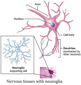
The human neural system is divided into two, the central neural system (CNS) and the peripheral neural system (PNS). The structural and functional units of the neural system are neurons that transmit nerve impulses. The non-nervous special cells called neuroglia form the supporting cells of the nervous tissue.

There are three functional classes of neurons. They are the afferent neurons that take sensory impulses to the Central Neural system (CNS) from the sensory organs; the efferent neurons that carry motor impulses from the CNS to the effector organs; and interneurons that lie entirely within the CNS between the afferent and efferent neurons.

The central neural system lacks connective tissue, so the interneuron space is filled by neuroglia. They perform several functions such as providing nourishment to the surrounding neurons; involving the memory process; repairing the injured tissues due to their dividing and regenerating capacity; and acting as phagocyte cells to engulf the foreign particles at the time of any injury to the brain.

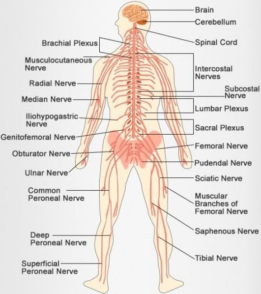
The human nervous system consists of two main parts: the central nervous system (CNS) and the peripheral nervous system (PNS). The CNS contains the brain and spinal cord. The PNS consists mainly of nerves, which are long fibers that connect the CNS to every other part of the body.

The central nervous system is made up of the brain and spinal cord, and the peripheral nervous system is made up of the Somatic and the Autonomic nervous systems.

The nervous system of vertebrates (including humans) is divided into the central nervous system (CNS) and the peripheral nervous system (PNS). The (CNS) is the major division, and consists of the brain and the spinal cord. The spinal canal contains the spinal cord, while the cranial cavity contains the brain.

The nervous system is the major controlling, regulatory, and communicating system in the body. It is the center of all mental activity including thought, learning, and memory. Together with the endocrine system, the nervous system is responsible for regulating and maintaining homeostasis.

The Four Main Functions of the Nervous System are:

Control of body’s internal environment to maintain ‘homeostasis’ An example of this is the regulation of body temperature. Programming of spinal cord reflexes. An example of this is the stretch reflex. Memory and learning. Voluntary control of movement.

The Nervous System has two main Parts:

The central nervous system is made up of the brain and spinal cord. The peripheral nervous system is made up of nerves that branch off from the spinal cord and extend to all parts of the body.

The nervous system includes the brain, nerves and spinal cord. It is the communication center for the body, sending and receiving messages, regulating body functions and serving as the control center for the five senses and for emotions, speech, coordination, balance, and learning.

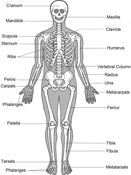
The 11 organ systems include the integumentary system, skeletal system, muscular system, lymphatic system, respiratory system, digestive system, nervous system, endocrine system, cardiovascular system, urinary system, and reproductive systems.
Human Neural System img 1

Neural System Definition, Function, Structure and its Types

Learninsta presents the core concepts of Biology with high-quality research papers and topical review articles.

Neural System Definition, Function, Structure and its Types

The neural system comprises of highly specialized cells called neurons, which can detect, receive, process and transmit different kinds of stimuli. Simple form of neural system as nerve net is seen in lower invertebrates. The neural system of higher animals are well developed and performs the following basic functions:

Sensory Functions:
It receives sensory input from internal and external environment.

Motor Functions:
It transmits motor commands from the brain to the skeletal and muscular system.

Autonomic Functions:
Reflex actions.

The nervous system is the part of an animal’s body that coordinates its behavior and transmits signals between different body areas. In vertebrates it consists of two main parts, called the central nervous system (CNS) and the peripheral nervous system (PNS). The CNS contains the brain and spinal cord.

The nervous system takes in information through our senses, processes the information and triggers reactions, such as making your muscles move or causing you to feel pain. For example, if you touch a hot plate, you reflexively pull back your hand and your nerves simultaneously send pain signals to your brain.

The central nervous system is made up of the brain and spinal cord, and the peripheral nervous system is made up of the Somatic and the Autonomic nervous systems.

The nervous system consists of the brain, spinal cord, sensory organs, and all of the nerves that connect these organs with the rest of the body.

The nervous system in a human is made of the brain, spinal cord, sensory organs and all the neurons that serve as communication channels between the various organs of the body.

The peripheral nervous system carries messages to and from the central nervous system. It sends information to the brain and carries out orders from the brain. Messages travel through the cranial nerves, those which branch out from the brain and go to many places in the head such as the ears, eyes and face.

The nervous system has three broad functions: sensory input, information processing, and motor output. In the PNS, sensory receptor neurons respond to physical stimuli in our environment, like touch or temperature, and send signals that inform the CNS of the state of the body and the external environment.

The neural or nervous system is a complex network of nerve cells or neurons. The nervous system is specialized to carry messages while the endocrine system provides chemical integration through hormones. To better understand the nervous system, one must realize the difference between a neuron and a nerve.

The Structure of a Neuron:

The above image shows the basic structural components of an average neuron, including the dendrite, cell body, nucleus, Node of Ranvier, myelin sheath, Schwann cell, and axon terminal.

The gap between two neurons called synapse, helps in quick transmission of impulses from one neuron to another. Always one-way communication i.e. unidirectional, transmitting from pre-synaptic to post-synaptic neurons. Can be used to calsculate timing of sensory inputs. Greater plasticity.

Neurons have specialized projections called dendrites and axons. The synapse contains a small gap separating neurons. The synapse consists of: a presynaptic ending that contains neurotransmitters, mitochondria and other cell organelles.
Neural Tissue img 1

Benefits of Regular Exercise

Learninsta presents the core concepts of Biology with high-quality research papers and topical review articles.

Benefits of Regular Exercise

Exercise and physical activity fall into four basic categories. Endurance, Strength, Balance and Flexibility. Endurance or aerobic activities increase the breathing and heart rate. They keep the circulatory system healthy and improve overall fitness.

Strength exercises make the muscles stronger. They help to stay independent and carry out everyday activities such as climbing stairs and carrying bags.

Balance exercises help to prevent falls which is a common problem in older adults. Many strengthening exercises also improves balance.

Flexibility exercises help to stretch body muscles for more freedom of joint movements. Regular exercises can produce the following beneficial physiological changes:

  • The muscles used in exercise grow larger and stronger.
  • The resting heart rate goes down.
  • More enzymes are synthesized in the muscle fibre.
  • Ligaments and tendons become stronger.
  • Joints become more flexible.
  • Protection from heart attack.
  • Influences hormonal activity.
  • Improves cognitive functions.
  • Prevents Obesity.
  • Promotes confidence, esteem.
  • Aesthetically better with good physique.
  • Over all well-being with good quality of life.
  • Prevents depression, stress and anxiety.

During muscular exercise, there is an increase in metabolism. The O2 need of the muscles is increased. This requirement is met with more oxygen rich RBCs available to the active sites. There is an increase in heart rate and cardiac output. Along with balanced diet, physical activity plays a significant role in strengthening the muscles and bones.
Benefits of Regular Exercise img 1

Disorders of Muscular and Skeletal System

Learninsta presents the core concepts of Biology with high-quality research papers and topical review articles.

Disorders of Muscular and Skeletal System

(a) Disorders of Muscular System

Myasthenia Gravis:

An autoimmune disorder affecting the action of acetylcholine at neuromuscular junction leading to fatigue, weakening and paralysis of skeletal muscles. Acetylcholine receptors on the sarcolemma are blocked by antibodies leading to weakness of muscles. When the disease progresses, it can make chewing, swallowing, talking and even breathing difficult.

Tetany:

Rapid muscle spasms occur in the muscles due to deficiency of parathyroid hormone resulting in reduced calcium levels in the body.

Muscle Fatigue:

Muscle fatigue is the inability of a muscle to contract after repeated muscle contractions. This is due to lack of ATP and accumulation of lactic acid by anaerobic breakdown of glucose.

Atrophy:

A decline or cessation of muscular activity results in the condition called atrophy which results in the reduction in the size of the muscle and makes the muscle to become weak, which occurs with lack of usage as in chronic bedridden patients.

Muscle Pull:

Muscle pull is actually a muscle tear. A traumatic pulling of the fibres produces a tear known as sprain. This can occur due to sudden stretching of muscle beyond the point of elasticity. Back pain is a common problem caused by muscle pull due to improper posture with static sitting for long hours.

Muscular Dystrophy:

The group of diseases collectively called the muscular dystrophy are associated with the progressive degeneration and weakening of skeletal muscle fires, leading to death from lung or heart failure. The most common form of muscular dystrophy is called Duchene Muscular Dystrophy (DMD).

(b) Disorders of Skeletal System

Arthritis and osteoporosis are the major disorders of skeletal system.

1. Arthritis:

Arthritis is an inflammatory (or) degenerative disease that damages the joints. There are several types of arthritis.

(i) Osteoarthritis:

The bone ends of the knees and other freely movable joints wear away as a person ages. The joints of knees, hip, fingers and vertebral column are affected.

(ii) Rheumatoid Arthritis:

The synovial membranes become inflamed and there is an accumulation of fluid in the joints. The joints swell and become extremely painful. It can begin at any age but symptoms usually emerge before the age of fifty.

(iii) Gouty Arthritis or Gout:

Inflammation of joints due to accumulation of uric acid crystals or inability to excrete it. It gets deposited in synovial joints.

2. Osteoporosis:

It occurs due to deficiency of vitamin D and hormonal imbalance. The bone becomes sof and fragile. It causes rickets in children and osteomalacia in adult females. It can be minimized with adequate calcium intake, vitamin D intake and regular physical activities.
Disorders of Muscular and Skeletal System img 1

Types of Joints in the Human Body

Learninsta presents the core concepts of Biology with high-quality research papers and topical review articles.

Types of Joints in the Human Body

Types of Joints

Joints are essential for all types of movements performed by the bony parts of the body. The joints are points of contact (Figure 9.11) between bones. Sometimes they are playing a protective role in the process. Force generated by the muscles are used to carry out the movement through joints which helps human functional activity of daily living and ambulation. The joint acts as a fulcrum of a lever.
Types of Joints img 1

(i) Fibrous Joints or Synarthroses:

They are immovable field joints in which no movement between the bones is possible. Sutures of the flat skull bones are firous joints.

(ii) Cartilaginous Joints or Amphiarthroses:

They are slightly movable joints in which the joint surfaces are separated by a cartilage and slight movement is only possible. E.g., Joints of adjacent vertebrae of the vertebral column.

(iii) Synovial Joints or Diarthroses Joints:

They are freely movable joints, the articulating bones are seperated by a cavity which is filled with synovial fluid.

There are six types of freely movable diarthrosis (synovial) joints:

Ball and socket joint. Permitting movement in all directions, the ball and socket joint features the rounded head of one bone sitting in the cup of another bone.

  • Hinge joint
  • Condyloid joint
  • Pivot joint
  • Gliding joint
  • Saddle joint

There are two basic structural types of joint: diarthrosis, in which fluid is present, and synarthrosis, in which there is no fluid. All the diarthroses (commonly called synovial joints) are permanent. Some of the synarthroses are transient; others are permanent.

With a force strength exceeding 350 kg (772 lbs), the iliofemoral ligament is not only stronger than the two other ligaments of the hip joint, the ischiofemoral and the pubofemoral, but also the strongest ligament in the human body and as such is an important constraint to the hip joint.

The Appendicular Skeleton Structure and its Types

Learninsta presents the core concepts of Biology with high-quality research papers and topical review articles.

The Appendicular Skeleton Structure and its Types

The bones of the upper and lower limbs along with their girdles constitute the appendicular skeleton. The appendicular skeleton is composed of 126 bones.

(a) The Pectoral Girdle

The upper limbs are attached to the pectoral girdles. These are very light and allow the upper limbs a degree of mobility not seen anywhere else in the body. The girdle is formed of two halves. Each half of the pectoral girdle (Figure 9.8) consists of a clavicle or collar bone and a scapula.

The scapula is a large, thin, triangular bone situated in the dorsal surface of the ribcage between the second and seventh ribs. It has a slightly elevated ridge called the spine which projects as a flat, expanded process called the acromion.
The Appendicular Skeleton img 1

The clavicle articulates with this process. Below the acromion is a depression called the glenoid cavity which articulates with the head of the humerus to form the shoulder joint. Each clavicle is a long slender bone with two curvatures which lies horizontally and connects axial skeleton with appendicular skeleton.

The Upper Limb

The upper limb consists of 30 separate bones and is specialized for mobility. The skeleton of the arm, the region between the shoulder and elbow is the humerus. The head of humerus articulates with the glenoid cavity of the scapula and forms the shoulder joint.

The distal end of humerus articulates with the two forearm bones the radius and ulna. The forearm is the region between the elbow and the wrist. Olecranon process is situated at the upper end of the ulna which forms the pointed portion of the elbow. The hand consists of carpals, metacarpals and phalanges.

Carpals,the wrist bones, 8 in number are arranged in two rows of four each. The anterior surface of the wrist has tunnellike appearance, due to the arrangement of carpals with the ligaments. This tunnel is termed as carpal tunnel. Metacarpals,the palm bones are 5 in number and phalanges the digits bones are 14 in number.

(b) Pelvic Girdle

The pelvic girdle is a heavy structure specialized for weight bearing. It is composed of two hip bones called coxal bones that secure the lower limbs to the axial skeleton (Figure 9.9). Together, with the sacrum and coccyx, the hip bones form the basin-like bony pelvis.

Each coxal bone consists of three fused bones, ilium, ischium and pubis. At the point of fusion of ilium, ischium, and pubis a deep hemispherical socket called the acetabulum is present on the lateral surface of the pelvis. It receives the head of the femur or thigh bone at the hip joint and helps in the articulation of the femur. Ventrally the two halves of the pelvic girdle meet and form the pubic symphysis containing firous
cartilage.
The Appendicular Skeleton img 2

The ilium is the superior floring portion of the hip bone. Each ilium forms a secure joint with the sacrum posteriorly. The ischium is a curved bar of bone. The V-shaped pubic bones articulate anteriorly at the pubic symphysis. The pelvis of male is deep and narrow with larger heavier bones and the female is shallow, wide and flexible in nature, and this helps during pregnancy which is influenced by female hormones.

The Lower Limb

The lower limb consists of 30 bones which carries the entire weight of the erect body and is subjected to exceptional forces when we jump or run. The bones of the lower limbs are thicker and stronger than the upper limbs.

The three segments of each lower limb are the thigh, the leg or the shank and the foot. The femur is the single bone of the thigh. It is the largest, longest and strongest bone in the body. The head of femur articulates with the acetabulum of the pelvis to form the hip joint.

Two parallel bones, the tibia and fibula, form the skeleton of the shank. A thick, triangular patella forms the knee cap, which protects the knee joint anteriorly and improves the leverage of thigh muscles acting across the knee.

The foot includes the bones of ankle, the tarsus, the metatarsus and the phalanges or toe bones. The foot supports our body weight and acts as a lever to propel the body forward, while walking and running. The tarsus is made up of seven bones called tarsals. The metatarsus consists of five bones called metatarsals. The arrangement of the metatarsals is parallel to each other. There are 14 phalanges in the toes which are smaller than those of the fingers.

Structure of a Typical Long Bone

A typical long bone has a diaphysis, epiphyses (singular-epiphysis) and membranes (Figure 9.10). A tubular diaphysis or shaft forms the long axis of the bone. It is constructed of a thick collar of compact bone that surrounds a central medullary cavity or marrow cavity.

The epiphyses are the bone ends. Compact bone forms the exterior of epiphyses and their interior contains spongy bone with red marrow. The region where the diaphysis and epiphyses meet is called the metaphysis.
The Appendicular Skeleton img 3

The external surface of the entire bone except the joint surface is covered by a doublelayered membrane called the periosteum. The outer firous layer is dense irregular connective tissue. The inner osteogenic layer consists of osteoblasts (bone- forming cells) which secrete bone matrix elements and osteoclasts (bone-destroying cells).

In addition, there are primitive stem cells, osteogenic cells, that give rise to the osteoblasts. The periosteum is richly supplied with nerve fires, lymphatic vessels and blood vessels. Internal bone surfaces are covered with a delicate connective tissue membrane called the endosteum. The endosteum covers the trabeculae of spongy bone and lines the canals that pass through the compact bone. It also contains both osteoblasts and osteoclasts. Between the epiphysis and diaphysis epiphyseal plate or growth plate is present.

The Skeletal System – The Axial Skeleton

Learninsta presents the core concepts of Biology with high-quality research papers and topical review articles.

The Skeletal System – The Axial Skeleton

Axial skeleton forms the main axis of the body. It consists of the skull, hyoid bone, vertebral column and thoracic cage.

(a) The Skull

The skull is composed of two sets of bones – cranial and facial bones. It consists of 22 bones of which 8 are cranial bones and 14 are facial bones (Figure 9.5). The cranial bones form the hard protective outer covering of the brain and called the brain box.

The capacity of the cranium is 1500 cm3. These bones are joined by sutures which are immovable. They are a paired parietal, paired temporal and individual bones such as the frontal, sphenoid, occipital and ethmoid.

The large hole in the temporal bone is the external auditory meatus. In the facial bones maxilla, zygomatic, palatine, lacrimal, nasal are paired bones whereas mandible or lower jaw and vomer are unpaired bones. They form the front part of the skull. A single U-shaped hyoid bone is present at the base of the buccal cavity.

It is the only one bone without any joint. Each middle ear contains three tiny bonesmalleus, incus and stapes collectively are called ear ossicles. The upper jaw is formed of the maxilla and the lower jaw is formed of the mandible. The upper jaw is fused with the cranium and is immovable.

The lower jaw is connected to the cranium by muscles and is movable. The most prominent openings in the skull are the orbits and the nasal cavity. Foramen magnum is a large opening found at the posterior base of the skull. Though this opening the medulla oblongata of the brain descends down as the spinal cord.
The Skeletal System - The Axial Skeleton img 1

(b) The Vertebral Column

Vertebral column is also called the back bone. It consists of 33 serially arranged vertebrae which are inter connected by cartilage known as intervertebral disc (Figure 9.6). The vertebral column extends from the base of the skull to the pelvis and forms the main frame work of the trunk.

The vertebral column has five major regions. They are, the Cervical, Thoracic, Lumbar, Sacrum (5 sacral vertebrae found in the infant which are fused to form one bone in the adult) and Coccyx (4 coccygeal vertebrae found in the infant which are fused to form one bone in the adult).
The Skeletal System - The Axial Skeleton img 2

Each vertebra has a central hollow portion, the neural canal, through which the spinal cord passes. The first vertebra is called as the atlas and the second vertebra is called as the axis. Atlas is articulated with the occipital condyles. The vertebral column protects the spinal cord, supports the head and serves as the point of attachment for the ribs and musculature of the back.

(c) The Sternum (Chest bone)

Sternum is a flat bone on the mid ventral line of the thorax. It provides space for the attachment of the thoracic ribs and abdominal muscles.

(d) The Rib Cage

There are 12 pairs of ribs (Figure 9.7). Each rib is a thin flat bone connected dorsally to the vertebral column and ventrally to the sternum. It has two articulation surfaces on its dorsal end, hence called bicephalic. The first seven pairs of ribs are called ‘true ribs’ or vertebro-sternal ribs.

Dorsally they are attached to the thoracic vertebrae and ventrally connected to the sternum with the help of hyaline cartilages. The 8th, 9th and 10th pairs of ribs do not articulate directly with the sternum but joined with the cartilaginous (hyaline cartilage) part of the seventh rib.

These are called ‘false ribs’ or vertebro-chondral ribs. The last 11th and 12th pairs of ribs are not connected ventrally. Therefore, they are called as ‘flating ribs’ or vertebral ribs. Thoracic vertebrae, ribs and sternum together form the ribcage. Rib cage protects the lungs, heart, liver and also plays a role in breathing.
The Skeletal System - The Axial Skeleton img 3

Mechanism of Muscle Contraction

Learninsta presents the core concepts of Biology with high-quality research papers and topical review articles.

Mechanism of Muscle Contraction

Sliding Filament Theory:

In 1954, Andrew F. Huxley and Rolf Niedergerke proposed the sliding-filament theory to explain muscle contraction. According to this theory, overlapping actin and myosin filaments of fixed length slide past one another in an energy requiring process, resulting in muscle contraction. The contraction of muscle fire is a remarkable process that helps in creating a force to move or to resist a load.

The force which is created by the contracting muscle is called muscle tension. The load is a weight or force that opposes contraction of a muscle. Contraction is the creation of tension in the muscle which is an active process and relaxation is the release of tension created by contraction. Muscle contraction is initiated by a nerve impulse sent by the central nervous system (CNS) through a motor neuron.

The junction between the motor neuron and the sarcolemma of the muscle fire is called the neuromuscular junction or motor end plate. When nerve impulse reaches a neuromuscular junction, acetylcholine is released. It initiates the opening of multiple gated channels in sarcolemma. The action potential travels along the T-tubules and triggers the release of calcium ions from the sarcoplasmic reticulum.

The released calcium ions bind to troponin on thin filaments. The tropomyosin uncovers the myosin-binding sites on thin filaments. Now the active sites are exposed to the heads of myosin to form a cross-bridge (Figure 9.3).

During cross-bridge formation acting and myosin form a protein complex called actomyosin. Utilizing the energy released from hydrolysis of ATP, the myosin head rotates until it forms a 90° angle with the long axis of the filament. In this position myosin binds to an actin and activates a contraction – relaxation cycle which is followed by a power stroke.
Mechanism of Muscle Contraction img 1

The power stroke (cross-bridge tilting) begins after the myosin head and hinge region tilt from a 90° angle to a 45° angle. The crossbridge transforms into strong, high-force bond which allows the myosin head to swivel. When the myosin head swivels it pulls the attached acting filament towards the centre of the A-band.

The myosin returns back to its relaxed state and releases ADP and phosphate ion. A new ATP molecule then binds to the head of the myosin and the cross-bridge is broken. At the end of each power stroke, each myosin head detaches from actin, then swivels back and binds to a new actin molecule to start another contraction cycle.

This movement is similar to the motion of an oar on a boat. At the end of each power stroke, each myosin head detaches from actin, then swivels back and binds to a new actin molecule to start another contraction cycle. The power stroke repeats many times until a muscle fire contracts.

The myosin heads bind, push and release actin molecules over and over as the thin filaments move toward the centre of the sarcomere. The repeated formation of cross-bridge cycles cause the sliding of the filaments only but there is no change in the lengths of either the thick or thin filaments.

The Z – discs attached to the actin filaments are also pulled inwards from both the sides, causing the shortening of the sarcomere (i.e. contraction). This process continues as long as the muscle receives the stimuli and with a steady flow of calcium ions.

When motor impulse stops, the calcium ions are pumped back into the sarcoplasmic reticulum which result in the masking of the active sites of the actin filaments. The myosin head fails to bind with the active sites of actin and these changes cause the return of Z – discs back to their original position, i.e. relaxation. (Figure 9.4)
Mechanism of Muscle Contraction img 2
Mechanism of Muscle Contraction img 3

Types of Skeletal Muscle Contraction

Learninsta presents the core concepts of Biology with high-quality research papers and topical review articles.

Types of Skeletal Muscle Contraction

There are two primary types of muscle contractions. They are isotonic contraction and isometric contraction. The types of contractions depend on the changes in the length and tension of the muscle fibres at the time of its contraction.

Isotonic contraction (iso – same, tonweight/resistance) In isotonic contraction the length of the muscle changes but the tension remains constant. Here, the force produced is unchanged. Example: lifting dumb bells and weightlifting. Isometric contraction (iso – same, metric – distance)

In isometric contraction the length of the muscle does not change but the tension of the muscle changes. Here, the force produced is changed. Example: pushing against a wall, holding a heavy bag.

Types of Skeletal Muscle Fibres

The muscle fibres can be classified on the basis of their rate of shortening, either fast or slow and the way in which they produce the ATP needed for contraction, either oxidative or glycolytic. Fibres containing myosin with high ATPase activity are classified as fast fibres and with lower ATPase activity are classified as slow fibres.

Fibres that contain numerous mitochondria and have a high capacity for oxidative phosphorylation are classified as oxidative fibres. Such fibres depend on blood flow to deliver oxygen and nutrients to the muscles. The oxidative fibres are termed as red muscle fibres.

Fibres that contain few mitochondria but possess a high concentration of glycolytic enzymes and large stores of glycogen are called glycolytic fibres. The lack of myoglobin gives pale colour to the fibres, so they are termed as white muscle fibres.

Skeletal muscle fibres are further classified into three types based on the above classification. They are slow – oxidative fibres, fast – oxidative fibres and fast – glycolytic fibres.

1. Slow – Oxidative Fibres

Have low rates of myosin ATP hydrolysis but have the ability to make large amounts of ATP. These fibres are used for prolonged, regular activity such as long distance swimming. Long – distance runners have a high proportion of these fibres in their leg muscles.

2. Fast – Oxidative Fibres

Have high myosin ATPase activity and can make large amounts of ATP. They are particularly suited for rapid actions.

3. Fast – Glycolytic Fibres

Have myosin ATPase activity but cannot make as much ATP as oxidative fires, because their source of ATP is glycolysis. These fibres are best suited for rapid, intense actions, such as short sprint at maximum speed.

Isometric:
A muscular contraction in which the length of the muscle does not change.

Isotonic:
A muscular contraction in which the length of the muscle changes.

Eccentric:
An isotonic contraction where the muscle lengthens.

Concentric:
An isotonic contraction where the muscle shortens. Types of Contractions. There are three types of muscle contraction: concentric, isometric, and eccentric.

Isometric:
Of or involving muscular contraction against resistance in which the length of the muscle remains the same.

Isotonic:
Of or involving muscular contraction against resistance in which the length of the muscle changes.

The process of muscular contraction occurs over a number of key steps, including: Depolarisation and calcium ion release.

Actin and myosin cross-bridge formation.
Sliding mechanism of actin and myosin filaments.
Sarcomere shortening (muscle contraction)

Isotonic contractions – These occur when a muscle contracts and changes length and there are two types:
Isotonic concentric contraction – This involves the muscle shortening.
Isotonic eccentric contraction – This involves the muscle lengthening whilst it is under tension.

The first step in the process of contraction is for Ca++ to bind to troponin so that tropomyosin can slide away from the binding sites on the actin strands. This allows the myosin heads to bind to these exposed binding sites and form cross-bridges.

Abstract. Skeletal muscle is one of the most dynamic and plastic tissues of the human body. In humans, skeletal muscle comprises approximately 40% of total body weight and contains 50-75% of all body proteins.

Skeletal muscles only pull in one direction. For this reason they always come in pairs. When one muscle in a pair contracts, to bend a joint for example, its counterpart then contracts and pulls in the opposite direction to straighten the joint out again.

This is a table of skeletal muscles of the human anatomy. There are around 650 skeletal muscles within the typical human body. Almost every muscle constitutes one part of a pair of identical bilateral muscles, found on both sides, resulting in approximately 320 pairs of muscles, as presented in this article.
Types of Skeletal Muscle Contraction img 1

Structure of Contractile Proteins

Learninsta presents the core concepts of Biology with high-quality research papers and topical review articles.

Structure of Contractile Proteins

Contraction of the muscle depends on the presence of contractile proteins (Figure 9.2) such as actin and myosin in the myofilaments. The thick filaments are composed of the protein myosin which are bundled together whose heads produce at opposite ends of the filament.

Each myosin molecule is made up of a monomer called meromyosin. The meromyosin has two regions, a globular head with a short arm and a tail. The short arm constitutes the heavy meromyosin (HMM).

The tail portion forms the light meromyosin (LMM). The head bears an actin-binding site and an ATP – binding site. It also contains ATP case enzyme that split ATP to generate energy for the contraction of muscle. The thin filaments are composed of two interwined actin molecules. Actin has polypeptide subunits called globular actin or G-actin and filamentous form or F-actin.

Each thin filament is made of two F-actins helically wound to each other. Each F-actin is a polymer of monomeric G-actins. It also contains a binding site for myosin. The thin filaments also contain several regulatory proteins like tropomyosin and troponin which help in regulating the contraction of muscles along with actin and myosin.

Structure of Contractile Proteins img 1

Structure of Contractile Proteins. Each actin (thin) filament is made of two ‘F’ (filamentous) actins helically wound to each other. Each ‘F’ actin is a polymer of monomeric ‘G’ (Globular) actins. Two filaments of another protein, tropomyosin also run close to the ‘F’ actins throughout its length.

The contractile proteins are myosin, the principal component of thick myofilaments, and actin, which is the principal component of thin myofilaments.

Contractile fibers are intracellular protein filament-based structures that are primarily composed of actin, myosin and tropomyosin.

Contractile proteins are proteins that mediate sliding of contractile fibres (contraction) of a cell’s cytoskeleton, and of cardiac and skeletal muscle.

Thick filaments contain myosin, thin filaments contain actin , troponin and tropomyosin. Scientists think that muscles contract by the two types of filament sliding over each other so that they overlap more.

Contractile function is a fundamental part of the CMR examination. Contractile function imaging is used for global and regional wall motion assessment and has been demonstrated to be highly accurate and reproducible for LV and right ventricular (RV) volume, ejection fraction, and mass measurements. Non-contractile (inert) tissues – joint capsules, ligaments, nerves and their sheaths, bursae, and cartilages.

Fiber-tracheids are intermediate forms between tracheids and libriform fibers. Tracheids are not fibers, as their major function is conducting water and the cell shape is not typical of a fiber, though they have relatively thick cell walls.

ATP is a nucleotide that consists of three main structures: the nitrogenous base, adenine; the sugar, ribose; and a chain of three phosphate groups bound to ribose. The phosphate tail of ATP is the actual power source which the cell taps.

Any metabolic process that requires oxygen to occur is referred to as aerobic. Humans, most other multicellular organisms, and some microorganisms require oxygen for the efficient capture of the chemical energy from food and its transformation into the cellular energy form known as ATP.

Skeletal Muscle (Voluntary Muscle) Definition and its Uses

Learninsta presents the core concepts of Biology with high-quality research papers and topical review articles.

Skeletal Muscle (Voluntary Muscle) Definition and its Uses

Skeletal muscle is attached to the bone by a bundle of collagen fibres known as tendon. Each muscle is made up of bundles of muscle fibres called fascicle. Each muscle fibre contains hundreds to thousands of rod-like structures called myofibrils that run parallel to its length.

The connective tissue covering the whole muscle is the epimysium, the covering around each fascicle is the perimysium and the muscle fibre is surrounded by the endomysium. They control the voluntary actions such as walking, running, swimming, writing hence termed as voluntary muscles.

Structure of a Skeletal Muscle Fibre

Each muscle fibre is thin and elongated. Most of them taper at one or both ends. Muscle fibre has multiple oval nuclei just beneath its plasma membrane or sarcolemma. The cytoplasm of the muscle fibre is called the sarcoplasm. It contains glycosomes, myoglobin and sarcoplasmic reticulum. Myoglobin is a red – coloured respiratory pigment of the muscle fibre.

It is similar to haemoglobin and contains iron group that has affinity towards oxygen and serves as the reservoir of oxygen. Glycosomes are the granules of stored glycogen that provide glucose during the period of muscle fibre activity. Actin and myosin are muscle proteins present in the muscle fibre.
Skeletal Muscle (Voluntary Muscle) img 1

Along the length of each myofibril there are a repeated series of dark and light bands (Figure 9.1). The dark A-bands (Anisotropic bands) and the light I-bands (Isotropic bands) are perfectly aligned with one another. This type of arrangement gives the cell a striated appearance.

Each dark band has a lighter region in its middle called the H-Zone (H-Helles: means clear). Each H-zone is bisected vertically by a dark line called the M-line (M-for middle). The light I-bands also have a darker mid line area called the Z-disc (from the German “Zwischenscheibe” the disc inbetween the I-bans).
Skeletal Muscle (Voluntary Muscle) img 2

The myofibrils contain the contractile element, the sarcomere which is the functional unit of the skeletal muscle. A Sarcomere is the region of a myofibril between two successive Z-discs. It contains an A-band with a half I-band at each end. Inside the sarcomere two types of filaments are present namely the thick and thin filaments.

The thick filaments extend the entire length of the A-band, the thin filaments extend across the I-band and partly into the A-band. The invagination of the sarcolemma forms transverse tubules (T-tubules) and they penetrate into the junction between the A and I-bands.

Muscle Terminology
Skeletal Muscle (Voluntary Muscle) img 3