Neural Tissue Definition and its Uses

Learninsta presents the core concepts of Biology with high-quality research papers and topical review articles.

Neural Tissue Definition and its Uses

Nervous tissue exerts the greatest control over the body’s responsiveness to changing conditions. Neurons, the unit of neural system are excitable cells (Figure 3.8).
Neural Tissue img 1

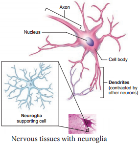
The neuroglial cells which constitute the rest of the neural system protect and support the neurons. Neuroglia makes up more than one-half of the volume of neural tissue in our body. When a neuron is suitably stimulated, an electrical disturbance is generated which swifty travels along its plasma membrane. Arrival of the disturbance at the neuron’s endings, or output zone, triggers events that may cause stimulation or inhibition of adjacent neurons and other cells (You will study in detail in Chapter 10)

Neurons, or nerves, transmit electrical impulses, while neuroglia do not; neuroglia have many other functions including supporting and protecting neurons.

Integration and communication are the two major functions of nervous tissue. Nervous tissue contains two categories of cells – neurons and neuroglia. Neurons are highly specialized nerve cells that generate and conduct nerve impulses.

Nervous tissue contains two major cell types, neurons and glial cells. Neurons are the cells responsible for communication through electrical signals.

Nervous tissue is made up of different types of neurons, all of which have an axon. Bundles of axons make up the nerves in the PNS and tracts in the CNS. Functions of the nervous system are sensory input, integration, control of muscles and glands, homeostasis, and mental activity.

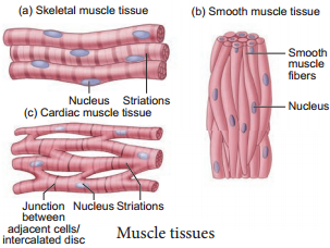
The function of muscle tissue (smooth, skeletal, and cardiac) is to contract, while nervous tissue is responsible for communication.

Brain, spinal cord and nerves constitute nervous tissue. Tendon is a fibrous connective tissue connecting bones to muscles. Nervous tissue is absent in tendon. These are made up of collagen.

Neurons, also known as nerve cells, send and receive signals from your brain. While neurons have a lot in common with other types of cells, they’re structurally and functionally unique. Specialized projections called axons allow neurons to transmit electrical and chemical signals to other cells.

Although the nervous system is very complex, there are only two main types of cells in nerve tissue. The actual nerve cell is the neuron. It is the “conducting” cell that transmits impulses and the structural unit of the nervous system. The other type of cell is neuroglia, or glial, cell.

Muscle Tissue Function and Its Types

Learninsta presents the core concepts of Biology with high-quality research papers and topical review articles.

Muscle Tissue Function and Its Types

Each muscle is made of many long, cylindrical fires arranged in parallel arrays. These fibres are composed of numerous fine firils, called myofirils. Muscle fires contract (shorten) in response to stimulation, then relax (lengthen) and return to their uncontracted state in a coordinated fashion. In general muscles play an active role in all the movements of the body.

Muscles are of three types, skeletal, smooth and cardiac. Skeletal muscle tissue is closely attached to skeletal bones. In a typical muscle such as the biceps, the striated (striped) skeletal muscle fires are bundled together in a parallel fashion. A sheath of tough connective tissue encloses several bundles of muscle fires (You will learn more about this in Chapter 9).

The smooth muscle fires taper at both ends (fusiform) and do not show striations (Figure 3.7). Cell junctions hold them together and they are bundled together in a connective tissue sheath. The walls of internal organs such as the blood vessels, stomach and intestine contain this type of muscle tissue. Smooth muscles are ‘involuntary’ as their functions cannot be directly controlled. Unlike the smooth muscles, skeletal muscles can be controlled by merely thinking.

Cardiac muscle tissue is a contractile tissue present only in the heart. Cell junctions fuse the plasma membranes of cardiac muscle cells and make them stick together. Communication junctions (intercalated discs) at some fusion points allow the cells to contract as a unit, i.e., when one cell receives a signal to contract, its neighbours are also stimulated to contract.
Muscle Tissue img 1

An Overview Of Connective Tissue

Learninsta presents the core concepts of Biology with high-quality research papers and topical review articles.

An Overview Of Connective Tissue

Connective tissue develops from the mesoderm and is widely distributed in the body. There are three main classes namely Loose connective tissue, Dense connective tissue and Specialized connective tissue. Major functions of connective tissues are binding, support, protection, insulation and transportation.
An Overview Of Connective Tissue img 1

Components of Connective Tissue

All connective tissues consist of three main components namely fires, ground substance and cells. The ‘Fibres’ of connective tissue provide support. Three types of fires are found in the connective tissue matrix. They are collagen, elastic and reticular fires.

Connective tissues are of three types namely, Loose connective tissues (Areolar, Adipose and Reticular) and Dense connective tissues (dense regular, dense irregular and elastic) and Specialized connective tissues (cartilage, bone and blood).

Loose Connective Tissues

In this tissue the cells and fires are loosely arranged in a semi fluid ground substances. For example the Areolar connective tissue beneath the skin acts as a support framework for epithelium and acts as a reservoir of water and salts for the surrounding body tissues, hence apply called tissue fluid. It contains firoblasts, macrophages, and mast cells (Figure 3.5).
An Overview Of Connective Tissue img 2

Adipose tissue is similar to areolar tissue in structure and function and located beneath the skin. Adipocytes commonly called adipose or fat cells predominate and account for 90% of this tissue mass. The cells of this tissue store fats and the excess nutrients which are not utilised immediately are converted to fats and are stored in tissues. Adipose tissue is richly vascularised indicating its high metabolic activity. While fasting, these cells maintain life by producing and supplying energy as fuel.

Adipose tissues are also found in subcutaneous tissue, surrounding the kidneys, eyeball, heart, etc. Adipose tissue is called ‘white fat’ or white adipose tissue. The adipose tissue which contains abundant mitochondria is called ‘Brown fat’ or Brown adipose tissue. White fat stores nutrients whereas brown fat is used to heat the blood stream to warm the body. Brown fat produces heat by nonshivering thermogenesis in neonates.

Reticular connective tissue resembles areolar connective tissue, but, the matrix is filled with firoblasts called reticular cells. It forms an internal framework (stroma) that supports the blood cells (largely lymphocytes) in the lymph nodes, spleen and bone marrow. Dense connective tissues (connective tissue proper) Fibres and firoblasts are compactly packed in the dense connective tissues. Orientation of fires show a regular or irregular pattern and is called dense regular and dense irregular tissues.

Dense regular connective tissues primarily contain collagen fires in rows between many parallel bundles of tissues and a few elastic fires. The major cell type is firoblast. It attaches muscles and bones and withstands great tensile stress when pulling force is applied in one direction. This connective tissue is present in tendons, that attach skeletal muscles to bones and ligaments attach one bone to another. Dense irregular connective tissues have bundles of thick collagen fires and firoblasts which are arranged irregularly.

The major cell type is the firoblast. It is able to withstand tension exerted in many directions and provides structural strength. Some elastic fires are also present. It is found in the skin as the leathery dermis and forms firous capsules of organs such as kidneys, bones, cartilages, muscles, nerves and joints.

Elastic connective tissue contains high proportion of elastic fires. It allows recoil of tissues following stretching. It maintains the pulsatile flow of blood through the arteries and the passive recoil of lungs following inspiration. It is found in the walls of large arteries; ligaments associated with vertebral column and within the walls of the bronchial tubes.

Specialised connective tissues are classified as cartilage, bones and blood. The intercellular material of cartilage is solid and pliable and resists compression. Cells of this tissue (chondrocytes) are enclosed in small cavities within the matrix secreted by them (Figure 3.6). Most of the cartilages in vertebrate embryos are replaced by bones in adults. Cartilage is present in the tip of nose, outer ear joints, ear pinna, between adjacent bones of the vertebral column, limbs and hands in adults.
An Overview Of Connective Tissue img 3

Bones have a hard and non-pliable ground substance rich in calcium salts and collagen fires which gives strength to the bones. It is the main tissue that provides structural frame to the body. Bones support and protect softer tissues and organs.

The bone cells (osteocytes) are present in the spaces called lacunae. Limb bones, such as the long bones of the legs, serve weightbearing functions. They also interact with skeletal muscles attached to them to bring about movements.

The bone marrow in some bones is the site of production of blood cells. Blood is the fluid connective tissue containing plasma, red blood cells (RBC), white blood cells (WBC) and platelets. It functions as the transport medium for the cardiovascular system, carrying nutrients, wastes, respiratory gases throughout the body. You will learn more about blood in Chapter 7.

Organisation of Epithelial Tissue

Learninsta presents the core concepts of Biology with high-quality research papers and topical review articles.

Organisation of Epithelial Tissue

Epithelial tissue is a sheet of cells that covers the body surface or lines the body cavity. It occurs in the body as a covering, as a lining epithelium and as glandular, epithelium. The functions of epithelium includes protection, absorption, filtration, excretion, secretion and sensory reception.

Based on the structural modification of the cells, the epithelial tissues are classified into simple epithelium and compound epithelium or stratified epithelium. Simple epithelium is composed of a single layer of cells. They are found in the organs of absorption, secretion and filtration. Simple epithelial tissue is further classified into squamous epithelium, cuboidal epithelium, columnar epithelium, ciliated epithelium and pseudostratifid epithelium (Figure 3.2).

The squamous epithelium is made of a single thin layer of flattened cells with irregular boundaries. They are found in the kidney glomeruli, air sacs of lungs, lining of heart, blood vessels and lymphatic vessels and are involved in functions like forming a diffusion boundary and filtration in sites where protection is not important.

The cuboidal epithelium is made of a single layer of cube like cells. This tissue is commonly found in the kidney tubules, ducts and secretory portions of small glands and surface of the ovary. Its main functions are secretion and absorption.

The columnar epithelium is composed of single layer of tall cells with round to oval nuclei at the base. It lines the digestive tract from the stomach to the rectum. The two modifications of this lining are the presence of microvilli on the apical surface of the absorptive cells and Goblet cell which secretes the protective lubricating mucus. The functions of this epithelium include absorption, secretion of mucus, enzymes and other substances.

If the columnar cells bear cilia on their free surfaces they are called ciliated epithelium. This ciliated type propels mucus by ciliary actions and it lines the small bronchioles, fallopian tubes and uterus. Nonciliated type lines most of the digestive tract, gall bladder and secretory ducts of glands.

Pseudo-stratified epithelial cells are columnar, but unequal in size. Although the epithelium is single layered yet it appears to be multi-layered because the nuclei lie at different levels in different cells. Hence, it is also called pseudostratified epithelium and its functions are protection, secretion and absorption. Ciliated forms line the trachea and the upper respiratory tract. The non ciliated forms, line the epididymis, large ducts of a glands and tracts of male urethra.
Epithelial Tissue img 1

Glandular Epithelium

Some of the cuboidal or columnar cells get specialized for secretion and are called glandular epithelium (Figure 3.3). They are mainly of two types: unicellular, consisting of isolated glandular cells (goblet cells of the alimentary canal), and multicellular, consisting of cluster of cells (salivary gland). On the basis of the mode of pouring of their secretions, glands are divided into two categories namely exocrine and endocrine
glands. Exocrine glands secrete mucus, saliva, earwax, oil, milk, digestive enzymes and other cell products.

These products are released through ducts or tubes. In contrast endocrine glands do not have ducts. Their secretions called hormones are secreted directly into the fluid bathing the gland. The exocrine glands are classified as unicellular and multicelluar glands.

The multicelluar glands are further classified based on the structure as simple and compound glands, based on their secretory units as tubular, alveolar (Acinus) and tubulo alveolar. Based on the mode of secretion exocrine glands are classified as merocrine, holocrine and apocrine.
Epithelial Tissue img 2

Compound epithelium is made of more than one layer (multi-layered) of cells and thus has a limited role in secretion and absorption(Figure 3.4). The compound epithelia may be stratified and transitional. Their main function is to provide protection against chemical and mechanical stresses. They cover the dry surface of the skin, the moist surface of buccal cavity, pharynx, inner lining of ducts of salivary glands and of pancreatic ducts.

There are four types of compound epithelium namely, stratified squamous epithelium, cuboidal epithelium, columnar epithelium and transitional epithelium. Stratified squamous epithelium is of two types called keratinized type which forms the dry epidermis of the skin and the non keratinized type forms the moist lining of the oesophagus, mouth, conjunctiva of the eyes and vagina.

Stratified cuboidal epithelium mostly found in the ducts of sweat glands and mammary glands. Stratified columnar epithelium has limited distribution in the body, found around the lumen of the pharynx, male urethra and lining of some glandular ducts.

Transitional Epithelium is found lining the ureters, urinary bladder and part of the urethra. This epithelium allows stretching and is protective in function. All cells of the epithelium are held together with little intercellular material. In most of the animal tissues, specialized junctions provide both structural and functional links between its individual cells.

Three types of cell junctions are found in the epithelium and other tissues. These are called as tight, adhering and gap junctions. Tight junctions help to stop substances from leaking across a tissue. Adhering junctions perform cementing to keep neighbouring cells together. Gap junctions facilitate the cells to communicate with each other by connecting the cytoplasm of adjoining cells, for rapid transfer of ions, small molecules and sometimes big molecules.

Tissue Level of Organisation – Animal Tissues

Learninsta presents the core concepts of Biology with high-quality research papers and topical review articles.

Tissue Level of Organisation – Animal Tissues

Animal tissues are classified according to the size, shape and function of the cells. There are four primary (basic) tissue types that interweave to form the ‘fabric’ of the body. They are, the epithelial tissue (covering), the connective tissue (support), the muscle tissue (movement) and the nervous tissue (control) (Figure 3.1).
Animal Tissues img 1

There are four types of animal tissues: epithelial tissue, connective tissue, muscle tissue and nervous tissue. Key Outcomes: Understand the differentiation of animal tissues and the relationship between structure and function of the various tissues. Know the location of the various tissues within the animal body.

Epithelial Tissue:
They cover the body, organs, blood vessels and all body cavities.

Muscular Tissue:
Smooth, Skeletal, and Cardiac Muscles.

Connective Tissue:
Connective tissues are made up of fibrous cells.

Nervous Tissue:
Neuron Structure.

Organs are composed of tissues, which are in turn composed of cells. Plants have three tissue types: ground, dermal, and vascular. Animals have four: epithelial, connective, muscle, and bone.

Supports an animal’s body – Connective tissue(supportive) Binds different tissues together – Fibrous connective tissue.

Connective tissue assists in support and protection of organs and limbs and depending on the location in the body it may join or separate organs or parts of the body. Muscle tissue enables various forms of movement, both voluntary and involuntary.

What are Parenchyma tissues? Parenchyma is a type of simple permanent tissue that makes a major part of ground tissues in plants, where other tissues like vascular tissues are embedded. They are non-vascular and composed of simple, living and undifferentiated cells, which are modified to perform various functions.

The nervous tissue is composed of two cell types: neurons and glia. The main function of nervous tissue is the processing of information coming from the external and internal environments, and then triggers a response.

Animal tissues are grouped into four basic types: connective, muscle, nervous, and epithelial. Collections of tissues joined in units to serve a common function compose organs.

Types of Tissues

  1. Simple squamous
  2. Simple cuboidal
  3. Cardiac muscle
  4. Skeletal muscle
  5. Bone
  6. Dense fibrous tissue
  7. Nerve
  8. Cartilage

Phylum: Chordata

Learninsta presents the core concepts of Biology with high-quality research papers and topical review articles.

Phylum: Chordata

(G. Chorda – string)

Chordata is the largest phylum with most familiar group of animals, such as fishes, amphibians, reptiles, birds and mammals and less known forms such as lancelets (Amphioxus) and tunicates (Ascidian). All chordates possess three fundamental distinct features at some stage of their life cycle (Figure 2.19), they are:
Phylum Chordata img 1

1. Presence of elongated rod like notochord below the nerve cord and above the alimentary canal. It serves as a primitive internal skeleton. It may persist throughout life in lancelets and lampreys. In adult vertebrates, it may be partially or completely replaced by backbone or vertebral column.

2. A dorsal hollow or tubular fluid filled nerve cord lies above the notochord and below the dorsal body wall. It serves to integrate and co-ordinate the body functions. In higher chordates, the anterior end of the nerve cord gets enlarged to form the brain and the posterior part becomes the spinal cord, protected inside the vertebral column.

3. Presence of pharyngeal gill slits or clefts in all chordates at some stage of their lifecycle. It is a series of gill slits or cleft that perforates the walls of pharynx and appears during the development of every chordate. In aquatic forms, pharyngeal gill slits are vascular, lamellar and form the gills for respiration. In terrestrial chordates, traces of non-functional gill cleft appear during embryonic developmental stages and disappear later.

Besides the above said features, chordates are bilaterally symmetrical, triploblastic, coelomates with organ system level of organisation; they possess post anal tail, closed circulatory system with a ventral myogenic heart except in Amphioxus.
Phylum Chordata img 2

Subphylum: Urochordata or Tunicata

(G. Oura – A tail; L. Chord – Cord)

They are exclusively marine and are commonly called sea squirts. Mostly sessile, some pelagic or free swimming, exist as solitary and colonial forms. Body is unsegmented and covered by a test or tunic. Adult forms are sac like. Coelom is absent, but has an atrial cavity surrounding the pharynx. Notochord is present only in the tail region of the larval stage, hence named urochordata. Alimentary canal is complete and circulatory system is of open type.

The heart is ventral and tubular. Respiration is through gill slits and cleft. Dorsal tubular nerve cord is present only in the larval stage and a single dorsal ganglion is present in the adults. Mostly hermaphrodites, development indirect and includes a free swimming tadpole larva with chordate characters. Retrogressive metamorphosis is seen (Figure 2.20). Examples: Ascidia, Salpa, Doliolum
Phylum Chordata img 3

Subphylum: Cephalochordata

(L. Cephalo – ‘Head’; G. Chorda ‘Cord’.)

Cephalochordates are marine forms, found in shallow waters, leading a burrowing mode of life. They are small fish like coelomate forms with chordate characters such us notochord, dorsal tubular nerve cord and pharyngeal gill slits throughout their life.

Closed type of circulatory system is seen without heart. Excretion is by protonephridia. Sexes are separate, Fertilization is external. Development is indirect and includes a free swimming larva (Figure 2.21). Example: Branchiostoma (Amphioxus or lancelet).
Phylum Chordata img 4

Subphylum-Vertebrata

(L. Vertebrus – Back Bone)

Vertebrates are also called higher chordates and they possess notochord during embryonic stage only. The notochord is replaced by a cartilaginous or bony vertebral column in the adult. Hence all vertebrates are chordates but all chordates are not vertebrates.

Vertebrates possess paired appendages such as fins or limbs. Skin is covered by protective skeleton comprising of scales, feathers, hairs, claws, nails, etc. Respiration is aerobic through gills, skin, buccopharyngeal cavity and lungs. Vertebrates have a ventral muscular heart with two, three or four chambers and kidneys for excretion and osmoregulation.

Subphylum Vertebrata is divided into two divisions, Agnatha and Gnathostomata. Agnatha includes jawless fish-like aquatic vertebrates without paired appendages. Notochord persists in the adult. Gnathostomata includes jawed vertebrates with paired appendages. Notochord is replaced partly or wholly by the vertebral column.

Agnatha includes one important class – Cyclostomata. Gnathostomata includes jawed fishes (Pisces) and Tetrapoda (amphibia, reptilia, aves and mammals). The superclass Pisces includes all fishes which are essentially aquatic forms with paired fish for swimming and gills for respiration. Pisces includes cartilaginous fishes (Chondrichthyes) and bony fishes (Osteichthyes).

Class: Cyclostomata

(G.cyklos – circle; stomata – mouth)

All members of cyclostomata are primitive, poikilothermic, jawless aquatic vertebrates and are ectoparasites on some fishes. Body is slender and eel-like bearing six to fiten pair of gill slits for respiration. Mouth is circular without jaws and suctorial. Heart is two chambered and circulation is of closed type. No paired appendages. Cranium and vertebral column are cartilaginous.

Cyclostomes are marine but migrate to fresh waters for spawning (anadromous migration). After spawning within a few days they die. The larvae (ammocoete) after metamorphosis returns to the ocean. Examples: Petromyzon (Lamprey) and Myxine (Hag fish) (Figure 2.22).
Phylum Chordata img 5

Class: Chondrichthyes

(G. chondros – cartilage; ichthys – fish)

They are marine fishes with cartilaginous endoskeleton. Notochord is persistent throughout life. Skin is tough covered by dermal placoid scales and the caudal fin is heterocercal (asymmetrical both externally and internally). Mouth is located ventrally and teeth are modified placoid scales which are backwardly directed. Their jaws are very powerful and are predaceous animals.

Respiration by lamelliform gills without operculum (gill cover). Excretory organs are mesonephric kidneys. Two chambered heart is present. Cartilaginous fishes are ureotelic and store urea in their blood to maintain osmotic concentration of body fluids. They are poikilothermic and viviparous. Sexes are separate. In males pelvic fish bear claspers to aid in internal fertilisation. Examples: Scoliodon (Shark), Trygon (Sting ray), Pristis (Saw fish) (Figure 2.23).
Phylum Chordata img 6

Class: Osteichthyes

(G. Osteon – Bone; Ichthys – Fish)

It includes both marine and freshwater fishes with bony endoskeleton and spindle shaped body. Skin is covered by ganoid, cycloid or ctenoid scales. Respiration is by four pairs of fiamentous gills and is covered by an operculum on either side.

Air bladder is present with or without a connection to the gut. It helps in gaseous exchange (lung fishes) and for maintaining buoyancy in most of the ray fined fishes. They have a ventrally placed two chambered heart. Excretory organs are mesonephric kidneys and are ammonotelic. Presence of well developed lateral line sense organ. Sexes are separate, external fertilization is seen and most forms are oviparous (Figure 2.24).
Phylum Chordata img 7

Examples: Exocoetus (Flying fish), Hippocampus (Sea horse), Labeo (Rohu), Catla (Catla), Echeneis (Sucker fish), Pterophyllum (Angel fish).

Class: Amphibia

(G. amphi – both; bios – life)

Amphibians are the first vertebrates and tetrapods to live both in aquatic as well as terrestrial habitats. They are poikilothermic. Their body is divisible into the head and trunk and most of them have two pairs of limbs; tail may or may not be present. Their skin is smooth or rough, moist, pigmented and glandular. Eyes have eyelids and the tympanum represents the ear.

Respiration is by gills, lungs and through the skin. Heart is three chambered. Kidneys are mesonephric. Sexes are separate and fertilization is external. They are oviparous and development is indirect. They show hibernation and aestivation. Examples: Bufo (Toad), Rana (Frog), Hyla (Tree frog), Salamandra (Salamander), lcthyophis (Limbless amphibians) (Figure 2.25).
Phylum Chordata img 8

Class: Reptilia

(L. Repere or Reptum – To Creep or Crawl)

They are mostly terrestrial animals and their body is covered by dry, and cornified skin with epidermal scales or scutes. Reptiles have three chambered heart but four chambered in crocodiles. All are cold blooded amniotes (poikilotherms).

Most reptiles lay cleidoic eggs with extraembryonic membranes like amnion, allantois, chorion and yolk sac. Excretion by metanephric kidneys and are uricotelic. Sexes are separate with well marked sexual dimorphism. Internal fertilization takes place and all are oviparous.

Examples: Chelone (Turtle), Testudo (tortoise), Hemidactylus (House lizard), Chameleon (Tree lizard), Calotes (Garden lizard), Draco (Flying lizard), Crocodilus (crocodile), Poisonous snakes – Naja (Cobra), Bangarus (Krait), Vipera (Viper) (Figure 2.26).
Phylum Chordata img 9

Class Aves (L. Avis – bird)

Aves are commonly known as birds. The characteristic feature of Aves is the presence of feathers and the ability to fly except for flightless birds (Eg. Ostrich, Kiwi, Penguin). The forelimbs are modified into wings, and the hind limbs are adapted for walking, running, swimming and perching.

The skin is dry and devoid of glands except the oil gland or preen gland at the base of the tail. The exoskeleton consists of epidermal feathers, scales, claws on legs and the horny covering on the beak. The endoskeleton is fully ossified (bony) and the long bones are hollow with air cavities (pneumatic bones). The pectoral muscles of flight (pectoralis major and pectoralis minor) are well developed. Respiration is by compact, elastic, spongy lungs that are continuous with air sacs to supplement respiration.

The heart is four chambered. Aves are homeothermic. Migration and parental care is well marked. Urinary bladder is absent. Sexes are separate with well marked sexual dimorphism. In males, the testes are paired but in females, only the lef ovary is well developed while the right ovary is atrophied. All birds are oviparous. Eggs are megalecithal and cleidoic. Fertilization is internal.

Examples Corvus (Crow), Columba (Pigeon),Psittacula (Parrot),Pavo (Peacock), Aptenodytes (Penguin), Neophron (Vulture), Chalcophaps indica (Tamilnadu state bird, Common Emerald Dove) (Figure 2.27).
Phylum Chordata img 10

Class: Mammalia

(L. Mamma – Breast)

They are found in a variety of habitats. Their body is covered by hair, a unique feature of mammals. Some of them are adapted to fly or live in water. Presence of mammary glands is the most unique feature of mammals.

They have two pairs of limbs adapted for walking, running, climbing, burrowing, swimming and fling. Their skin is glandular in nature, consisting of sweat glands, scent glands and sebaceous glands. Exoskeleton includes horny epidermal horns, spines, scales, claws, nails, hooves and bony dermal plates. Teeth are thecodont, heterodont and diphyodont. External ears or pinnae are present. The heart is four chambered and possess a left systematic arch.

Mature RBCs are circular, biconcave and non nucleated. Mammals have a large brain when compared to other animals. They show greatest intelligence among all animals. Their kidneys are metanephric and are ureotelic. All are homeothermic, sexes are separate and fertilization is internal.

Examples OviparousOrnithorhynchus (Platypus), ViviparousMacropus (Kangaroo), Pteropus (Flying fox), Macaca (Monkey), Canis (Dog), Felis (Cat), Elephas (Elephant), Equus (Horse), Delphinus (Common dolphin) Balaenoptera (Blue whale), Panthera tigris (Tiger), Panther leo (Lion), Homo sapiens (Human) Bos (Cattle) (Figure 2.28).
Phylum Chordata img 11

Non Chrodates

Learninsta presents the core concepts of Biology with high-quality research papers and topical review articles.

Non Chrodates

Phylum: Porifera (L. poros-pore; ferre-to bear)

These pore bearing animals are commonly called sponges. They are aquatic, mostly marine, asymmetrical and a few species live in freshwaters. They are primitive, multicellular, sessile animals with cellular level of organisation in which the cells are loosely arranged. They are either radially symmetrical or asymmetrical animals.

They possess a water transport system or canal system where water enters through minute pores called ostia lining the body wall through which the water enters into a central cavity (spongocoel) and goes out through the osculum. This water transport system is helpful in food gathering, circulation, respiration and removal of waste. Choanocytes or collar cells are special flagellated cells lining the spongocoel and the canals.

The body is supported by a skeleton made up of calcareous and siliceous spicules or spongin or both. Nutrition is holozoic and intracellular. All sponges are hermaphrodites (i.e.) the ova and sperms are produced by the same individual.

They also reproduce asexually by fragmentation or gemmule formation and sexually by the formation of gametes. Development is indirect with different types of larval stages such as parenchymula and amphiblastula. Examples: Sycon (Scypha), Spongilla (fresh water sponge), Euspongia (bath sponge) Euplectella (Venus flower basket) (Figure 2.9).
Non Chrodates img 1

Phylum: Cnidaria (G. knode -needle or sting cells)

Cnidarians (were previously called Coelenterata), are aquatic, sessile or free swimming, solitary or colonial forms with radial symmetry. The name Cnidaria is derived from cnidocytes or cnidoblasts with stinging cells or nematocyst on tentacles. Cnidoblasts are used for anchorage, defense, and to capture the prey. Cnidarians are the first group of animals to exhibit tissue level organisation and are diploblastic.

They have a central vascular cavity or coelenteron (serves both digestion and circulatory function) with a single opening called mouth or hypostome, which serves the process of ingestion and egestion. Digestion is both extracellular and intracellular. The nervous system is primitive and is formed of diffused nerve net. Cnidarians like corals have a skeleton made up of calcium carbonate.

Cnidarians exhibit two basic body forms, polyp and medusa. The polyp forms are sessile and cylindrical (e.g. Hydra, Adamsia), whereas the medusa are umbrella shaped and free swimming. Cnidarians which exist in both forms, also exhibit alternation of generations in their life cycle (Metagenesis).

The polyp represents the asexual generation and medusa represents the sexual generation. Polyps produce medusa asexually and medusa forms polyps sexually. Development is indirect and includes a free swimming ciliated planula larva. Examples: Physalia (Portugese man of war), Adamsia (Sea anemone), Pennatula (Sea pen), Meandrina (Brain coral) (Figure 2.10).
Non Chrodates img 2

Phylum: Ctenophora (G. Ktenos – comb; phoros – bearing)

Ctenophora are exclusively marine, biradially symmetrical, diploblastic animals with tissue level of organisation. Though they are diploblastic, their mesoglea is different from that of cnidaria. It contains amoebocytes and smooth muscle cells. They have eight external rows of ciliated comb plates (comb jellies) which help in locomotion, hence commonly called comb jellies or sea walnuts.

Bioluminescence (the ability of a living organism to emit light) is well marked in ctenophores. They lack nematocysts but possess special cells called lasso cells or colloblasts which help in food capture. Digestion is both extracellular and intracellular. Sexes are not separate (monoecious). They reproduce only by sexual means. Fertilization is external and development is indirect and includes a larval stage called cydippid larva. e.g., Pleurobrachia (Figure 2.11).
Non Chrodates img 3

Phylum: Platyhelminthes (Flatworms)

(G. Platy -broad or flat; helmin-worm)

They have a dorsoventrally flattened body and hence called flatworms. These animals are bilaterally symmetrical, triploblastic, acoelomate with organ system level of organisation. They show moderate cephalization and unidirectional movement. They are, mostly endoparasites of animals including human beings. Hooks and suckers are present in the parasitic forms and serve as organs of attachment.

Their body is not segmented, but some exhibit pseudosegmentation. Some of the parasitic flatworms absorb nutrients directly from the host through their body surface. However, flatworms like liver fluke have an incomplete digestive system. Specialized excretory cells called flame cells help in osmoregulation and excretion.

Sexes are not separate (monoecious); fertilisation is internal and development is through larval stages (miracidium, sporocyst, redia, cercaria). Polyembryony is common in some flatworms (Liver flukes). Some members like Planaria show high regeneration capacity (Figure 2.12). Examples: Taenia solium (Tape worm), Fasciola hepatica (Liver fluke), Schistosoma (Blood fluke).
Non Chrodates img 4

Phylum: Aschelminthes (Round Worms)

(G. Askes – cavity; helminths – worms)

Previously called Nematoda, this phylum is now named as Aschelminthes. The body of these worms is circular (round) in cross section and hence are called round worms. They are free living or parasitic on aquatic and terrestrial plants and animals. They are bilaterally symmetrical, triploblastic and pseudocoelomate animals with organ system level of organisation.

The body is unsegmented and covered by a transparent, tough and protective collagenous layer called cuticle. The alimentary canal is complete with a well developed mouth, muscular pharynx and anus. Excretory system consists of renette glands. Sexes are separate; and exhibit sexual dimorphism; often females are longer than males. Fertilisation is internal; majority are oviparous (e.g. Ascaris) few are ovoviviparous (Wuchereria). Development may be direct or indirect.

Examples. Ascaris lumbricoides (Round worm), Enterobius vermicularis (Pin worm), Wuchereria bancroft (Filarial worm), Ancylostomaa deuodenale (Hook worm) (Figure 2.13).
Non Chrodates img 5

Phylum: Annelida (Segmented worm)

(L. annulus – a ring, and G. edios – form)

Annelids were the first segmented animals to evolve. They are aquatic or terrestrial, free living but some are parasitic. They are triploblastic, bilaterally symmetrical, schizocoelomates and exhibit organ system level of body organisation. The coelom with coelomic fluid creates a hydrostatic skeleton and aids in locomotion. Their elongated body is metamerically segmented and the body surface is divided into segment or metameres.

Internally the segments are divided from one another by partitions called septa. This phenomenon is known as metamerism. The longitudinal and circular muscles in the body wall help in locomotion. Aquatic annelids like Nereis have lateral appendages called parapodia, which help in swimming. Chitinous setae in Earthworms, and suckers in Leech help in locomotion. The circulatory system is of closed type and the respiratory pigments are haemoglobin and chlorocruorin.

Nervous system consists of paired ganglion connected by the lateral nerves to the double ventral nerve cord. They reproduce sexually. Development is direct or indirect and includes a trochophore larva. Some are monoecious (earthworms) while some are dioecious (Neries and Leech). (Figure 2.14) Examples: Lampito mauritii (earthworm), Neries (sand worm), Hirudinaria (leech).
Non Chrodates img 6

Phylum: Arthropoda

(G. arthros – jointed; podes – feet)

This is the largest phylum of the Kingdom Animalia and includes the largest class called insecta (total species ranges from 2-10 million). They are bilaterally symmetrical, segmented, triploblastic and schizocoelomate animals with organ system grade of body organisation.

They have jointed appendages which are used for locomotion, feeding and are sensory in function. Body is covered by chitinous exoskeleton for protection and to prevent water loss, It is shed of periodically by a process called moulting or ecdysis. The body consists of a head, thorax, and abdomen with a body cavity called haemocoel. Respiratory organs are gills, book gills, book lungs and trachea.

Circulatory system is of open type. Sensory organs like antennae, eyes (compound and simple), statocysts (organs of balance/ equilibrium) are present. Excretion takes place through malpighian tubules, green glands, coxal glands, etc.

They are mostly dioecious and oviparous; fertilization is usually internal. Development may be direct or indirect. Life history includes many larval stages followed by metamorphosis. Examples: Limulus (King crab, a living fossil), Palamnaeus (Scorpion), Eupagarus (Hermit crab), Apis (Honey bee), Musca (House fly), Vectors – Anopheles, Culex, Aedes (mosquitoes), Economically important insects – Apis – (Honey bee), Bombyx (Silk worm), Laccifer (Lac insects), Gregarious pest – Locusta (Locust) (Figure 2.15)
Non Chrodates img 7

Phylum: Mollusca (L. molluscs – soft bodied)

This is the second largest animal phylum. Molluscs are terrestrial and aquatic (marine or fresh water) and exhibit organ system level of body organisation. They are bilaterally symmetrical (except univalves eg. apple snail) triploblastic and coelomate animals. Body is covered by a calcareous shell and is unsegmented with a distinct head, muscular foot and a visceral hump or visceral mass.

A soft layer of skin forms a mantle over the visceral hump. The space between the visceral mass and mantle (pallium) is called the mantle cavity in which a number of feather like gills (ctenidia) are present, which are respiratory in function.

The digestive system is complete and mouth contains a rasping organ called radula with transverse rows of chitinous teeth for feeding (radula is absent in bivalves). The sense organs are tentacles, eyes and osphraidium (to test the purity of water and present in bivalves and gastropods).

Excretory organs are nephridia. Open type of circulatory system is seen except for cephalopods such as squids, cuttle fishes and octopus. Blood contains haemocyanin, a copper containing respiratory pigment. They are dioecious and oviparous. Development is indirect with a veliger larva (a modifid trochophore larva). Examples: Pila (Apple snail), Lamellidens (Mussel), Pinctada (Pearl oyster), Sepia (Cuttle fish), Loligo (Squid), Octopus (Devil fish) (Figure 2.16).
Non Chrodates img 8

Phylum Echinodermata

(G. Echinos – spiny; dermos – skin)

All Echinoderms are marine animals. The adults are radially symmetrical but the larvae are bilaterally symmetrical. These animals have a mesodermal endoskeleton of calcareous ossicles and hence the name Echinodermata (spiny skin).

They are exclusively marine with organ system level of organisation. The most distinctive feature of echinoderms is the presence of the water vascular system or ambulacral system with tube feet or podia, which helps in locomotion, capture and transport of food and respiration.

The digestive system is complete with mouth on ventral side and anus on the dorsal side. Excretory organs are absent. The nervous system and sensory organs are poorly developed. The circulatory system is open type without heart and blood vessels.

Sexes are separate. Reproduction is sexual and fertilization is external. Development is indirect with free swimming bilaterally symmetrical larval forms. Some echinoderms exhibit autotomy with remarkable powers of regeneration. e.g. Star fish. (Figure 2.17) Examples: Asterias (Starfish or Sea star), Echinus (Sea-urchin), Antedon (Sealily), Cucumaria (Sea-cucumber), Ophiura (Brittle star).
Non Chrodates img 9

Phylum: Hemichordata

(G.hemi –half; chorda-string)

Hemichordates were earlier treated as a subphylum of Chordata (or Prochordata). They are now regarded to be an independent phylum of invertebrates, close to Echinodermata. The animals of this group possess the characters of invertebrates as well as chordates.

This phylum consists of a small group of worm-like, soft marine animals, mostly tubiculous and commonly called the ‘acorn worms’ or ‘tongue worms’. They are bilaterally symmetrical, triploblastic and coelomate animals with organ system level of organisation. Their body is cylindrical and is divided into three regions, the anterior proboscis, a short collar and a long trunk.

Most hemichordates are ciliary feeders. Their circulatory system is simple and open or lacune type with a dorsal heart. Respiration is through paired gill slits opening into the pharynx. Excretion is by a single proboscis gland or glomerulus situated in the proboscis.

Nervous system is primitive. Sexes are separate and exhibit sexual mode of reproduction; Fertilization is external. Development is indirect with a free swimming tornaria larva. Examples: Balanoglossus, Saccoglossus, Ptychodera flava (Indian Hemichordate found in Kurusadai islands in Tamilnadu). (Figure 2.18).
Non Chrodates img 10

Classification of Kingdom Animalia

Learninsta presents the core concepts of Biology with high-quality research papers and topical review articles.

Classification of Kingdom Animalia

Animal kingdom is divided into two sub-kingdoms, the Parazoa and Eumetazoa based on their organisation.

1. Parazoa:

These include the multicellular sponges and their cells are loosely aggregated and do not form tissues or organs.

2. Eumetazoa:

These include multicellular animals with well defined tissues, which are organised as organs and organ systems. Eumetazoans includes two taxonomic levels called grades. They include Radiata and Bilateria.

Grade 1: Radiata

Among the eumetazoa, a few animals have an organisation of two layers of cells, the outer ectoderm and inner endoderm, separated by a jelly like mesoglea. They are radially symmetrical and are diploblastic. Examples: Cnidarians (sea anemone, jelly fish) and Ctenophores (comb jellies).

Grade 2: Bilateria

The eumetazoans other than Radiata, show organ level of organisation and are bilaterally symmetrical and triploblastic. The grade Bilateria includes two taxonomic levels called Division.

Division 1: Protostomia (Proto: first; stomium: mouth)

Protostomia includes the eumetazoans in which the embryonic blastopore develops into mouth. This division includes three subdivisions namely acoelomata, pseudocoelomata and schizocoelomata.

Division 2: Deuterostomia (deuteron: secondary; stomium: mouth)

Eumetazoans in which anus is formed from or near the blastopore and the mouth is formed away from the blastopore. It includes only one subdivision Enterocoelomata. They have a true coelom called enterocoel, formed from the archenteron.
Classification of Kingdom Animalia img 1

Basis of Classification

Learninsta presents the core concepts of Biology with high-quality research papers and topical review articles.

Basis of Classification

Multicellular organisms are structurally and functionally different but yet they possess certain common fundamental features such as the arrangement of cell layers, the levels of organisation, nature of coelom, the presence or absence of segmentation, notochord and the organisation of the organ system.

Levels of Organisation

All members of Kingdom Animalia are metazoans (multicellular animals) and exhibit different patterns of cellular organisation. The cells of the metazoans are not capable of independent existence and exhibit division of labour. Among the metazoans, cells may be functionally isolated or similar kinds of cells may be grouped together to form tissues, organ and organ systems.

Cellular Level of Organisation

This basic level of organisation is seen in sponges. The cells in the sponges are arranged as loose aggregates and do not form tissues, i.e. they exhibit cellular level of organisation. There is division of labour among the cells and different types of cells are functionally isolated.

In sponges, the outer layer is formed of pinacocytes (platelike cells that maintain the size and structure of the sponge) and the inner layer is formed of choanocytes. These are flagellated collar cells that create and maintain water flow through the sponge thus facilitating respiratory and digestive functions.

Tissue Level of Organisation

In some animals, cells that perform similar functions are aggregated to form tissues. The cells of a tissue integrate in a highly coordinated fashion to perform a common function, due to the presence of nerve cells and sensory cells. This tissue level of organisation is exhibited in diploblastic animals like cnidarians. The formation of tissues is the first step towards evolution of body plan in animals (Hydra – Coelenterata).

Organ Level of Organisation

Different kinds of tissues aggregate to form an organ to perform a specific function. Organ level of organisation is a further advancement over the tissue level of organisation and appears for the first time in the Phylum Platyhelminthes and seen in other higher phyla.

Organ System Level of Organisation

The most efficient and highest level of organisation among the animals is exhibited by flatworms, nematodes, annelids, arthropods, molluscs, echinoderms and chordates. The evolution of mesoderm in these animals has led to their structural complexity. The tissues are organised to form organs and organ systems. Each system is associated with a specific function and show organ system level of organisation.

Highly specialized nerve and sensory cells coordinate and integrate the functions of the organ systems, which can be very primitive and simple or complex depending on the individual animal. For example, the digestive system of Platyhelminthes has only a single opening to the exterior which serves as both mouth and anus, and hence called an incomplete digestive system. From Aschelminthes to Chordates, all animals have a complete digestive system with two openings, the mouth and the anus.

Similarly, the circulatory system is of two types, the open type: in which the blood remains filled in tissue spaces due to the absence of blood capillaries. (arthropods, molluscs, echinoderms and urochordates) and the closed type: in which the blood is circulated through blood vessels of varying diameters (arteries, veins and capillaries) as in annelids, cephalochordates and vertebrates.

Diploblastic and Triploblastic Organisation

During embryonic development, the tissues and organs of animals originate from two or three embryonic germ layers. On the basis of the origin and development, animals are classified into two categories: Diploblastic and Triploblastic.

Animals in which the cells are arranged in two embryonic layers (Figure 2.1), the external ectoderm, and internal endoderm are called diploblastic animals. In these animals the ectoderm gives rise to the epidermis (the outer layer of the body wall) and endoderm gives rise to gastrodermis (tissue lining the gut cavity). An undiffrentiated layer present between the ectoderm and endoderm is the mesoglea. (Corals, Jellyfih, Sea anemone)

Animals in which the developing embryo has three germinal layers are called triploblastic animals and consists of outer ectoderm (skin, hair, neuron, nail, teeth, etc), inner endoderm (gut, lung, liver) and middle mesoderm (muscle, bone, heart). Most of the triploblastic animals show organ system level of organisation (Flat worms to Chordates).
Basis of Classification img 1

Patterns of Symmetry

Symmetry is the body arrangement in which parts that lie on opposite side of an axis are identical. An animal’s body plan results from the animal’s pattern of development. The simplest body plan is seen in sponges (Figure 2.2).

They do not display symmetry and are asymmetryical. Such animals lack a definite body plan or are irregular shaped and any plane passing through the centre of the body does not divide them into two equal halves (Sponges). An asymmetrical body plan is also seen in adult gastropods (snails).
Basis of Classification img 2

Symmetrical animals have paired body parts that are arranged on either side of a plane passing through the central axis. When any plane passing through the central axis of the body divides an organism into two identical parts, it is called radial symmetry.

Such radially symmetrical animals have a top and bottom side but no dorsal (back) and ventral (abdomen) side, no right and left side. They have a body plan in which the body parts are organised in a circle around an axis. It is the principal symmetry in diploblastic animals. Cnidarians such as sea anemone and corals (Figure 2.3) are radially symmetrical.
Basis of Classification img 3

However, triploblastic animals like echinoderms (e.g., starfish) have five planes of symmetry and show Pentamerous radial symmetry. Animals which possess two pairs of symmetrical sides are said to be biradially symmetrical (Figure 2.4).

Biradial symmetry is a combination of radial and bilateral symmetry as seen in ctenophores. There are only two planes of symmetry, one through the longitudinal and sagittal axis and the other through the longitudinal and transverse axis. (e.g., Comb jellyfish – Pleurobrachia)
Basis of Classification img 4

Animals which have two similar halves on either side of the central plane show bilateral symmetry (Figure 2.5). It is an advantageous type of symmetry in triploblastic animals, which helps in seeking food, locating mates and escaping from predators more efficiently. Animals that have dorsal and ventral sides, anterior and posterior ends, right and left sides are bilaterally symmetrical and exhibit cephalisation, in which the sensory and brain structures are concentrated at the anterior end of the animal.
Basis of Classification img 5

Coelom

The presence of body cavity or coelom is important in classifying animals. Most animals possess a body cavity between the body wall and the alimentary canal, and is lined with mesoderm (Figure 2.6)

Animals which do not possess a body cavity are called acoelomates. Since there is no body cavity in these animals their body is solid without a perivisceral cavity, this restricts the free movement of internal organs. (e.g., Flatworms)
Basis of Classification img 6

In some animals, the body cavity is not fully lined by the mesodermal epithelium, but the mesoderm is formed as scattered pouches between the ectoderm and endoderm. Such a body cavity is called a pseudocoel and is filed with pseudocoelomic fluid.

Animals that possess a pseudocoel are called pseudocoelomates e.g., Round worms. The pseudocoelomic fluid in the pseudocoelom acts as a hydrostatic skeleton and allows free movement of the visceral organs and for circulation of nutrients.

Eucoelom or true coelom is a fluidfilled cavity that develops within the mesoderm and is lined by mesodermal epithelium called peritoneum. Such animals with a true body cavity are called coelomates or eucoelomates.

Based on the mode of formation of coelom, the eucoelomates are classified into two types, Schizocoelomates – in these animals the body cavity is formed by splitting of mesoderm. (e.g., annelids, arthropods, molluscs). In Enterocoelomate animals the body cavity is formed from the mesodermal pouches of archenteron. (e.g., Echinoderms, hemichordates and chordates) (Figure 2.7).
Basis of Classification img 7

Segmentation and Notochord

In some animals, the body is externally and internally divided into a series of repeated units called segments with a serial repetition of some organs (Metamerism). The simplest form of segmentation is found in Annelids in which each unit of the body is very similar to the next one. But in arthropods (cockroach), the segments may look different and has different functions.

Animals which possess notochord at any stage of their development are called chordates. Notochord is a mesodermally derived rod like structure formed on the dorsal side during embryonic development in some animals. Based on the presence or absence of notochord, animals are classified as chordates (Cephalochordates, Urochordates, Pisces to Mammalia) and nonchordates (Porifera to Hemichordata).

Tools for Study of Taxonomy

Learninsta presents the core concepts of Biology with high-quality research papers and topical review articles.

Tools for Study of Taxonomy

Tools and taxonomical aids may be different for the study of plants and animals. Herbarium and Botanical garden may be used as tools for the study of plant taxonomy. In the case of animal studies, the classical tools are Museum, Taxonomical Keys and Zoological and Marine parks.

The important components of the taxonomical tools are field visits, survey, identifiation, classification, preservation and documentation. Many tools are being used for taxonomical studies, amongst them some of the important tools are discussed below:

The Classical Taxonomical Tools

Taxonomical Keys:

Keys are based on comparative analysis of the similarities and dissimilarities of organisms. There are separate keys for different taxonomic categories.

Museum:

Biological museums have collection of preserved plants and animals for study and ready reference. Specimens of both extinct and living organisms can be studied.

Zoological Parks:

These are places where wild animals are kept in protected environments under human care. It enables us to study their food habits and behaviour.

Marine Parks:

Marine organisms are maintained in protected enviroments. Printed taxonomical tools consist of identifiation cards, description, field guides and manuals.

Molecular Taxonomical Tools

Technological advancement has helped to evolve molecular taxonomical tools from classical tools to molecular tools. The accuracy and authenticity is more significant in the molecular tools. The following methods are being used for taxonomical classification.

Molecular techniques and approaches such as DNA barcoding (short genetic marker in an organism’s DNA to identify it as belonging to a particular species), DNA hybridization (measures the degree of genetic similarity between pools of DNA sequences), DNA fingerprinting (to identify an individual from a sample of DNA by looking at unique patterns in their DNA).

Restriction Fragment Length Polymorphisms (RFLP) analysis (difference in homologous DNA sequences that can be detected by the presence of fragments of different lengths after digestion of the DNA samples), and Polymerase Chain Reaction (PCR) sequencing (to amplify a specific gene, or portion of gene) are used as taxonomical tools.

Automated Species Identifiation Tools

It consists of Cyber tools. For example:
ALIS, DAISY, ABIS, SPIDA, Draw wing, etc.

ALIS → Automated Leafhpper Identifiation System.
DAISY → Digital Automated Identifiation System.
ABIS → Automatic Bee Identifiation System.
SPIDA → Species Identifid Automatically (spiders, wasp and bee wing characters).
Draw wing → Honey bee wing identifiation.

Neo Taxonomical Tools:

This is based on Electron Microscopy images to study the molecular structures of cell organelles.

Ethology of Taxonomical Tools:

Based on the behaviour of the organisms it can be classified. For example sound of birds, bioluminescence, etc.

e-Taxonomic Resources:

INOTAXA is an electronic resource for digital images and description about the species which was developed by Natural History Museum, London. INOTAXA means Integrated Open taxonomic Access.

Concept of Species

Learninsta presents the core concepts of Biology with high-quality research papers and topical review articles.

Concept of Species

Species is the basic unit of classification. The term species was coined by John Ray, and in his book “Historia Generalis Plantarum” (3 volumes) in 1693 described species as a group of morphologically similar organisms arising from a common ancestor. Carolus Linnaeus in his book “Systema naturae” considered species as the basic unit of classification.

Species can be defined as a group of organisms that have similar morphology and physiology and can interbreed to produce fertile offsprings. In 1859 Charles Darwin in his book Origin of species explains the evolutionary connection of species by the process of natural selection.

The concept of species is an important but difficult one in biology, and is sometimes referred to the “species problem”. Some major species concepts are: Typological (or Essentialist, Morphological, Phenetic) species concept. Typology is based on morphology/phenotype.

Linnaeus (1707-1778), nearly 50 years later whose work was the most eminent and momentous in the taxonomy field, adopting a broader concept gave a new definition of species.

The biological species concept relies on behavioral data and emphasizes reproductive isolation between groups. The lineage species concept relies on genetic data and emphasizes distinct evolutionary trajectories between groups, which result in distinct lineages (branches on a phylogenetic tree).

Typological or Essentialist Species

Concept 2. Nominalistic Species
Concept 3. Biological Species
Concept 4. Evolutionary Species

Although the biological species concept has long been accepted by many evolutionary biologists (especially zoologists) as the best species concept, these kinds of problems have led to increasing attacks.

The natural world contains about 8.7 million species, according to a new estimate described by scientists as the most accurate ever. But the vast majority have not been identified – and cataloguing them all could take more than 1,000 years.

Organisms may appear to be alike and be different species. For example, Western meadowlarks (Sturnella neglecta) and Eastern meadowlarks (Sturnella magna) look almost identical to one another, yet do not interbreed with each other – thus, they are separate species according to this definition.