Gene As The Functional Unit Of Inheritance

Learninsta presents the core concepts of Biology with high-quality research papers and topical review articles.

Gene As The Functional Unit Of Inheritance

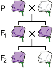
A gene is a basic physical and functional unit of heredity. The concept of the gene was first explained by Gregor Mendel in 1860’s. He never used the term ‘gene’. He called it ‘factor’. In 1909, the Danish biologist Wilhelm Johannsen, coined the term ‘gene’, that was referred to discrete determiners of inherited characteristics.

According to the classical concept of gene introduced by Sutton in 1902, genes have been defined as discrete particles that follow Mendelian rules of inheritance, occupy a definite locus in the chromosome and are responsible for the expression of specific phenotypic character. They show the following properties:

  • Number of genes in each organism is more than the number of chromosomes; hence several genes are located on the same chromosome.
  • The genes are arranged in a single linear order like beads on a string.
  • Each gene occupies a specific position called locus.
  • Genes may exist in several alternate forms called alleles.
  • Genes may undergo sudden change in positions and composition called mutations.
  • Genes are capable of self-duplication producing their own copies.
    Gene As The Functional Unit Of Inheritance img 1

Chromosomal Abnormalities

Learninsta presents the core concepts of Biology with high-quality research papers and topical review articles.

Chromosomal Abnormalities

Each human diploid (2n) body cell has 46 chromosomes (23 pairs). Chromosomal disorders are caused by errors in the number or structure of chromosomes. Chromosomal anomalies usually occur when there is an error in cell division. Failure of chromatids to segregate during cell division resulting in the gain or loss of one or more chromosomes is called aneuploidy.

It is caused by nondisjunction of chromosomes. Group of signs and symptoms that occur together and characterize a particular abnormality is called a syndrome. In humans, Down’s syndrome, Turner’s syndrome, Klinefelter’s syndrome, Patau’s syndrome are some of the examples of chromosomal disorders.

a. Autosomal aneuploidy in human beings

Several autosomal aneuploidies have been reported in human beings. eg. Down’s syndrome (21-Trisomy), Patau’s syndrome (13-Trisomy).

1. Down’s Syndrome/Trisomy – 21

Trisomic condition of chromosome – 21 results in Down’s syndrome. It is characterized by severe mental retardation, defective development of the central nervous system, increased separation between the eyes, flttened nose, ears are malformed, mouth is constantly open and the tongue protrudes.

2. Patau’s Syndrome/Trisomy-13

Trisomic condition of chromosome 13 results in Patau’s syndrome. Meiotic non disjunction is thought to be the cause for this chromosomal abnormality. It is characterized by multiple and severe body malformations as well as profound mental deficiency. Small head with small eyes, clef palate, malformation of the brain and internal organs are some of the symptoms of this syndrome.

b. Allosomal abnormalities in human beings

Mitotic or meiotic non-disjunction of sex chromosomes causes allosomal abnormalities. Several sex chromosomal abnormalities have been detected. Eg. Klinefelter’s syndrome and Turner’s syndrome.

1. Klinefelter’s Syndrome (XXY Males)

This genetic disorder is due to the presence of an additional copy of the X chromosome resulting in a karyotype of 47,XXY. Persons with this syndrome have 47 chromosomes (44AA+XXY). They are usually sterile males, tall, obese, with long limbs, high pitched voice, under developed genitalia and have feeble breast (gynaecomastia) development.

2. Turner’s Syndrome (XO Females)

This genetic disorder is due to the loss of a X chromosome resulting in a karyotype of 45, X. Persons with this syndrome have 45 chromosomes (44 autosomes and one X chromosome) (44AA+XO) and are sterile females. Low stature, webbed neck, under developed breast, rudimentary gonads lack of menstrual cycle during puberty, are the main symptoms of this syndrome.

Mendelian Disorders | Thalassemia | Phenylketonuria | Albinism | Huntington’s Chorea

Learninsta presents the core concepts of Biology with high-quality research papers and topical review articles.

Mendelian Disorders | Thalassemia | Phenylketonuria | Albinism | Huntington’s Chorea

Alteration or mutation in a single gene causes Mendelian disorders. These disorders are transmitted to the offsprings on the same line as the Mendelian pattern of inheritance. Some examples for Mendelian disorders are Thlassemia, albinism, phenylketonuria, sickle cell anaemia, Huntington’s chorea, etc., These disorders may be dominant or recessive and autosomal or sex linked.

Thlassemia

Thlassemia is an autosomal recessive disorder. It is caused by gene mutation resulting in excessive destruction of RBC’s due to the formation of abnormal haemoglobin molecules. Normally haemoglobin is composed of four polypeptide chains, two alpha and two beta globin chains. Thlassemia patients have defects in either the alpha or beta globin chain causing the production of abnormal haemoglobin molecules resulting in anaemia.

Thlassemia is classified into alpha and beta based on which chain of haemoglobin molecule is affected. It is controlled by two closely linked genes HBA1 and HBA2 on chromosome 16. Mutation or deletion of one or more of the four alpha gene alleles causes Alpha Thlassemia.

In Beta Thlassemia, production of beta globin chain is affected. It is controlled by a single gene (HBB) on chromosome 11. It is the most common type of Thlassemia and is also known as Cooley’s anaemia. In this disorder the alpha chain production is increased and damages the membranes of RBC.

Phenylketonuria

It is an inborn error of Phenylalanine metabolism caused due to a pair of autosomal recessive genes. It is caused due to mutation in the gene PAH (phenylalanine hydroxylase gene) located on chromosome 12 for the hepatic enzyme “phenylalanine hydroxylase” This enzyme is essential for the conversion of phenylalanine to tyrosine.

Affected individual lacks this enzyme, so phenylalanine accumulates and gets converted to phenylpyruvic acid and other derivatives. It is characterized by severe mental retardation, light pigmentation of skin and hair. Phenylpyruvic acid is excreted in the urine.
Mendelian Disorders img 1

Albinism

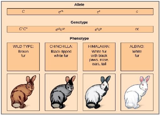
Albinism is an inborn error of metabolism, caused due to an autosomal recessive gene. Melanin pigment is responsible for skin colour. Absence of melanin results in a condition called albinism. A person with the recessive allele lacks the tyrosinase enzyme system, which is required for the conversion of dihydroxyphenyl alanine (DOPA) into melanin pigment inside the melanocytes. In an albino, melanocytes are present in normal numbers in their skin, hair, iris, etc., but lack melanin pigment.
Mendelian Disorders img 2

Huntington’s chorea

It is inherited as an autosomal dominant lethal gene in man. It is characterized by involuntary jerking of the body and progressive degeneration of the nervous system, accompanied by gradual mental and physical deterioration. The patients with this disease usually die between the age of 35 and 40.

Pedigree Analysis – Genetic Disorders

Learninsta presents the core concepts of Biology with high-quality research papers and topical review articles.

Pedigree Analysis – Genetic Disorders

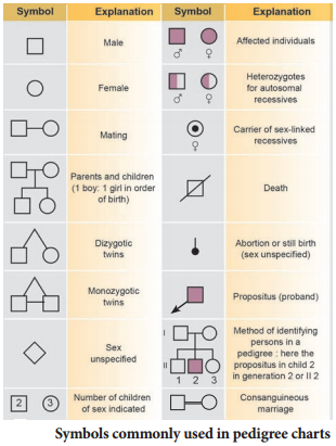
Pedigree is a “family tree”, drawn with standard genetic symbols, showing the inheritance pathway for specific phenotypic characters.(Fig. 4.6). Pedigree analysis is the study of traits as they have appeared in a given family line for several past generations.
Pedigree Analysis img 1

Genetic Disorders

A genetic disorder is a disease or syndrome that is caused by an abnormality in an individual DNA. Abnormalities can range from a small mutation in a single gene to the addition or subtraction of an entire chromosome or even a set of chromosomes. Genetic disorders are of two types namely, Mendelian disorders and chromosomal disorders.

A pedigree shows relationships between family members and indicates which individuals have certain genetic pathogenic variants, traits, and diseases within a family as well as vital status. A pedigree shows relationships between family members and patterns of inheritance for certain traits and diseases.

Pedigrees are drawn using standard symbols and formatting. Males are represented by squares and females by circles. Individuals who are deceased have a slash through the symbol representing them. Symbols for individuals affected by a particular disorder are shaded.

It can be simply called as a “family tree” Pedigrees use a standardized set of symbols, squares represent males and circles represent females.

The modes of inheritance are autosomal dominant , autosomal recessive, and X-linked. To simplify the discussion of these different forms, the trait used in the following text will be a hereditary disease.

In this family pedigree, black squares indicate the presence of a particular trait in a male, and white squares represent males without the trait. White circles are females. A trait in one generation can be inherited, but not outwardly apparent before two more generations (compare black squares).

There are five basic modes of inheritance for single-gene diseases: autosomal dominant, autosomal recessive, X-linked dominant, X-linked recessive, and mitochondrial. Genetic heterogeneity is a common phenomenon with both single-gene diseases and complex multi-factorial diseases.

The study of an inherited trait in a group of related individuals to determine the pattern and characteristics of the trait, including its mode of inheritance, age of onset, and phenotypic variability.

Karyotyping Preparation and Its Applications

Learninsta presents the core concepts of Biology with high-quality research papers and topical review articles.

Karyotyping Preparation and Its Applications

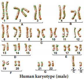
Karyotyping is a technique through which a complete set of chromosomes is separated from a cell and the chromosomes are arranged in pairs. An idiogram refers to a diagrammatic representation of chromosomes.

Preparation of Karyotype

Tjio and Levan (1960) described a simple method of culturing lymphocytes from the human blood. Mitosis is induced followed by addition of colchicine to arrest cell division at metaphase stage and the suitable spread of metaphase chromosomes is photographed. The individual chromosomes are cut from the photograph and are arranged in an orderly fashion in homologous pairs. This arrangement is called a karyotype. Chromosome banding permits structural definitions and diffrentiation of chromosomes.

Applications of Karyotyping:

  • It helps in gender identification.
  • It is used to detect the chromosomal aberrations like deletion, duplication, translocation, nondisjunction of chromosomes.
  • It helps to identify the abnormalities of chromosomes like aneuploidy.
  • It is also used in predicting the evolutionary relationships between species.
  • Genetic diseases in human beings can be detected by this technique.
    Karyotyping Preparation and Its Applications img 1

Human Karyotype

Depending upon the position of the centromere and relative length of two arms, human chromosomes are of three types: Metacentric, sub metacentric and acrocentric. The photograph of chromosomes are arranged in the order of descending length in groups from A to G (Fig. 4.5).

Sex Linked Inheritance

Learninsta presents the core concepts of Biology with high-quality research papers and topical review articles.

Sex Linked Inheritance

The inheritance of a trait that is determined by a gene located on one of the sex chromosomes is called sex linked inheritance. Genes present on the differential region of X or Y chromosomes are called sex linked genes. The genes present in the differential region of “X” chromosome are called X linked genes. The X-linked genes have no corresponding alleles in the Y chromosome. The genes present in the differential region of Y chromosome are called Y – linked or holandric genes.

The Y linked genes have no corresponding allele in X chromosome. The Y linked genes inherit along with Y chromosome and they phenotypically express only in the male sex. Sex linked inherited traits are more common in males than females because, males are hemizygous and therefore express the trait when they inherit one mutant allele. The X – linked and Y – linked genes in the differential region (non-homologus
region) do not undergo pairing or crossing over during meiosis. The inheritance of X or Y linked genes is called sex-linked inheritance.

Inheritance of X – linked genes

Red-green colour blindness or daltonism, haemophilia and Duchenne’s muscular dystrophy are examples of X-linked gene inheritance in humans.

1. Haemophilia

Haemophilia is commonly known as bleeder’s disease, which is more common in men than women. This hereditary disease was first reported by John Cotto in 1803. Haemophilia is caused by a recessive X-linked gene.

A person with a recessive gene for haemophilia lacks a normal clotting substance (thromboplastin) in blood, hence minor injuries cause continuous bleeding, leading to death. The females are carriers of the disease and would transmit the disease to 50% of their sons even if the male parent is normal. Haemophilia follows the characteristic criss – cross pattern of inheritance.

2. Colour blindness

In human beings a dominant X – linked gene is necessary for the formation of colour sensitive cells, the cones. The recessive form of this gene is incapable of producing colour sensitive cone cells. Homozygous recessive females (XcXc) and hemizygous recessive males (XcY) are unable to distinguish red and green colour. The inheritance of colour blindness can be studied in the following two types of marriages.

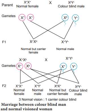
(i) Marriage between colour blind man and normal visioned woman

A marriage between a colour blind man and a normal visioned woman will produce normal visioned male and female individuals in F1 generation but the females are carriers. The marriage between a F1 normal visioned carrier woman and a normal visioned male will produce one normal visioned female, one carrier female, one normal visioned male and one colour blind male in F2 generation.

Colour blind trait is inherited from the male parent to his grandson through carrier daughter, which is an example of criss-cross pattern of inheritance (Fig. 4.3).
Sex Linked Inheritance img 1

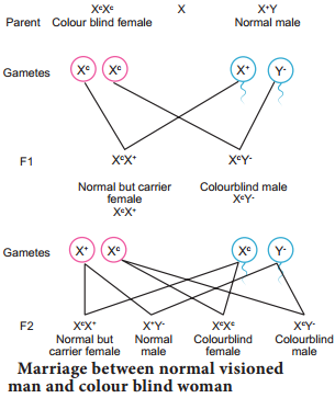
(ii) Marriage between normal visioned man and colour blind woman

If a colour blind woman (XcXc) marries a normal visioned male (XcXc), all F1 sons will be colourblind and daughters will be normal visioned but are carriers. Marriage between F1 carrier female with a colour blind male will produce normal visioned carrier daughter, colourblind daughter, normal visioned son and a colourblind son in the F2 generation (Fig. 4.4).
Sex Linked Inheritance img 2

Inheritance of Y-linked genes

Genes in the non-homologous region of the Y-chromosome are inherited directly from male to male. In humans, the Y-linked or holandric genes for hypertrichosis (excessive development of hairs on pinna of the ear) are transmitted directly from father to son, because males inherit the Y chromosome from the father. Female inherits only X chromosome from the father and are not affected.

Sex Determination – An Overview

Learninsta presents the core concepts of Biology with high-quality research papers and topical review articles.

Sex Determination – An Overview

Sex determination is the method by which the distinction between male and female is established in a species. Sex chromosomes determine the sex of the individual in dioecious or unisexual organisms. The chromosomes other than the sex chromosomes of an individual are called autosomes.

Sex chromosomes may be similar (homomorphic) in one sex and dissimilar (heteromorphic) in the other. Individuals having homomorphic sex chromosomes produce only one type of gametes (homogametic) whereas heteromorphic individuals produce two types of gametes (heterogametic).

Chromosomal basis of sex determination

Heterogametic Sex Determination:

In heterogametic sex determination one of the sexes produces similar gametes and the other sex produces dissimilar gametes. The sex of the offspring is determined at the time of fertilization.

Heterogametic Males

In this method of sex determination the males are heterogametic producing dissimilar gametes while females are homogametic producing similar gametes. It is of two kinds XX-XO type (e.g. Bugs, cockroaches and grasshoppers) and XX-XY type (e.g. Human beings and Drosophila).

Heterogametic Females

In this method of sex determination the females are heterogametic producing dissimilar gametes while males are homogametic producing similar gametes. To avoid confusion with the XX-XO and XX-XY types of sex determination, the alphabets ‘Z’ and ‘W’ are used here instead of X and Y respectively. Heterogametic females are of two types, ZO-ZZ type (eg. Moths, butterflies and domestic chickens) and ZW-ZZ type (eg. Gypsy moth, fihes, reptiles and birds).

Sex determination in human beings

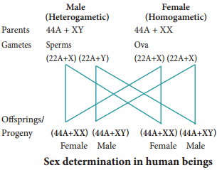
Genes determining sex in human beings are located on two sex chromosomes, called allosomes. In mammals, sex determination is associated with chromosomal differences between the two sexes, typically XX females and XY males. 23 pairs of human chromosomes include 22 pairs of autosomes (44A) and one pair of sex chromosomes (XX or XY).

Females are homogametic producing only one type of gamete (egg), each containing one X chromosome while the males are heterogametic producing two types of sperms with X and Y chromosomes. An independently evolved XX: XY system of sex chromosomes also exist in Drosophila (Fig. 4.2).
Sex Determination img 1

The Y Chromosome and Male Development

Current analysis of Y chromosomes has revealed numerous genes and regions with potential genetic function; some genes with or without homologous counterparts are seen on the X. Present at both ends of the Y chromosome are the pseudoautosomal regions (PARs) that are similar with regions on the X chromosome which synapse and recombine during meiosis.

The remaining 95% of the Y chromosome is referred as the Non – combining Region of the Y (NRY). The NRY is divided equally into functional genes (euchromatic) and non functional genes (heterochromatic). Within the euchromatin regions, is a gene called Sex determining region Y (SRY). In humans, absence of Y chromosome inevitably leads to female development and this SRY gene is absent in X chromosome. The gene product of SRY is the testes determining factor (TDF) present in the adult male testis.

Dosage compensation Barr Body

In 1949, Barr and Bertram first observed a condensed body in the nerve cells of female cat which was absent in the male. This condensed body was called sex chromatin by them and was later referred as Barr body. In the XY chromosomal system of sex determination, males have only one X chromosome, whereas females have two. A question arises: how does the organism compensate for this dosage differences between the sexes? In mammals the necessary dosage compensation is accomplished by the inactivation of one of the X chromosome in females so that both males and females have only one functional X chromosome per cell.

Mary Lyon suggested that Barr bodies represented an inactive chromosome, which in females becomes tightly coiled into a heterochromatin, a condensed and visible form of chromatin (Lyon’s hypothesis). The number of Barr bodies observed in cell was one less than the number of X-Chromosome. XO females have no Barr body, whereas XXY males have one Barr body.

Haplodiploidy in Honeybees

In hymenopteran insects such as honeybees, ants and wasps a mechanism of sex determination called haplodiploidy mechanism of sex determination is common. In this system, the sex of the offspring is determined by the number of sets of chromosomes it receives.

Fertilized eggs develop into females (Queen or Worker) and unfertilized eggs develop into males (drones) by parthenogenesis. It means that the males have half the number of chromosomes (haploid) and the females have double the number (diploid), hence the name haplodiplody for this system of sex determination.

This mode of sex determination facilitates the evolution of sociality in which only one diploid female becomes a queen and lays the eggs for the colony. All other females which are diploid having developed from fertilized eggs help to raise the queen’s eggs and so contribute to the queen’s reproductive success and indirectly to their own, a phenomenon known as Kin Selection. The queen constructs their social environment by releasing a hormone that suppresses fertility of the workers.

Genetic Control of Rh Factor

Learninsta presents the core concepts of Biology with high-quality research papers and topical review articles.

Genetic Control of Rh Factor

Fisher and Race Hypothesis:

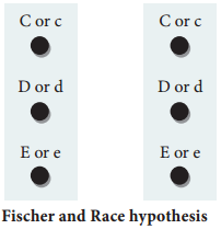
Rh factor involves three different pairs of alleles located on three different closely linked loci on the chromosome pair. This system is more commonly in use today, and uses the ‘Cde’ nomenclature.
Genetic Control Of Rh Factor img 1

In the above Fig. 4.1, three pairs of Rh alleles (Cc, Dd and Ee) occur at 3 different loci on homologous chromosome pair. The possible genotypes will be one C or c, one D or d, one E or e from each chromosome. For e.g. CDE/cde; CdE/cDe; cde/cde; CDe/CdE etc., All genotypes carrying a dominant ‘D’ allele will produce Rh positive phenotype and double recessive genotype ‘dd’ will give rise to Rh negative phenotype.

Wiener Hypothesis

Wiener proposed the existence of eight alleles (R1, R2, RO, Rz, r, r1, r11, ry) at a single Rh locus. All genotypes carrying a dominant ‘R allele’ (R1, R2, RO, Rz) will produce Rh positive phenotype and double recessive genotypes (rr, r1r1, r11r11, ry ry) will give rise to Rh negative phenotype.

Incompatibility of Rh Factor – Erythroblastosis foetalis

Rh incompatability has great signifiance in child birth. If a woman is Rh negative and the man is Rh positive, the foetus may be Rh positive having inherited the factor from its father. The Rh negative mother becomes sensitized by carrying Rh positive foetus within her body. Due to damage of blood vessels, during child birth, the mother’s immune system recognizes the Rh antigens and gets sensitized. The sensitized mother produces Rh antibodies.

The antibodies are IgG type which are small and can cross placenta and enter the foetal circulation. By the time the mother gets sensitized and produce anti ‘D’ antibodies, the child is delivered.

Usually no effects are associated with exposure of the mother to Rh positive antigen during the first child birth, subsequent Rh positive children carried by the same mother, may be exposed to antibodies produced by the mother against Rh antigen, which are carried across the placenta into the foetal blood circulation. This causes haemolysis of foetal RBCs resulting in haemolytic jaundice and anaemia. This condition is known as Erythoblastosis foetalis or Haemolytic disease of the new born (HDN).

Prevention of Erythroblastosis Foetalis

If the mother is Rh negative and foetus is Rh positive, anti D antibodies should be administered to the mother at 28th and 34th week of gestation as a prophylactic measure. If the Rh negative mother delivers Rh positive child then anti D antibodies should be administered to the mother soon after delivery. This develops passive immunity and prevents the formation of anti D antibodies in the mothers blood by destroying the Rh foetal RBC before the mother’s immune system is sensitized. This has to be done whenever the woman attains pregnancy.

Human Blood Groups | ABO Blood Groups and Its Types

Learninsta presents the core concepts of Biology with high-quality research papers and topical review articles.

Human Blood Groups | ABO Blood Groups and Its Types

Multiple allelism occurs in humans, particularly in the inheritance of different types of blood groups. The blood group inheritance in human can be understood by learning about antigens and antibodies. The composition of blood, different types of blood groups (ABO) the blood antigens and antibodies were discussed in chapter 7 of class XI.

ABO blood types

Multiple allele inheritance of ABO blood groups Blood differs chemically from person to person. When two different incompatible blood types are mixed, agglutination (clumping together) of erythrocytes (RBC) occurs.

The basis of these chemical differences is due to the presence of antigens (surface antigens) on the membrane of RBC and epithelial cells. Karl Landsteiner discovered two kinds of antigens called antigen ‘A’ and antigen ‘B’ on the surface of RBC’s of human blood.

Based on the presence or absence of these antigens three kinds of blood groups, type ‘A’, type ‘B’, and type ‘O’ (universal donor)were recognized. The fourth and the rarest blood group ‘AB’ (universal recipient) was discovered in 1902 by two of Landsteiner’s students Von De Castelle and Sturli.

Bernstein in 1925 discovered that the inheritance of different blood groups in human beings is determined by a number of multiple allelic series. The three autosomal alleles located on chromosome 9 are concerned with the determination of blood group in any person.

The gene controlling blood type has been labeled as ‘L’ (after the name of the discoverer, Landsteiner) or I (from isoagglutination). The I gene exists in three allelic forms, IA, IB and IO. IA specifies A antigen. IB allele determines B antigen and IO allele specifies no antigen. Individuals who possess these antigens in their fluids such as the saliva are called secretors.

Each allele (IA and IB) produces a transferase enzyme. IA allele produces N-acetyl galactose transferase and can add N-acetyl galactosamine (NAG) and IB allele encodes for the enzyme galactose transferase that adds galactose to the precursor (i.e., H substances) In the case of IO/IO allele no terminal transferase enzyme is produced and therefore called “null” allele and hence cannot add NAG or galactose to the precursor.

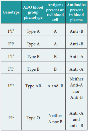
From the phenotypic combinations it is evident that the alleles IA and IB are dominant to IO, but co-dominant to each other (IA = IB). Their dominance hierarchy can be given as (IA = IB > IO). A child receives one of three alleles from each parent, giving rise to six possible genotypes and four possible blood types (phenotypes). The genotypes are IAIA, IA IO, IBIB, IBIO, IAIB and IO IO.
Human Blood Groups img 1
Genetic basis of the human ABO blood groups

Rhesus or Rh Factor

The Rh factor or Rh antigen is found on the surface of erythrocytes. It was discovered in 1940 by Karl Landsteiner and Alexander Wiener in the blood of rhesus monkey, Macaca rhesus and later in human beings. The term ‘Rh factor’ refers to “immunogenic D antigen of the Rh blood group system. An individual having D antigen are Rh D positive (Rh+) and those without D antigen are Rh D negative (Rh)”.

Rhesus factor in the blood is inherited as a dominant trait. Naturally occurring Anti D antibodies are absent in the plasma of any normal individual. However if an Rh (Rh negative) person is exposed to Rh+ (Rh positive) blood cells (erythrocytes) for the first time, anti D antibodies are formed in the blood of that individual. On the other hand, when an Rh positive person receives Rh negative blood no effect is seen.

Principles of Inheritance and Variation Multiple Alleles

Learninsta presents the core concepts of Biology with high-quality research papers and topical review articles.

Principles of Inheritance and Variation Multiple Alleles

The genetic segregations in Mendelian inheritance reveal that all genes have two alternative forms – dominant and recessive alleles e.g. tall versus dwarf (T and t). The former is the normal allele or wild allele and the latter the mutant allele.

A gene can mutate several times producing several alternative forms. When three or more alleles of a gene that control a particular trait occupy the same locus on the homologous chromosome of an organism, they are called multiple alleles and their inheritance is called multiple allelism.

Multiple alleles exist in a population when there are many variations of a gene present. In organisms with two copies of every gene, also known as diploid organisms, each organism has the ability to express two alleles at the same time. They can be the same allele, which is called a homozygous genotype.

Multiple alleles refer to the occurrence of three or more than three alleles for a particular gene. Alleles are different or contrasting forms of a gene. For example, for the gene encoding for height, one allele can be for tallness, whereas the other can be for dwarfness.

Mendel implied that only two alleles, one dominant and one recessive, could exist for a given gene. The variant may be recessive or dominant to the wild-type allele. An example of multiple alleles is the ABO blood-type system in humans.

Polygenic Inheritance: Human skin color is a good example of polygenic (multiple gene) inheritance. A genotype with all “dominant” capital genes (AABBCC) has the maximum amount of melanin and very dark skin. A genotype with all “recessive” small case genes (aabbcc) has the lowest amount of melanin and very light skin.

Multiple alleles exist in a population when there are many variations of a gene present. In both haploid and diploid organisms, new alleles are created by spontaneous mutations. These mutations can arise in a variety of ways, but the effect is a different sequence of nucleic acid bases in the DNA.

Multiple alleles are present at the same locus of the chromosomes. A classical example of multiple alleles is found in ABO blood group system of humans. Despite the multiple alleles of any gene, an individual possess and can have only two alleles at a time.
Multiple Alleles img 1

Detection of Foetal Disorders During Early Pregnancy

Learninsta presents the core concepts of Biology with high-quality research papers and topical review articles.

Detection of Foetal Disorders During Early Pregnancy

Ultrasound scanning Ultrasound has no known risks other than mild discomfort due to pressure from the transducer on the abdomen or vagina. No radiation is used during this procedure. Ultrasonography is usually performed in the first trimester for dating, determination of the number of foetuses, and for assessment of early pregnancy complications.

Amniocentesis

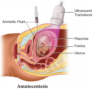
Amniocentesis involves taking a small sample of the amniotic fluid that surrounds the foetus to diagnose for chromosomal abnormalities (Fig. 3.1).
Detection Of Foetal Disorders During Early Pregnancy img 1

Amniocentesis is generally performed in a pregnant woman between the 15th and 20th weeks of pregnancy by inserting a long, thin needle through the abdomen into the amniotic sac to withdraw a small sample of amniotic fluid. The amniotic fluid contains cells shed from the foetus.

Chorionic villus sampling (CVS)

CVS is a prenatal test that involves taking a sample of the placental tissue to test for chromosomal abnormalities.

Foetoscope

Foetoscope is used to monitor the foetal heart rate and other functions during late pregnancy and labour. The average foetal heart rate is between 120 and 160 beats per minute. An abnormal foetal heart rate or pattern may mean that the foetus is not getting enough oxygen and it indicates other problems.

A hand-held doppler device is often used during prenatal visits to count the foetal heart rate. During labour, continuous electronic foetal monitoring is often used.