Exchange of Gases in Respiratory Pigments, Methaemoglobin

Learninsta presents the core concepts of Biology with high-quality research papers and topical review articles.

Exchange of Gases in Respiratory Pigments, Methaemoglobin

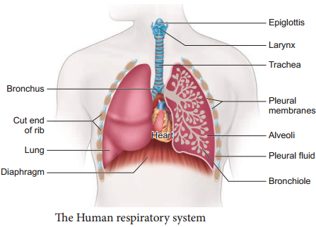
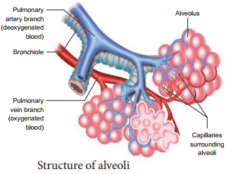
The primary site for the exchange of gases is the alveoli. The uptake of O2 and the release of CO2 occur between the blood and tissues by simple diffusion driven by partial pressure gradient of O2 and CO2. Partial pressure is the pressure contributed by an individual gas in a mixture of gases.

It is represented as pO2 for oxygen and pCO2 for carbon-dioxide. Due to pressure gradients, O2 from the alveoli enters into the blood and reaches the tissues. CO2 enters into the blood from the tissues and reaches alveoli for elimination. As the solubility of CO2 is 20-25 times higher than that of O2, the partial pressure of CO2 is much higher than that of O2 (Table 6.1 and Figure 6.6).

Respiratory Pigments

Haemoglobin

Haemoglobin belongs to the class of conjugated protein. The iron containing pigment portion haem constitutes only 4% and the rest colourless protein globin belongs to histone classs. Haemoglobin has a molecular weight of 68,000 daltons and contains four atoms of iron, each of which can combine with a molecule of oxygen.

Methaemoglobin

If the iron component of the haem moieties is in the ferric state, than the normal ferrous state, it is called methaemoglobin. Methaemoglobin does not bind O2. Normally RBC contains less than 1% methaemoglobin.
Exchange of Gases img 1
Exchange of Gases img 2
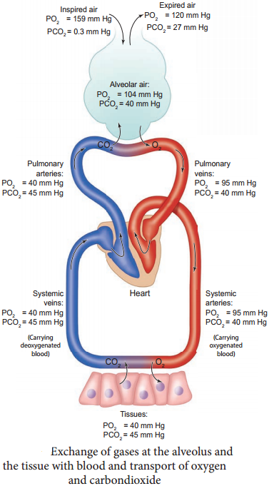
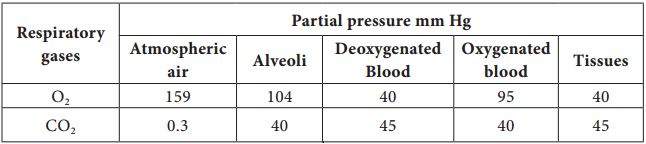
Table 6.1 Partial pressure of Oxygen and Carbon dioxide (in mmHg) in comparison to those gases in the atmosphere.

Mechanism of Breathing in Human Beings

Learninsta presents the core concepts of Biology with high-quality research papers and topical review articles.

Mechanism of Breathing in Human Beings

The movement of air between the atmosphere and the lungs is known as ventilation or breathing. Inspiration and expiration are the two phases of breathing. Inspiration is the movement of atmospheric air into the lungs and expiration is the movement of alveolar air that diffuse out of the lungs. (Figure 6.4)
Muscle Tissue img 1

Lungs do not contain muscle fires but expands and contracts by the movement of the ribs and diaphragm. The diaphragm is a sheet of tissue which separates the thorax from the abdomen. In a relaxed state, the diaphragm is domed shaped. Ribs are moved by the intercostal muscles.

External and internal intercostal muscles found between the ribs and the diaphragm helps in creating pressure gradients. Inspiration occurs if the pressure inside the lungs (intrapulmonary pressure) is less than the atmospheric pressure.
Muscle Tissue img 2

Inspiraton is initiated by the contraction of the diaphragm muscles and external intercostal muscles, which pulls the ribs and sternum upwards and outwards and increases the volume of the thoracic chamber in the dorso-ventral axis, forcing the lungs to expand the pulmonary volume.

The increase in pulmonary volume and decrease in the intrapulmonary pressure forces the fresh air from outside to enter the air passages into the lungs to equalize the pressure. This process is called inspiration.

Relaxation of the diaphragm allows the diaphragm and sternum to return to its dome shape and the internal intercostal muscles contract, pulling the ribs downward reducing the thoracic volume and pulmonary volume. This results in an increase in the intrapulmonary pressure slightly above the atmospheric pressure causing the expulsion of air from the lungs. This process is called expiration.

On an average, a healthy human breathes 12-16 times/minute. An instrument called Spirometer is used to measure the volume of air involved in breathing movements for clinical assessment of a person’s pulmonary function.

Respiratory Volumes and Capacities

The volume of air present in various phases of respiration is denoted as

Respiratory Volumes: (Figure 6.5)

Tidal Volume (TV)

Tidal volume is the amount of air inspired or expired with each normal breath. It is approximately 500 mL., i.e. a normal human adult can inspire or expire approximately 6000 to 8000mL of air per minute. During vigorous exercise, the tidal volume is about 4-10 times higher.

Inspiratory Reserve Volume (IRV)

Additional volume of air a person can inspire by forceful inspiration is called Inspiratory Reserve Volume. The normal value is 2500-3000 mL.

Expiratory Reserve Volume (ERV)

Additional volume of air a person can forcefully exhale by forceful expiration is called Expiratory Reserve Volume. The normal value is 1000-1100 mL.

Residual Volume (RV)

The volume of air remaining in the lungs after a forceful expiration. It is approximately 1100-1200 mL.

Respiratory Capacities:

Vital capacity (VC) the maximum volume of air that can be moved out during a single breath following a maximal inspiration. A person first inspires maximally then expires maximally. VC = ERV + TV + IRV

Inspiratory Capacity (IC)

The total volume of air a person can inhale after normal expiration. It includes tidal volume and inspiratory reserve volume. IC = TV + IRV

Expiratory Capacity (EC)

The total volume of air a person can exhale after normal inspiration. It includes tidal volume and expiratory reserve volume. EC = TV + ERV
Muscle Tissue img 3

Total Lung Capacity (TLC)

The total volume of air which the lungs can accommodate after forced inspiration is called Total Lung Capacity. This includes the vital capacity and the residual volume. It is approximately 6000mL. TLC = VC + RV

Minute Respiratory Volume

The amount of air that moves into the respiratory passage per minute is called minute respiratory volume.
Normal TV = 500mL; Normal respiratory rate = 12 times/minute Therefore, minute respiratory volume = 6 Litres/minute (for a normal healthy man).

Dead Space

Some of the inspired air never reaches the gas exchange areas but fills the respiratory passages where exchange of gases does not occur. This air space is called dead space. Dead space is not involved in gaseous exchange. It amounts to approximately 150mL.

Respiratory Function, Facts Organs & Anatomy

Learninsta presents the core concepts of Biology with high-quality research papers and topical review articles.

Respiratory Function, Facts Organs & Anatomy

The five primary functions of the respiratory system are: –

  • To exchange O2 and CO2 between the atmosphere and the blood.
  • To maintain homeostatic regulation of body pH.
  • To protect us from inhaled pathogens and pollutants.
  • To maintain the vocal cords for normal communication (vocalization).
  • To remove the heat produced during cellular respiration.

Your lungs are part of the respiratory system, a group of organs and tissues that work together to help you breathe. The respiratory system’s main job is to move fresh air into your body while removing waste gases.

There are Five Functions of the Respiratory System

Gas Exchange – oxygen and carbon dioxide.
Breathing – movement of air.
Sound Production.
Oldfactory Assistance – sense of smell.
Protection – from dust and microbes entering body through mucus production, cilia, and coughing.

Allows you to talk and to smell. Brings air to body temperature and moisturizes it to the humidity level your body needs. Delivers oxygen to the cells in your body. Removes waste gases, including carbon dioxide, from the body when you exhale.

Respiratory failure is a serious condition that develops when the lungs can’t get enough oxygen into the blood. Buildup of carbon dioxide can also damage the tissues and organs and further impair oxygenation of blood and, as a result, slow oxygen delivery to the tissues.

Inside the lungs, oxygen is exchanged for carbon dioxide waste through the process called external respiration. This respiratory process takes place through hundreds of millions of microscopic sacs called alveoli. Oxygen from inhaled air diffuses from the alveoli into pulmonary capillaries surrounding them.

The bronchial tubes divide into smaller air passages called bronchi, and then into bronchioles. The bronchioles end in tiny air sacs called alveoli, where oxygen is transferred from the inhaled air to the blood. After absorbing oxygen, the blood leaves the lungs and is carried to the heart.

The oxygen we inhale is used to breakdown glucose into carbon dioxide and water. Energy is released in the process. The breakdown of glucose occurs in the cells of an organism (cellular respiration). If the breakdown occurs without the use of oxygen, the respiration is called anaerobic respiration.

Respiratory Organs in Various Organisms

Learninsta presents the core concepts of Biology with high-quality research papers and topical review articles.

Respiratory Organs in Various Organisms

Different animals have different organs for exchange of gases, depending upon their habitats and levels of organization. The amount of dissolved oxygen is very low in water compared to the amount of oxygen in the air. So the rate of breathing in aquatic organisms is much faster than land animals.

In animals like sponges, coelenterates and flatworms exchange of gases takes place through the body surface by simple diffusion. Earthworms use their moist skin, whereas insects have tracheal tubes. Gills are used as respiratory organs in most of the aquatic Arthropods and Molluscs.

Among vertebrates, fishes use gills whereas amphibians, reptiles, birds and mammals have well vascularised lungs. Frogs spend most of their time in water and also use their moist skin for respiration along with lungs and bucco pharynx.

Human Respiratory System

The respiratory system includes external nostrils, nasal cavity, pharynx, larynx, trachea, bronchi and bronchioles and lungs which contain alveoli (Figure 6.1). The parts starting from the external nostrils up to the terminal bronchioles constitute the conducting zone, whereas the alveoli and the ducts are called the respiratory zone. The parts of the conducting zone, humidifies and warms the incoming air.
Respiratory Organs in Various Organisms img 1

In human beings, air enters the upper respiratory tract through the external nostrils. The air passing through the nostrils is filtered by fine hairs and mucus lining the passage. The external nostrils lead to the nasal chamber which opens into the nasopharynx which opens through the glottis of the larynx region into the trachea. The ciliated epithelial cells lining the trachea, bronchi and bronchioles secrete mucus.

Mucus membrane lining the airway contains goblet cells which secrete mucus, a slimy material rich in glycoprotein. Microorganisms and dust particles attach in the mucus films and are carried upwards to pass down the gullet during normal swallowing. During swallowing a thin elastic flap called epiglottis prevents the food from entering into the larynx and avoids choking of food.

The trachea is semiflexible tube supported by multiple cartilaginous rings which extends up to the midthoracic cavity and at the level of the 5th thoracic vertebra where it divides into right and left primary bronchi, one bronchus to each lung. Within the lungs the bronchi divides repeatedly into secondary and tertiary bronchi and further divides into terminal bronchioles and respiratory bronchioles.

Bronchi have ‘C’ shaped curved cartilage plates to ensure that the air passage does not collapse or burst as the air pressure changes during breathing. The bronchioles are without cartilaginous rings and have rigidity that prevent them from collapsing but are surrounded by smooth muscle which contracts or relaxes to adjust the diameter of these airways.

The fine respiratory bronchioles terminate into highly vascularised thin walled pouch like air sacs called alveoli meant for gaseous exchange (Figure 6.2, 6.3). The diffusion membrane of alveolus is made up of three layers – the thin squamous epithelial cells of the alveoli, the endothelium of the alveolar capillaries and the basement substance found in between them. The thin squamous epithelial cells of the alveoli are composed of Type I and Type II cells. Type I cells are very thin so that gases can diffuse rapidly through them. Type II cells are thicker, synthesize and secrete a substance called Surfactant.

The lungs are light spongy tissues enclosed in the thoracic cavity surrounded by an airtight space. The thoracic cavity is bound dorsally by the vertebral column and ventrally by the sternum, laterally by the ribs and on the lower side by the dome shaped diaphragm.

The lungs are covered by double walled pleural membrane containing a several layers of elastic connective tissues and capillaries, which encloses the pleural fluid. Pleural fluid reduces friction when the lungs expand and contract.
Respiratory Organs in Various Organisms img 2
Respiratory Organs in Various Organisms img 3

Characteristic Features of Respiratory Surface:

  • Surface area must be very large and richly supplied with blood vessels
  • Should be extremely thin and kept moist
  • Should be in direct contact with the environment
  • Should be permeable to respiratory gases

The Steps Involved in Respiration are:-

  • The exchange of air between the atmosphere and the lungs.
  • The exchange of O2 and CO2 between the lungs and the blood.
  • Transport of O2 and CO2 by the blood.
  • Exchange of gases between the blood and the cells.
  • Uptake of O2 by the cells for various activities and the release of CO2.

Nutritional and Digestive Disorders

Learninsta presents the core concepts of Biology with high-quality research papers and topical review articles.

Nutritional and Digestive Disorders

Intestinal tract is more prone to bacterial, viral and parasitic worm infections. This infection may cause inflammation of the inner lining of colon called colitis. The most common symptoms of colitis are rectal bleeding, abdominal cramps, and diarrhoea.

Protein Energy Malnutrition: (PEM)

Growing children require more amount of protein for their growth and development. Protein deficient diet during early stage of children may lead to protein energy malnutrition such as Marasmus and Kwashiorkor. Symptoms are dry skin, pot-belly, oedema in the legs and face, stunted growth, changes in hair colour, weakness and irritability.

Marasmus is an acute form of protein malnutrition. This condition is due to a diet with inadequate carbohydrate and protein. Such children are suffer from diarrhoea, body becomes lean and weak (emaciated) with reduced fat and muscle tissue with thin and folded skin.

Indigestion:

It is a digestive disorder in which the food is not properly digested leading to a feeling of fullness of stomach. It may be due to inadequate enzyme secretion, anxiety, food poisoning, over eating, and spicy food.

Constipation:

In this condition, the faeces are retained within the rectum because of irregular bowel movement due to poor intake of fire in the diet and lack of physical activities.

Vomiting:

It is reverse peristalsis. Harmful substances and contaminated food from stomach are ejected through the mouth. This action is controlled by the vomit centre located in the medulla oblongata. A feeling of nausea precedes vomiting.

Jaundice:

It is the condition in which liver is affected and the defective liver fails to break down haemoglobin and to remove bile pigments from the blood. Deposition of these pigments changes the colour of eye and skin yellow. Sometimes, jaundice is caused due to hepatitis viral infections.

Liver Cirrhosis:

Chronic disease of liver results in degeneration and destruction of liver cells resulting in abnormal blood vessel and bile duct leading to the formation of fibrosis. It is also called deserted liver or scarred liver. It is caused due to infection, consumption of poison, malnutrition and alcoholism.

Gall Stones:

Any alteration in the composition of the bile can cause the formation of stones in the gall bladder. The stones are mostly formed of crystallized cholesterol in the bile. The gall stone causes obstruction in the cystic duct, hepatic duct and also hepato-pancreatic duct causing pain, jaundice and pancreatitis.

Appendicitis:

It is the inflammation of the vermiform appendix, leading to severe abdominal pain. The treatment involves the removal of appendix by surgery. If treatment is delayed the appendix may rupture and results in infection of the abdomen, called peritonitis.

Hiatus Hernia (Diaphragmatic hernia):

It is a structural abnormality in which superior part of the stomach protrudes slightly above the diaphragm. The exact cause of hiatus hernias is not known. In some people, injury or other damage may weaken muscle tissue, by applying too much pressure (repeatedly) on the muscles around the stomach while coughing, vomiting, and straining during bowel movement and lifting heavy object.

Heart burn is also common in those with a hiatus hernia. In this condition, stomach contents travel back into the oesophagus or even into oral cavity and causes pain in the centre of the chest due to the eroding nature of acidity (Figure 5.10).
Nutritional and Digestive Disorders img 1

Diarrhoea:

It is the most common gastrointestinal disorder worldwide. It is sometimes caused by bacteria or viral infections through food or water. When the colon is infected, the lining of the intestine is damaged by the pathogens, thereby the colon is unable to absorb fluid.

The abnormal frequency of bowel movement and increased liquidity of the faecal discharge is known as diarrhoea. Unless the condition is treated, dehydration can occur. Treatment is known as oral hydration therapy. This involves drinking plenty of fluids – sipping small amounts of water at a time to rehydrate the body.

Peptic Ulcer:

It refers to an eroded area of the tissue lining (mucosa) in the stomach or duodenum. Duodenal ulcer occurs in people in the age group of 25 – 45 years. Gastric ulcer is more common in persons above the age of 50 years.

Ulcer is mostly due to infections caused by the bacterium Helicobacter pylori. It may also be caused due to uncontrolled usage of aspirin or certain antiinflammatory drugs. Ulcer may also be caused due to smoking, alcohol, caffine and psychological stress.

Obesity:

It is caused due to the storage of excess of body fat in adipose tissue. It may induce hypertension, atherosclerotic heart disease and diabetes. Obesity may be genetic or due to excess intake of food, endocrine and metabolic disorders.

Degree of obesity is assessed by body mass index (BMI). A normal BMI range for adult is 19-25 above 25 is considered as obese. BMI is calculated as body weight in Kg, divided by the square of body height in meters. For example, a 50 Kg person with a height of 160 cms would have a BMI of 19.5. That is BMI = 50/(1.6)2 = 19.5

Caloric Value of Carbohydrates, Proteins and Fats

Learninsta presents the core concepts of Biology with high-quality research papers and topical review articles.

Caloric Value of Carbohydrates, Proteins and Fats

We obtain 50% energy from carbohydrates 35% from fats and 15% from proteins. We require about 400 to 500 gm of carbohydrates, 60 to 70 gm of fats and 65 to 75 gm of proteins per day. Balanced diet of each individual will vary according to their age, gender, level of physical activity and others conditions such as pregnancy and lactation.

Carbohydrates are sugar and starch. These are the major source of cellular fuel which provides energy. The caloric value of carbohydrate is 4.1 Kcal per gram and its physiological fuel value is 4 Kcal per gram.

Lipids are fats and derivatives of fats, are also the best reserved food stored in our body which is used for production of energy. Fat has a caloric value of 9.45 Kcal and a physiological fuel value of 9 Kcal per gram. Proteins are source of amino acids required for growth and repair of body cells.

They are stored in the body only to a certain extent large quantities are excreted as nitrogenous waste. The
caloric value and physiological fuel value of one gram of protein are 5.65 Kcal and 4 Kcal respectively. According to ICMR (Indian Council of Medical Research and WHO (World Health Organization), the daily requirement of protein for an average Indian is 1gm per 1 kg body weight.

Carbohydrates provide 4 calories per gram, protein provides 4 calories per gram, and fat provides 9 calories per gram. You can view this information on the bottom of the Nutrition Facts Panel on food packages.

Ethanol and fats have the highest amount of calorific value per gram i.e. 29 and 37 kilojoules per gram or 6.9 and 8.8kcal/g respectively and proteins and most carbohydrates both have about 17kJ/g or 4kcal/g.

Fat has more than twice as many calories per gram as carbohydrates and proteins. A gram of fat has about 9 calories, while a gram of carbohydrate or protein has about 4 calories. In other words, you could eat twice as much carbohydrates or proteins as fat for the same amount of calories.

Carbohydrates, proteins, and fats are digested in the intestine, where they are broken down into their basic units: Carbohydrates into sugars. Proteins into amino acids. Fats into fatty acids and glycerol. Which hormone regulates carbohydrate, protein and fat metabolism in the body. Thyroxin is the hormone that regulates carbohydrate, protein and fat metabolism in the body.

Absorption and Assimilation of Proteins, Carbohydrates and Fats

Learninsta presents the core concepts of Biology with high-quality research papers and topical review articles.

Absorption and Assimilation of Proteins, Carbohydrates and Fats

Absorption is a process by which the end product of digestion passes through the intestinal mucosa into the blood and lymph. The villi in the lumen of ileum are the absorbing units, consisting of a lacteal duct in the middle surrounded by fine network of blood capillaries.

The process of absorption involves active, passive and facilitated transport. Small amounts of glucose, amino acids and electrolytes like chloride ions are generally absorbed by simple diffusion.

The passage of these substances into the blood depends upon concentration gradients. However, some of the substances like fructose are absorbed with the help of the carrier ions like Na+. This mechanism is called facilitated transport.

Nutrients like amino acids, glucose and electrolytes like Na+ are absorbed into the blood against the concentration gradient by active transport.

The insoluble substances like fatty acids, glycerol and fat soluble vitamins are first incorporated into small, spherical water soluble droplets called micelles and are absorbed into the intestinal mucosa where they are re-synthesized into protein coated fat globules called chylomicrons which are then transported into the lacteals within the intestinal villi and eventually empty into lymphatic duct.

The lymphatic ducts ultimately release the absorbed substances into the blood stream. While the fatty acids are absorbed by the lymph duct, other materials are absorbed either actively or passively by the capillaries of the villi (Figure 5.9). Water soluble vitamins are absorbed by simple diffusion or active transport. Transport of water depends upon the osmotic gradient.
Absorption and Assimilation of Proteins, Carbohydrates and Fats img 1

Absorption of substances in the alimentary canal takes place in mouth, stomach, small intestine and large intestine. However maximum absorption takes place in the small intestine. Absorption of simple sugars, alcohol and medicines takes place in the stomach.

Certain drugs are absorbed by blood capillaries in the lower side of the tongue and mucosa of mouth. Large intestine is also involved in absorption of more amounts of water, vitamins, some minerals and certain drugs.

Absorbed substances are transported through blood and lymph to the liver through the hepatic portal system. From the liver, nutrients are transported to all other regions of the body for utilization. All the body tissues utilize the absorbed substance for their activities and incorporate into their protoplasm, this process is called assimilation.

Digestive System Function and its Types

Learninsta presents the core concepts of Biology with high-quality research papers and topical review articles.

Digestive System Function and its Types

The process of digestion involves intake of the food (Ingestion), breakdown of the food into micromolecules (Digestion), absorption of these molecules into the blood stream (Absorption), the absorbed substances becoming components of cells (Assimilation) and elimination of the undigested substances (Egestion). Digestive system includes the alimentary canal and associated digestive glands.

Structure of the Alimentary Canal

The alimentary canal is a continuous, muscular digestive tract that begins with an anterior opening, the mouth and opens out posteriorly through the anus. The alimentary canal consists of mouth, buccal cavity, pharynx, oesophagus, stomach, intestine, rectum and anus (Figure 5.1).

The mouth is concerned with the reception of food and leads to the buccal cavity or oral cavity (Figure 5.2). Mechanical digestion is initiated in the buccal cavity by chewing with the help of teeth and tongue. Chemical digestion is through salivary enzymes secreted by the salivary glands.
Digestive System img 1
Digestive System img 2

Each tooth is embedded in a socket in the jaw bone this type of attachment is called thecodont. Human beings and many mammals form two sets of teeth during their life time, a set of 20 temporary milk teeth (deciduous teeth) which gets replaced by a set of 32 permanent teeth (adult teeth).

This type of dentition is called diphyodont. The permanent teeth are of four different types (heterodont), namely, Incisors (I) chisel like cutting teeth, Canines (C) dagger shaped tearing teeth, Pre molars (PM) for grinding, and Molars (M) for grinding and crushing. Arrangement of teeth in each half of the upper and lower jaw, in the order of I, C, PM and M can be represented by a dental formula, in human the dental formula is \(\frac{2123}{2123}\) × 2

Mineral salts like calcium and magnesium are deposited on the teeth and form a hard layer of ‘tartar’ or calculus called plaque. If the plaque formed on teeth is not removed regularly, it would spread down the tooth into the narrow gap between the gums and enamel and causes inflammation called gingivitis, which leads to redness and bleeding of the gums and to bad smell. The hard chewing surface of the teeth is made of enamel and helps in mastication of food.

Tongue is a freely movable muscular organ attached at the posterior end by the frenulum to the floor of the buccal cavity and is free in the front. It acts as a universal tooth brush and helps in intake food, chew and mix food with saliva, to swallow food and also to speak. The upper surface of the tongue has small projections called papillae with taste buds.

The oral cavity leads into a short common passage for food and air called pharynx. The oesophagus and the trachea (wind pipe) open into the pharynx. Food passes into the oesophagus through a wide opening called gullet at the back of the pharynx. A cartilaginous flap called epiglottis prevents the entry of food into the glottis (opening of trachea) during swallowing. Two masses of lymphoid tissue called tonsils are also located at the sides of the pharynx.

Oesophagus is a thin long muscular tube concerned with conduction of the food to a ‘J’ shaped stomach passing through the neck, thorax and diaphragm. A cardiac sphincter (gastro oesphageal sphincter) regulates the opening of oesophagus into the stomach (Figure. 5.1). If the cardiac sphincter does not contract properly during the churning action of the stomach the gastric juice with acid may flow back into the oesophagus and cause heart burn, resulting in GERD (Gastero Oesophagus Reflex Disorder).
Digestive System img 1

The stomach functions as the temporary storage organ for food and is located in the upper left portion of the abdominal cavity. It consists of three parts – a cardiac portion into which the oesophagus opens a fundic portion and a pyloric portion that opens into the duodenum.

The opening of the stomach into the duodenum is guarded by the pyloric sphincter. It periodically allows partially digested food to enter the duodenum and also prevents regurgitation of food. The inner wall of stomach has many folds called gastric rugae which unfolds to accommodate a large meal.

The small intestine assists in the final digestion and absorption of food. It is the longest part of the alimentary canal and has three regions, a ‘U’ shaped duodenum (25 cm long), a long coiled middle portion jejunum (2.4 m long) and a highly coiled ileum (3.5 m long).

The wall of the duodenum has Brunner’s glands which secrete mucus and enzymes. Ileum is the longest part of the small intestine and opens into the caecum of the large intestine. The ileal mucosa has numerous vascular projections called villi which are involved in the process of absorption and the cells lining the
villi produce numerous microscopic projections called microvilli giving a brush border appearance that increase the surface area enormously.

Along with villi, the ileal mucosa also contain mucus secreting goblet cells and lymphoid tissue known as Peyer’s patches which produce lymphocytes. The wall of the small intestine bears crypts between the base of villi called crypts of Leiberkuhn (Figure.5.3).
Digestive System img 4

The large intestine consists of caecum, colon and rectum. The caecum is a small blind pouch like structure that opens into the colon and it possesses a narrow finger like tubular projection called vermiform appendix. Both caecum and vermiform appendix are large in herbivorous animal and act as an important site for cellulose digestion with the help of symbiotic bacteria. The colon is divided into four regions – an ascending, a transverse, a descending part and a sigmoid colon.

The colon is lined by dilations called haustra (singular – haustrum) (Figure 5.4). The “S” shaped sigmoid colon (pelvic colon) opens into the rectum. Rectum is concerned with temporary storage of faeces. The rectum open out through the anus.

The anus is guarded by two anal sphincter muscles. The anal mucosa is folded into several vertical folds and contains arteries and veins called anal columns. Anal column may get enlarged and causes piles or haemorrhoids.
Digestive System img 5

Histology of the Gut

The wall of the alimentary canal from oesophagus to rectum consists of four layers (Figure 5.5) namely serosa, muscularis, sub-mucosa and mucosa. The serosa (visceral peritoneum) is the outermost layer and is made up of thin squamous epithelium with some connective tissues.

Muscularis is made of smooth circular and longitudinal muscle fibres with a network of nerve cells and parasympathetic nerve fibres which controls peristalsis. The submucosal layer is formed of loose connective tissue containing nerves, blood, lymph vessels and the sympathetic nerve fibres that control the secretions of intestinal juice. The innermost layer lining the lumen of the alimentary canal is the mucosa which secretes mucous.
Digestive System img 6

Digestive Glands

Digestive glands are exocrine glands which secrete biological catalysts called enzymes. The digestive glands associated with the alimentary canal are salivary glands, liver and pancreas. Stomach wall has gastric glands that secrete gastric juice and the intestinal mucosa secretes intestinal juice.

Salivary Glands

There are three pairs of salivary glands in the mouth. They are the largest parotids gland in the cheeks, the sub-maxillary/sub-mandibular in the lower jaw and the sublingual beneath the tongue. These glands have ducts such as Stenson’s duct, Wharton’s duct and Bartholin’s duct or duct of Rivinis respectively (Figure. 5.6). The salivary juice secreted by the salivary glands reaches the mouth through these ducts. The daily secretion of saliva from salivary glands ranges from 1000 to 1500mL.
Digestive System img 7

Gastric Glands

The wall of the stomach is lined by gastric glands. Chief cells or peptic cells or zymogen cells in the gastric glands secrete gastric enzymes and Goblet cells secrete mucus. The Parietal or oxyntic cells secrete HCl and an intrinsic factor responsible for the absorption of Vitamin B12 called Castle’s intrinsic factor.

Liver

The liver, the largest gland in our body is situated in the upper right side of the abdominal cavity, just below the diaphragm. The liver consists of two major lef and right lobes and two minor lobes. These lobes are connected with diaphragm. Each lobe has many hepatic lobules (functional unit of liver) and is covered by a thin connective tissue sheath called the Glisson’s capsule.

Liver cells (hepatic cells) secrete bile which is stored and concentrated in a thin muscular sac called the gall bladder. The duct of gall bladder (cystic duct) along with the hepatic duct from the liver forms the common bile duct. The bile duct passes downwards and joins with the main pancreatic duct to form a common duct called hepato-pancreatic duct.

The opening of the hepato-pancreatic duct into the duodenum is guarded by a sphincter called the sphincter of Oddi (Figure 5.7). Liver has high power of regeneration and liver cells are replaced by new ones every 3-4 weeks.

Apart from bile secretion, the liver also performs several functions

  • Destroys aging and defective blood cells
  • Stores glucose in the form of glycogen or disperses glucose into the blood stream with the help of pancreatic hormones.
  • Stores fat soluble vitamins and iron
  • Detoxifies toxic substances.
  • Involves in the synthesis of nonessential amino acids and urea.

Digestive System img 8

Pancreas

The second largest gland in the digestive system is the Pancreas, which is a yellow coloured, compound elongated organ consisting of exocrine and endocrine cells. It is situated between the limbs of the ‘U’ shaped duodenum.

The exocrine portion secretes pancreatic juice containing enzymes such as pancreatic amylase, trypsin and pancreatic lipase and the endocrine part called Islets of Langerhans, secretes hormones such as insulin and glucagon. The pancreatic duct directly opens into the duodenum.

Frog – Rana Hexadactyla

Learninsta presents the core concepts of Biology with high-quality research papers and topical review articles.

Frog – Rana Hexadactyla

About 360 million years ago, amphibians were the first vertebrates to live on land. Amphibians are diverse, widespread, and abundant group since the early diversification. There are about 4,500 species of amphibians. Frog is an amphibian and hence placed in the class Amphibia [Greek. Amphi – Both, bios –
life]. The largest order, with more than 3,900 species, is Anura, which includes the frogs and toads.

Rana hexadactyla is placed in the order Anura. Frogs live in fresh water ponds, streams and in moist places. They feed on small animals like insects, worms, small fishes, slugs, snails, etc. During its early development a frog is fully aquatic and breathes like a fish with gills. It is poikilothermic, i.e., their body temperature varies with the varying environmental temperature.

Morphology of Frog

Frog - Rana Hexadactyla img 1

The body of a frog is streamlined to help in swimming. It is dorso-ventrally flattened and is divisible into head and trunk. Body is covered by a smooth, slimy skin loosely attached to the body wall. The skin is dark green on the dorsal side and pale ventrally. The head is almost triangular in shape and has an apex which forms the snout. The mouth is at the anterior end and can open widely.

Differences between a Frog and Toad
Frog - Rana Hexadactyla img 2

External nostrils are present on the dorsal surface of the snout, one on each side of the median line (Figure 4.15). Eyes are large and project above the general surface of the body. They lie behind the external nostrils and are protected by a thin movable lower eyelid, thick immovable upper eyelid and a third transparent eyelid called nictitating membrane.

This membrane protects the eye when the frog is under water. A pair of tympanic membranes forms the ear drum behind the eyes on either side. Frogs have no external ears, neck and tail are absent. Trunk bears a pair of fore limbs and a pair of hind limbs. At the posterior end of the dorsal side, between the hind limbs is the cloacal aperature.
Frog - Rana Hexadactyla img 3

This is the common opening for the digestive, excretory and reproductive systems. Fore limbs are short, stumpy, and helps to bear the weight of the body. They are also helpful for the landing of the frog after leaping. Each forelimb consists of an upper arm, fore arm and a hand. Hand bears four digits. Hind limbs are large, long and consist of thigh, shank and foot.

Foot bears five long webbed toes and one small spot called the sixth toe. These are adaptations for leaping and swimming. When the animal is at rest, the hind limbs are kept folded in the form of letter ‘Z’. Sexual dimorphism is exhibited clearly during the breeding season.

The male frog has a pair of vocal sacs and a copulatory or nuptial pad on the ventral side of the first digit of each forelimb (Figure 4.16). Vocal sacs assist in amplifying the croaking sound of frog. Vocal sacs and nuptial pads are absent in the female frogs.
Frog - Rana Hexadactyla img 4

Anatomy

The Digestive System

The alimentary canal consists of the buccal cavity, pharynx, oesophagus, duodenum, ileum and the rectum which leads to the cloaca and opens outside by the cloacal aperture. The wide mouth opens into the buccal cavity.

On the floor of the buccal cavity lies a large muscular sticky tongue. The tongue is attached in front and free behind. The free edge is forked. When the frog sights an insect it flicks out its tongue and the insect gets
glued to the sticky tongue.

The tongue is immediately withdrawn and the mouth closes. A row of small and pointed maxillary teeth is found on the inner region of the upper jaw (Figure. 4.17) In addition vomerine teeth are also present as two groups, one on each side of the internal nostrils.

The lower jaw is devoid of teeth. The mouth opens into the buccal cavity that leads to the oesophagus through the pharynx. Oesophagus is a short tube that opens into the stomach and continues as the intestine, rectum and finally opens outside by the cloaca (Figure 4.18). Liver secretes bile which is stored in the gall bladder. Pancreas, a digestive gland produces pancreatic juice containing digestive enzymes.
Frog - Rana Hexadactyla img 5
Frog - Rana Hexadactyla img 6

Food is captured by the bifid tongue. Digestion of food takes place by the action of Hydrochloric acid and gastric juices secreted from the walls of the stomach. Partially digested food called chyme is passed from the stomach to the first part of the intestine, the duodenum.

The duodenum receives bile from the gall bladder and pancreatic juices from the pancreas through a common bile duct. Bile emulsifies fat and pancreatic juices digest carbohydrates, proteins and lipids. Final digestion takes place in the intestine. Digested food is absorbed by the numerous finger-like folds in the inner wall of intestine called villi and microvilli. The undigested solid waste moves into the rectum and passes out through the cloaca.

Respiratory System

Frog respires on land and in the water by two different methods. In water, skin acts as aquatic respiratory organ (cutaneous respiration). Dissolved oxygen in the water gets, exchanged through the skin by diffusion. On land, the buccal cavity, skin and lungs act as the respiratory organs. In buccal respiration on land, the mouth remains permanently closed while the nostrils remain open.

The floor of the buccal cavity is alternately raised and lowered, so air is drawn into and expelled out of the buccal cavity repeatedly through the open nostrils. Respiration by lungs is called pulmonary respiration. The lungs are a pair of elongated, pink coloured saclike structures present in the upper part of the trunk region (thorax). Air enters through the nostrils into the buccal cavity and then to the lungs. During aestivation and hibernation gaseous exchange takes place through skin.

The Blood-Vascular System

Blood vascular system consists of a heart with three chambers, blood vessels and blood. Heart is covered by a double-walled membrane called pericardium. There are two thin walled anterior chambers called auricles (Atria) and a single thick walled posterior chamber called ventricle.

Sinus venosus is a large, thin walled, triangular chamber, which is present on the dorsal side of the heart. Truncus arteriosus is a thick walled and cylindrical structure which is obliquely placed on the ventral surface of the heart. It arises from the ventricle and divides into right and left aortic trunk, which is further divided into three aortic arches namely carotid, systemic and pulmo-cutaneous.

The Carotid trunk supplies blood to the anterior region of the body. The Systemic trunk of each side is joined posteriorly to form the dorsal aorta. They supply blood to the posterior part of the body. Pulmo-cutaneous trunk supplies blood to the lungs and skin.

Sinus venosus receives the deoxygenated blood from the body parts by two anterior precaval veins and one post caval vein. It delivers the blood to the right auricle; at the same time left auricle receives oxygenated blood through the pulmonary vein. Renal portal and hepatic portal systems are seen in frog (Figure. 4.19 and 4.20).
Frog - Rana Hexadactyla img 7
Frog - Rana Hexadactyla img 8

The blood consists of plasma [60%] and blood cells [40 %] includes red blood cells, white blood cells, and platelets. RBCs are loaded with red pigment, nucleated and oval in shape. Leucocytes are nucleated, and circular in shape (Figure 4.21).
Frog - Rana Hexadactyla img 9

The Nervous System

The Nervous system is divided into the Central Nervous System [CNS], the Peripheral Nervous System [PNS] and the Autonomous Nervous System [ANS]. Peripheral Nervous System consists of 10 pairs of cranial nerves and 10 pairs of spinal nerves. Autonomic Nervous System is divided into sympathetic and parasympathetic nervous system. They control involuntary functions of visceral organs. CNS consists of the Brain and Spinal cord.

Brain is situated in the cranial cavity and covered by two meninges called piamater and duramater. The brain is divided into forebrain, midbrain and hindbrain. Fore brain (Prosencephalon) is the anterior most and largest part consisting of a pair of olfactory lobes and cerebral hemisphere (as Telencephalon) and a diencephalon. Anterior part of the olfactory lobes is narrow and free but is fused posteriorly. The olfactory
lobes contain a small cavity called olfactory ventricle.

The mid brain (Mesencephalon) includes two large, oval optic lobes and has cavities called optic ventricles. The hind brain (Rhombencephalon) consists of the cerebellum and medulla oblongata. Cerebellum is a narrow, thin transverse band followed by medulla oblongata. The medulla oblongata passes out through the foramen magnum and continues as spinal cord, which is enclosed in the vertebral column (Figure 4.22).
Frog - Rana Hexadactyla img 10

Excretory system

Elimination of nitrogenous waste and salt and water balance are performed by a well developed excretory system. It consists of a pair of kidneys, ureters, urinary bladder and cloaca. Kidneys are dark red, long, flat organs situated on either sides of the vertebral column in the body cavity. Kidneys are Mesonephric. Several nephrons are found in each kidney.

They separate nitrogenous waste from the blood and excrete urea, so frogs are called ureotelic organisms. A pair of ureters emerges from the kidneys and opens into the cloaca. A thin walled unpaired urinary bladder is present ventral to the rectum and opens into the cloaca.

Reproductive system

The male frog has a pair of testes which are attached to the kidney and the dorsal body wall by folds of peritonium called mesorchium. Vasa efferentia arise from each testis. They enter the kidneys on both side and open into the bidder’s canal. Finally, it communicates with the urinogenital duct that comes out of kidneys and opens into the cloaca (Figure 4.23).
Frog - Rana Hexadactyla img 11

Female reproductive system (Figure 4.24) consists of paired ovaries, attached to the kidneys, and dorsal body wall by folds of peritoneum called mesovarium. There is a pair of coiled oviducts lying on the sides of the kidney.

Each oviduct opens into the body-cavity at the anterior end by a funnel like opening called ostia. Unlike the male frog, the female frog has separate genital ducts distinct from ureters. Posteriorly the oviducts dilated to form ovisacs before they open into cloaca. Ovisacs store the eggs temporarily before they are sent out through the cloaca. Fertilization is external.
Frog - Rana Hexadactyla img 12

Within few days of fertilization, the eggs hatch into tadpoles. A newly hatched tadpole lives off the yolk stored in its body. It gradually grows larger and develops three pairs of gills. The tadpole grows and metamorphosis into an air – breathing carnivorous adult frog (Figure 4.25). Legs grow from the body, and the tail and gills disappear. The mouth broadens, developing teeth and jaws, and the lungs become functional.
Frog - Rana Hexadactyla img 13

Economic Importance of Frog

  • Frog is an important animal in the food chain; it helps to maintain our ecosystem. So ‘frogs should be protected’.
  • Frog are beneficial to man, since they feed on insects and helps in reducing insect pest population.
  • Frogs are used in traditional medicine for controlling blood pressure and for its anti aging properties.
  • In USA, Japan, China and North East of India, frogs are consumed as delicious food as they have high nutritive value.

Cockroach – Periplaneta Americana

Learninsta presents the core concepts of Biology with high-quality research papers and topical review articles.

Cockroach – Periplaneta Americana

Cockroach is a typical cosmopolitan insect and exhibits all the fundamental characteristics of Class Insecta. Generally cockroaches are reddish brown or black bodied with a light brown margin in the first thoracic segment.

They are omnivores, nocturnal, living in damp and warm places and are quite common in kitchens, hotels, bakeries, restaurants, warehouse, sewage and public places. Periplaneta is a cursorial (swift runner) animal. It is dioecious and oviparous and exhibits parental care. They carry with them harmful germs of various bacterial diseases like cholera, diarrhoea, tuberculosis, and typhoid and hence are known as “Vectors”.
Cockroach - Periplaneta Americana img 1

Morphology

The adult cockroaches are about 2 to 4 cm in length and about 1cm in width. The body of the cockroach is compressed dorso-ventrally, bilaterally symmetrical, segmented and is divisible into three distinct regions – head, thorax and abdomen. The entire body is covered by a hard, brown coloured, chitinous exoskeleton.

In each segment, exoskeleton has hardened plates called sclerites, which are joined together by a delicate and elastic articular membrane or arthrodial membrane. The sclerites of the dorsal side are called tergites, those on the ventral side are called sternites and those of lateral sides are called pleurites.

The head of cockroach is small, triangular lies at right angle to the longitudinal body axis the mouth parts are directed downwards so it is hypognathous. It is formed by the fusion of six segments and shows great mobility in all directions due to a flexible neck (Figure 4.8).

The head capsule bears a pair of large, sessile, and reniform compound eyes, a pair of antennae and appendages around the mouth. Antennae have sensory receptors that help in monitoring the environment. The appendages form the mouth parts which are of biting and chewing type (Mandibulate or Orthopterus type).
Cockroach - Periplaneta Americana img 2

The mouth parts consist of a labrum (upper lip), a pair of mandibles, a pair of maxillae, a labium (lower lip) and a hypopharynx (tongue) or lingua (Figure 4.9). The thorax consists of three segments – Prothorax, Mesothorax and Metathorax. The prothoracic segment is the largest. The head is connected with thorax by a short extension of the prothorax called as the neck or cervicum. Each thoracic segment bears a pair of walking legs.
Cockroach - Periplaneta Americana img 3

Due to the presence of three pairs of walking legs it is also called hexapoda (hexa-six, poda-feet) All the three pairs of walking legs are similar and each leg consists of five segments – coxa (large), trochanter (small), femur (long and broad), tibia (long and thick) and tarsus. The last segment of the leg – tarsus has five movable joints or podomeres or tarsomeres. Cockroach has two pairs of wings, the first pair arises from mesothorax and protects the hind wings when at rest, and is called elytra or tegmina.

The second pair of wings arises from the metathorax and are used in flight. The abdomen in both male and female consists of 10 segments. Each segment is covered by the dorsal tergum, the ventral sternum and between them a narrow membranous pleuron on each side. In females, the 7th sternum is boat shaped and together with the 8th and 9th sterna forms a brood or genital pouch whose anterior parts contains female gonopore, spermathecal pores, collaterial glands and posterior parts constitutes the oothecal chamber in which the cocoons are formed.

In males, the genital pouch lies at the hind end of the abdomen bound dorsally by 9th and 10th terga and ventrally by the 9th sternum. It contains the dorsal anus and ventral male genital pore. In both the sexes, genital apertures are surrounded by sclerites called gonapophysis. Male bears a pair of short and slender anal styles in the 9th sternum which are absent in the female.

In both sexes, the 10th segment bears a pair of jointed filamentous structures called anal cerci and bears a sense organ that is receptive to vibrations in air and land. The 7th sternum of male has a pair of large and oval apical lobes or gynovalvular plates which form a keel like structure which distinguishes the male from the female.

Differences between male and female cockroach
Cockroach - Periplaneta Americana img 4

Anatomy

Digestive System

The digestive system of cockroach consists of the alimentary canal and digestive glands. The alimentary canal is present in the body cavity and is divided into three regions: foregut, midgut and hindgut (Figure 4.10). The foregut includes pre-oral cavity, mouth, pharynx and oesophagus.

This in turn opens into a sac like structure called the crop which is used for storing food. The crop is followed by the gizzard or proventriculus which has an outer layer of thick circular muscles and thick inner cuticle forming six highly chitinous plates called “teeth”. Gizzard helps in the grinding of the food particles.
Cockroach - Periplaneta Americana img 5

Digestive glands of cockroach consist of the salivary glands, the glandular cells and hepatic caecae. A pair of salivary glands is found on either side of the crop in the thorax. The glandular cells of the midgut and hepatic or gastric caecae produce digestive juices.

Respiratory System

The respiratory system of cockroach is well developed compared with other terrestrial insects (Figure 4.11). Branched tubes known as trachea open through 10 pairs of small holes called spiracles or stigmata, present on the lateral side of the body.

Terminal branches of tracheal tubes are called tracheoles which carry oxygen to the entire body. The spiracles open and close by valves regulated by sphincter or spiracular muscles. Each tracheole is filled with a watery fluid through which exchange of gases takes place. During high muscular activity, a part of the fluid is drawn into the tissues to enable more oxygen intake and rapid diffusion. The passage of air in the tracheal system is:
Cockroach - Periplaneta Americana img 6
Cockroach - Periplaneta Americana img 7

Circulatory System

Periplaneta has an open type of circulatory system (Figure 4.12) Blood vessels are poorly developed and opens into the haemocoel in which the blood or haemolymph flows freely. Visceral organs located in the haemocoel are bathed in blood. The haemolymph is colourless and consists of plasma and haemocytes which are ‘phagocytic’ in nature. Heart is an elongated tube with muscular wall lying mid dorsally beneath the thorax.

The heart consists of 13 chambers with ostia on either side. The blood from the sinuses enters the heart through the ostia and is pumped anteriorly to sinuses again. The triangular muscles that are responsible for blood circulation in the cockroach are called alary muscles (13 pairs). One pair of these muscles is found in each segment on either side of the heart. In cockroach, there is an accessory pulsatile vesicle at the base of each antenna which also pumps blood.
Cockroach - Periplaneta Americana img 8

Nervous System

The nervous system of cockroach consists of a nerve ring and a ganglionated double ventral nerve cord, suboesophageal ganglion, circum – oesophageal connectives and double ventral nerve cord (Figure 4.13). The nerve ring is present around the oesophagus in the head capsule and is formed by the supra-oesophagial ganglion called the ‘brain’, The brain is mainly a sensory and an endocrine centre and lies above the oesophagus.

Sub-oesophageal ganglion is the motor centre that controls the movements of the mouth parts, legs and wings. It lies below the oesophagus and formed by the fusion of the paired gangalia of mandibular, maxillary and labial segments of the head. A pair of circum – oesophageal connectives is present around the oesophagus, connecting the supra-oesophageal ganglia with the suboesophageal ganglion.

The double ventral nerve cord is solid, ganglionated and arises from the sub-oesophageal ganglion and extends up to the 7th abdominal segment. Three thoracic ganglia are present, one in each thoracic segment and six abdominal ganglia in the abdomen. In cockroach, the sense organs are antennae, compound eyes, labrum, maxillary palps, labial palps and anal cerci.

The receptor for touch (thigmo receptors) is located in the antenna, maxillary palps and cerci. The receptor for smell (olfactory receptors) is found on the antennae. The receptor for taste (gustatory receptors) is found on the palps of maxilla and labium.
Cockroach - Periplaneta Americana img 9

Thermoreceptors are found on the first four tarsal segments on the legs. The receptor chordotonal is found on the anal cerci which respond to air or earth borne vibrations. The photoreceptors of the cockroach consists of a pair of compound eyes at the dorsal surface of the head. Each eye is formed of about 2000 simple eyes called the ommatidia (singular: ommatidium), through which the cockroach can receive several images of an object. This kind of vision is known as mosaic vision with more sensitivity but less resolution.

Excretory System

The Malpighian tubules are the main excretory organs of cockroach which help in eliminating the nitrogenous wastes from the body in the form of uric acid. Cockroach excretes uric acid, so it is uricotelic. In addition, fat body, nephrocytes, cuticle, and urecose glands are also excretory in function. The malpighian tubules are thin, long, filamentous, yellow coloured structures attached at the junction of midgut and hindgut.

These are about 100-150 in number and are present in 6-9 bundles. Each tubule is lined by glandular and ciliated cells and the waste is excreted out through the hindgut. The glandular cells of the malpighian tubules absorb water, salts, and nitrogenous wastes from the haemolymph and transfer them into the lumen of the tubules. The cells of the tubules reabsorb water and certain inorganic salts. By the contraction of the tubules nitrogenous waste is pushed into the ileum, where more water is reabsorbed. It moves into the rectum and almost solid uric acid is excreted along with the faecal matter.

Reproductive System

Cockroach is dioecious or unisexual. They have well developed reproductive organs. The male reproductive system consists of a pair of testes, vasa deferentia, an ejaculatory duct, utricular gland, phallic gland and the external genitalia. A pair of three lobed testes lies on the lateral side of the 4th and 6th abdominal segments. From each testis arises a thin vas deferens, which opens into the ejaculatory duct through the seminal vesicles.

The ejaculatory duct is an elongated duct which opens out by the male gonopore lying ventral to the anus. A utricular or mushroom shaped gland is a large accessory reproductive gland, which opens into the anterior part of the ejaculatory duct. The seminal vesicles are present on the ventral surface of the ejaculatory duct.

These sacs store the sperms in the form of bundles called spermatophores. The duct of phallic or conglobate gland also opens near the gonopore, whose function is uncertain. Surrounding the male genital opening are few chitinous and asymmetrical structures called phallomeres or gonapophyses which help in copulation.

The female reproductive system of cockroach consists of a pair of ovaries, vagina, genital pouch, collaterial glands, spermathecae and the external genitalia. A pair of ovaries lies laterally in the 22nd and 6th abdominal segment. Each ovary is formed of a group of eight ovarian tubules or ovarioles, containing a chain of developing ova. The lateral oviducts of each ovary unite into a broad median common oviduct known as
vagina, which opens into the genital chamber.

The vertical opening of the vagina is the female genital pore. A pair of spermathecae is present in the 6th segment, which opens by a median aperture in the dorsal wall of the genital pouch. During copulation, the ova descend to the genital chamber, where they are fertilized by the sperms. A pair of white and branched collaterial glands present behind the ovaries forms a hard egg case called Ootheca around the eggs. Genital pouch is formed by the 7th, 8th and 9th abdominal sterna.

The genital pouch has two chambers, a genital chamber into which the vagina opens and an oothecal chamber where oothecae are formed. Three pairs of plate like chitinous structures called gonapophyses are present around the female genital aperture. These gonapophyses guide the ova into the ootheca as ovipositors. (Figure 4.14).
Cockroach - Periplaneta Americana img 10

Ootheca is a dark reddish to blackish brown capsule about 12mm long which contains nearly 16 fertilized eggs and dropped or glued to a suitable surface, usually in crack or crevice of high relative humidity near a food source. On an average, each female cockroach produces nearly 15 – 40 oothecae in its life span of about one to two years. The embryonic development occurs in the ootheca, which takes nearly 5 – 13 weeks.

The development of cockroach is gradual through nymphal stages (paurometabolus). The nymph resembles the adult and undergoes moulting. The nymph grows by moulting or ecdysis about 13 times to reach the adult form. Many species of cockroaches are wild. About 30 cockroach species out of 4,600 are associated with human habitats.

About four species are well known as pests. They destroy food and contaminate with their offensive odour. The mere presence of cockroaches is a sign of unhygienic condition and they are also known to be carriers of a number of bacterial diseases. The cockroach allergen can cause asthma to sensitive people.

Earthworm – Lampito Mauritii

Learninsta presents the core concepts of Biology with high-quality research papers and topical review articles.

Earthworm – Lampito Mauritii

Earthworm is a terrestrial invertebrate that inhabits the upper layers of the moist soil, rich in decaying organic matter. It is nocturnal and during the day it lives in burrows made by burrowing and swallowing the soil. In gardens, they can be traced by their faecal deposits known as worm castings on the soil surface.
Earthworm - Lampito mauritii img 1

Earthworms are considered as “Friends of Farmers”. The common Indian earthworms are Lampito mauritii (Syn. Megascolex mauritii), Perioynx excavatus and Metaphire posthuma (Syn. Pheretima posthuma). Earthworms are also conveniently classified based on their ecological strategies as epigeics, anecics and endogeics (Figure 4.1).

Epigeics (Greek for “up on the earth”) are surface dwellers, eg. Perionyx excavatus and Eudrilus eugeniae. Anecics (Greek for “outer layer of the earth”) are found in upper layers of the soil, eg. Lampito mauritii, Lumbricus terrestris. Endogeics (Greek for “within the earth”) are found in deeper layers of the soil eg. Octochaetona thurstoni.
Earthworm - Lampito mauritii img 2

Morphology

Lampito mauritii is commonly found in Tamil Nadu. It has a long and cylindrical narrow body which is bilaterally symmetrical. L. mauritii is 80 to 210 mm in length with a diameter of 3.5 – 5 mm, and is light brown in colour, with purplish tinge at the anterior end. This colour of the earthworm is mainly due to the presence of porphyrin pigment.

The body of the earthworm is encircled by a large number of grooves which divides it into a number of compartments called segments or metameres (Figure 4.2). L. mauritii consists of about 165 – 190 segments. The dorsal surface of the body is marked by a dark mid dorsal line (dorsal blood vessel) along the longitudinal axis of the body.

The ventral surface is distinguished by the presence of genital openings. The mouth is found in the centre of the first segment of the body, called the peristomium. Overhanging the mouth is a small flap called the upper lip or prostomium. The last segment has the anus called the pygidium. In mature worms, segments 14 to 17 may be found swollen with a glandular thickening of the skin called the clitellum. This helps in the formation of the cocoon.

Due to the presence of clitellum, the body of an earthworm is divided into pre clitellar region (1st – 13th segments), clitellar region (14th – 17th segments) and the post – clitellar region (after the 17th segment). In all the segments of the body except the first, last and clitellum, there is a ring of chitinous body setae. This body setae arises from a setigerous sac of the skin and it is curved as S – shaped. Setae can be protruded or retracted and their principal role is in locomotion.

The external apertures are the mouth, anus, dorsal pores, spermathecal openings, genital openings and nephridiopores. The dorsal pores are present from the 10th segment onwards. The coelomic fluid communicates to the exterior through these pores and keeps the body surface moist and free from harmful microorganisms.

Spermathecal openings are three pairs of small ventrolateral apertures lying intersegmentally between the grooves of the segments 6/7, 7/8 and 8/9.  A pair of female genital apertures lie on the ventral side in the 14th segment and a pair of male genital apertures are situated latero-ventrally in the 18th segment. Nephridiopores are numerous and found throughout the body of the earthworm except a few anterior segments, through which the metabolic wastes are eliminated.
Earthworm - Lampito mauritii img 3
Earthworm - Lampito mauritii img 4

Anatomy

The body wall of the earthworm is very moist, thin, soft, skinny, elastic and consists of the cuticle, epidermis, muscles and coelomic epithelium. The epidermis consists of supporting cells, gland cells, basal cells and sensory cells.

A spacious body cavity called the coelom is seen between the alimentary canal and the body wall. The coelom contains the coelomic fluid and serves as a hydrostatic skeleton, in which the coelomocytes are known to play a major role in regeneration, immunity and wound healing. The coelomic fluid of the earthworm is milky and alkaline, which consists of granulocytes or eleocytes, amoebocytes, mucocytes and leucocytes.

Digestive System

The digestive system of the earthworm consists of the alimentary canal and the digestive glands. The alimentary canal runs as a straight tube throughout the length of the body from the mouth to anus (Figure 4.3).
Earthworm - Lampito mauritii img 5

The mouth opens into the buccal cavity which occupies the 1st and 22nd segments. The buccal cavity leads
into a thick muscular pharynx, which occupies the 3rd and 4th segments and is surrounded by the pharyngeal glands. A small narrow tube, oesophagus lies in the 5th segment and continues into a muscular gizzard in the 6th segment.

The gizzard helps in the grinding of soil particles and decaying leaves. Intestine starts from the 7th segment and continues till the last segment. The dorsal wall of the intestine is folded into the cavity as the typhlosole. This fold contains blood vessels and increases the absorptive area of the intestine. The inner epithelium consists of columnar cells and glandular cells. The alimentary canal opens to the exterior through the anus.

The ingested organic rich soil passes through the digestive tract where digestive enzymes breakdown complex food into smaller absorbable units. The simpler molecules are absorbed through the intestinal membrane and are utilized.

The undigested particles along with earth are passed out through the anus, as worm castings or vermicasts. The pharyngeal or salivary gland cells and the glandular cells of the intestine are supposed to be the digestive glands which secrete digestive enzymes for digestion of food.

Respiratory System

The earthworm has no special respiratory organs like lungs or gills. Respiration takes place through the body wall. The outer surface of the skin is richly supplied with blood capillaries which aid in the diffusion of gases. Oxygen diffuses through the skin into the blood while carbon dioxide from the blood diffuses out. The skin is kept moist by mucous and coelomic fluid and facilitates exchange of gases.

Circulatory System

Lampito mauritii exhibits a closed type of blood vascular system consisting of blood vessels, capillaries and lateral hearts (Figure 4.4). Two median longitudinal vessels run above and below the alimentary canal as dorsal and ventral vessels of the earthworm. There are paired valves in the dorsal vessels which prevent the backward flow of the blood.

The ventral vessel has no valves and is non contractile, allowing the backward flow of blood. In the anterior part of the body the dorsal vessel is connected with the ventral vessel by eight pairs of commissural vessels or the lateral hearts lying in the 6th to 13th segments. These vessels run on either side of the alimentary canal and pump blood from the dorsal vessel to the ventral vessel.

The dorsal vessel receives blood from various organs in the body. The ventral vessel supplies blood to the various organs. Blood glands are present in the anterior segments of the earthworm. They produce blood cells and haemoglobin which is dissolved in the plasma and gives red colour to the blood.
Earthworm - Lampito mauritii img 6

Nervous System

The bilobed mass of nervous tissue called supra – pharyngeal ganglia, lies on the dorsal wall of the pharynx in the 3rd segment, is referred as the “brain”. The ganglion found below the pharynx in the 4th segment is called the sub-pharyngeal ganglion (Figure. 4.4). The brain and the sub – pharyngeal ganglia are connected by a pair of circum-pharyngeal connectives.
Earthworm - Lampito mauritii img 7

They run one on each side of the pharynx. Thus a nerve ring is formed around the anterior region of the alimentary canal. The double ventral nerve cord runs backward from the sub – pharyngeal ganglion. The brain along with other nerves in the ring integrates sensory inputs and command muscular responses of the body.

The earthworm’s receptors are stimulated by a group of slender columnar cells connected with nerves. The Photoreceptors (sense of light) are found on the dorsal surface of the body. Gustatory (sense of taste) and oldfactory receptors (sense of smell) are found in the buccal cavity. Tactile receptors (sense of touch), chemoreceptors (detect chemical changes) and thermoreceptors (changes in temperature) are present in
the prostomium and the body wall.

Excretory System

Excretion is the process of elimination of metabolic waste products from the body. In earthworm, excretion is effected by segmentally arranged, minute coiled, paired tubules called nephridia. There are three types of nephridia; (i) pharyngeal or tufted nephridia – present as paired tufts in the 5th – 9th segments (ii) Micronephridia or Integumentary nephridia – attached to the lining of the body wall from the 14th segment to the last which open on the body surface (iii) Meganephridia or septal nephridia – present as pair on both sides of intersegmental septa of the 19th segment to the last and open into intestine (Figure 4.5).
Earthworm - Lampito mauritii img 8

The meganephridium has an internal funnel like opening called the nephrostome, which is fully ciliated. The nephrostome is in the preceding segment and the rest of the tube is in the succeeding segment. This tube consists of three distinct divisions, the ciliated, the glandular and the muscular region.

The waste material collected through the ciliated funnel is pushed into the muscular part of nephridium by the ciliated region. The glandular part extracts the waste from the blood and finally the wastes exit out through the nephridiopore.

Besides nephridia, special cells on the coelomic wall of the intestine, called chloragogen cells are present. They extract the nitrogenous waste from the blood of the intestinal wall, into the body cavity to be sent out through the nephridia.

Reproductive System

Earthworms are hermaphrodites or monoecious i.e. male and female reproductive organs are found in the same individual (Figure 4.6). Self fertilization is avoided because two sex organs mature at different times, which means the sperm develops earlier than the production of ova (Protandrous). Thus cross fertilization takes place.
Earthworm - Lampito mauritii img 9

In the male reproductive system, two pairs of testes are present in the 10th and 11th segments. The testes
give rise to the germ cells or spermatogonia, which develops into spermatozoa in the two pairs of seminal vesicles. Two pairs of seminal funnels called ciliary rosettes are situated in the same segments as the testes.

The ciliated funnels of the same side are connected to a long tube called vas deferens. The vasa deferentia run upto the 18th segment where they open to the exterior through the male genital aperture. The male genital aperture contains two pairs of penial setae for copulation. A pair of prostate glands lies in the 18th – 19th segments. The secretion of the prostate gland serves to cement the spermatozoa into bundles known as spermatophores.

The female reproductive system consists of a pair of ovaries lying in the 13th segment. Each ovary has finger like projections which contain ova in linear series. Ovarian funnels are present beneath the ovaries which continue into the oviducts.

They open as a pair of genital apertures on the ventral side of the 14th segment. Spermathecae or seminal receptacles are three pairs lying in segments 7th, 8th and 9th, opening to the exterior on the ventral side between 6th & 7th, 7th & 8th and 8th & 9th segments. They receive spermatozoa from the partner and store during copulation.

A mutual exchange of sperms occurs between two worms during mating. One worm has to find another worm and they mate juxtaposing opposite gonadal openings, exchanging the sperms. Mature egg cells in the nutritive fluid are deposited in the cocoons produced by the gland cells of the clitellum which also collects the partner’s sperms from the spermthecae. Fertilization and development occurs within the cocoons, which are deposited in the soil. After about 2 – 3 weeks, each cocoon produces baby earthworms. Development is direct and no larva is formed during development.

Life Cycle

Lampito mauritii begins its life cycle, from the fertilized eggs. The eggs are held in a protective cocoon. These cocoons have an incubation period of about 14 – 18 days after which they hatch to release juveniles (Figure 4.7).

The juveniles undergo changes into non-clitellate forms in phase – I after about 15 days, which then develops a clitellum, called the clitellate at the end of the growth phase – II taking 15 – 17 days to complete. During the reproductive stage, earthworms copulate, and later shed their cocoons in the soil after about 10 days. The life cycle of Lampito mauritii takes about 60 days to complete.
Earthworm - Lampito mauritii img 10