Types of Immunization/Vaccination

Learninsta presents the core concepts of Microbiology with high-quality research papers and topical review articles.

Types of Immunization/Vaccination

Father of Immunology is Edward Jenner. He produced the vaccine for small pox from cow pox virus. Vaccine is a substance that is introduced into the body to prevent the disease produced by certain pathogens. Vaccines consist of dead pathogens or live but attenuated (artificially weakened) organisms.

Immunization programmes and the development of new vaccines play an important role in protecting individuals against illness. Vaccination works by safely exposing individuals to a specific pathogenic microbe, artificially increasing their immunity to it.

Vaccines are made from

  • Live micro-organisms that have been ‘treated’ so that they are weakened (attenuated) and are unable to cause disease.
  • Dead micro-organisms.
  • Some part or product of the microorganism that can produce an immune response.

Vaccine Types

Live attenuated vaccines:
These vaccines contain modified strains of a pathogen that have been weakened but are able to multiply within the body and remain antigenic enough to induce a strong immune response. Example: Oral Polio vaccine

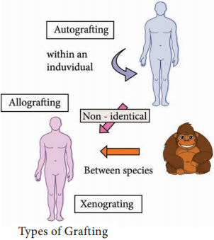
Heterologous vaccine:
These are a group of live attenuated vaccines produced from the strains that are pathogenic in animals and not in humans. It is a vaccine that confers protective immunity against a pathogen that shares cross-reacting antigens with the microorganisms in the vaccine. Example: Cow pox virus that protects against small pox in humans.

Killed inactivated vaccines:
These groups of vaccine are produced either by killing or inactivating the bacteria or virus by chemical treatment or heat. Example; Polio virus.

Sub unit vaccine:
The antigenic determinant / epitope (the very specific part of the microbe) is used to prepare the vaccine.
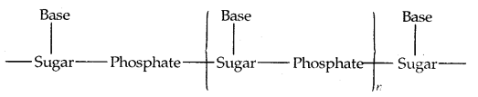
DNA Vaccines:
When the genes for microbe’s antigens are introduced into the body some cells will take up the DNA. The DNA then instructs those cells to make the antigen molecules. The cells secrete the antigens and display them on their surfaces. The body’s own cells become vaccine generating factories.

Routes of Administration

  • Deep subcutaneous or intramuscular route – most vaccines
  • Oral route – Oral BCG vaccine
  • Intradermal route – BCG vaccine
  • Scarification – Small pox vaccine
  • Intranasal route – Live attenuated influenza virus

Types of Immunization

Immunization is of two types:

  1. Passive Immunization
  2. Active Immunization

1. Passive Immunization

  • Passive immunization is produced without challenging the immune system of the body. It is done by administration of serum or gamma globulins from a person who is already immunized to a non-immune person.
  • Passive immunization is the administration of preformed antibodies either intravenously or intramuscularly.
  • It is used to provide rapid protection in certain infections such as diphtheria or tetanus or in the event of accidental exposure to certain pathogens such as hepatitis B.
  • It is also used to provide protection in immune compromised individuals.

Passive natural immunization:

Acquired from the mother before and after birth. Before birth, immunity is transferred from mother to the fetus in the form of maternal antibodies through placenta. After birth, the antibodies (Ig A) are transferred through breast milk (Table 11.2).

Table 11.2: Passive Immunization

Infection

Source of Antiserum

Indications

Tetanus Immune human; horse Post exposure (Plus vaccine)
Diptheria Horse Post-exposure
Gas gangrene Horse Psot-exposure
Botulism Horse Post-exposure
Varicella-Zoster Immune human Post-exposure in immunodeficiency
Rabies Immune human Post exposure (Plus vaccine)
Hepatitis B Immune human Post-exposure prophylaxis
Hepatisis A Pooled human Ig Prophylaxis
Measles Immune human Prophylaxis
Snakebite Horse Post-bite
Some autoimmune disease Pooled human ig Acute thrombocytopenia and neutropenia

Passive artificial immunization:

Developed by injecting previously prepared antibodies using serum from humans or animals. This type of immunity is useful for providing immediate protection against acute infections like tetanus, measles etc.

2. Active Immunization

Active immunization is the administration of vaccines containing microbial products with or without adjuvants in order to obtain long term immunological protection against the offending microbe.

At present the normal route of vaccination in most instances is either intramuscular or subcutaneous. Oral immunization is the method of choice for polio and Salmonella typhi vaccines. However, there is an increasing awareness that this route of immunization may be the best for most immunizations since nearly all infectious agents gain entrance through the mucosal surfaces.

Active natural immunization involves activation of immune system in the body to produce antibodies. It is achieved in both clinical and subclinical infections. Active artificial immunization is achieved by the administration of vaccines or toxoids.

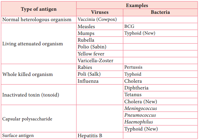
Antigen preparations

Most vaccines consist of attenuated organisms, killed organisms, inactivated toxins, or sub cellular fragments and more recently genes for antigens in viral ‘vectors’, and DNA itself. Thus, vaccines must be capable of targeting the immune system appropriately i.e. cellular/or humoral mechanisms (Table 11.3).
Table 11.3: Antigen Preparation Used in Vaccines.
Vaccination img 1

Adjuvants

Nonliving vaccines, especially those consisting of small molecules require the inclusion of agents to enhance their effectiveness.

These adjuvants include microbial, synthetic and endogenous preparations having adjuvant activity, but at present only aluminium or calcium salts are generally used in humans.

Adjuvants should enable antigens to be slowly released, preserve antigen integrity, target antigen presenting cells and induce cytotoxic lymphocytes.

Overview of an Transplantation

Learninsta presents the core concepts of Microbiology with high-quality research papers and topical review articles.

Overview of an Transplantation

Transfer of living cells, tissues or organs from one part of the body to another or from one individual to another is known as transplantation. A tissue or organ that is removed from one site and placed to another site usually in a same or different individual is called graft. The individual who provides the graft is called donor and the individual who receives the graft is called host or recipient.

If the graft is placed into its normal anatomic location, the procedure is called orthotopic transplantation. If the graft is placed in a different site it is called heterotopic transplantation. Transplantation is the only form of treatment for most end-stage organ failure.

In clinical practice, transplantation is used to overcome a functional and anatomic deficit in the recipient. Transplantation of kidneys, hearts, livers, lungs, pancreas and bone marrow are widely done today.

Methods of Transplantation

Auto grafting:
The transfer of self tissue from one body site to another in the same individual

Allografting:
The transfer of organs or tissues from human to human

Xenografting:
The transfer of tissue from one species to another (Figure 11.11).
Overview of an Transplantation img 1

Graft Acceptance

When transplantation is made between genetically identical individuals the graft survives and lives as healthy as it is in the original places. When the graft tissue remains alive, it is said to be accepted and the process is called graft acceptance.

Graft Rejection

When transplantation is made between genetically distinct individual the graft tissue dies and decays. When the graft tissue dies, the graft is said to be rejected and the process is called graft rejection. It is of two types. They are:-

  1. Host Verses Graft Reaction
  2. Graft Verses Host Rejection.

Host Verses Graft Reaction (HVG)

The graft tissue antigens induce an immune response in the host. This type of immune response is called host versus graft reaction.

Allograft Rejection

Types of allograft rejection

  • Acute rejection-Quick graft rejection. It is due to stimulation of thymocytes and B lymphocytes
  • Hyperacute rejection-It is a very quick rejection. It is due to pre-existing humoral antibodies in the serum of the host as a result of presensitization with previous grafts.
  • Insidious rejection-It is a secret rejection due to deposition of immune complex on the tissues like glomerulus membrane that can be demonstrated in kidney by immune fluorescence.

Mechanism of Allograft Rejection

Immunological contact

When tissue is implanted as graft, its antigen can pass into local lymph nodes of the host. The graft antigens then make contact with the lymphocytes of the host. Production of sensitized T cells and cytotoxic antibodies are produced in the host. This brings about graft rejection.

First set rejection

When the graft is made between genetically different individuals, the graft gets blood supply from the host and it appears to be normal for the first 3 days. But on the 5th day, sensitized T cells, macrophages and a few plasma cells invade the graft. Inflammation starts in the graft. This leads to necrosis. It is similar to the primary immune response to an antigen.

Second set rejection

When a graft is implanted in an individual who has already rejected a graft is second set rejection. This is similar to the secondary immune response of our body.

Cell mediated cytotoxic reaction

The 1st set of rejection of allograft is brought about mainly by CMI response. In this process the cells involved in the cytotoxic mediated immunity involves. On stimulation of these cells interferon causes the lysis of the graft.

Antibody mediated cytotoxic reaction

The 22nd set rejection of graft is brought about mainly by HMI response. This is one of the hyperacute rejection brought about by the antibodies. Complement, macrophages, mast cells, platelets, B cells bring about this reaction.

Graft versus Host Rejection (GVH)

Sometimes the graft tissue elicits an immune response against the host antigens. This immune response is called graft versus host reaction. It occurs when:

  • Graft remains inside the host and the host should not reject the graft.
  • The graft should have immune competent T cells.
  • The transplantation antigens of the host should be different from that of the graft.

Mechanism of the graft rejection

The graft lymphocytes aggregate in the host lymphoid organs and are stimulated by the lymphocytes of the host. The stimulated lymphocytes produce lymphokines. Lymphocytes in turn activate the host T cell. Activated T cell further activates the B cells. The stimulated B cell reacts with the self antigen and causes the damage.

How to prevent graft rejection?

Before transplantation the following things should be done to avoid graft rejection.

  • Perform blood grouping and Rh grouping
  • HLA typing should be done
  • Immuno suppressive drugs should be administered
  • Suitable donor should be chosen

Hypersensitivity Types and its Classification

Learninsta presents the core concepts of Microbiology with high-quality research papers and topical review articles.

Hypersensitivity Types and its Classification

Hypersensitivity is defined as the exaggerated immunological response leading to severe symptoms and even death in a sensitized individual when exposed for the second time. It is commonly termed as allergy. The substances causing allergic/hypersensitivity is known as allergens. Example: Drugs, food stuffs, infectious microorganisms, blood transfusion and contact chemicals.

Classification of Hypersensitivity (Coombs and Gell Classification)

Type I:
Immediate (Atopic or anaphylactic) Hypersensitivity

Type II:
Antibody-dependent Hypersensitivity

Type III:
Immune complex mediated Hypersensitivity

Type IV:
Cell mediated or delayed Hypersensitivity

Type I:
Immediate (Atopic or anaphylactic) Hypersensitivity

This type of hypersensitivity is an allergic reaction provoked by the re-exposure to a specific antigen. The antigen can make its entry through ingestion, inhalation, injection or direct contact. The reaction may involve skin, eyes, nasopharynx and gastrointestinal tract. The reaction is mediated by IgE antibodies (Figure 11.7).
Hypersensitivity Types and its Classification img 1

IgE has very high affinity for its receptor on mast cells and basophils. Cross linking of IgE receptor is important in mast cell trigerring. Mast cell degranulation is preceded by increased Ca++ influx.

Basophils and mast cells release pharmacologically active substances such as histamines and tryptase. This causes inflammatory response. The response is immediate (within seconds to minutes). Hence, it is termed as immediate hypersensitivity. The reaction is either local or systemic.

Hay Fever

Allergic rhinitis is commonly known as hay fever. Allergic rhinitis develops when the body’s immune system becomes sensitized and overreacts to something in the environment like pollen grains, strong odour of perfumes, dust etc that typically causes no problem in most people. When a sensitive person inhales an allergen the body’s immune system may react with the symptoms such as sneezing, cough and
puffy swollen eyelids.

Type II Hypersensitivity: Antibody dependent hypersensitivity

In this type of hypersensitivity reactions the antibodies produced by the immune response binds to antigens on the patient’s own cell surfaces. It is also known as cytotoxic hypersensitivity and may affect variety of organs or tissues. Ig G and Ig M antibodies bind to these antigens and form complexes. This inturn activates the classical complement pathway and eliminates the cells presenting the foreign antigen. The reaction takes hours to day (Figure 11.8).
Hypersensitivity Types and its Classification img 2

Drug induced haemolytic anaemia Certain drugs such as penicillin, cephalosporin and streptomycin can absorb non-specifically to protein on surface of RBC forming complex similar to hapten-carrier complex. In some patients these complex induce formation of antibodies, which binds to drugs on RBC and induce complement mediated lysis of RBC and thus produce progressive anaemia. This drug induced haemolytic anaemia is an example of Type II hypersensitivity reaction.

Type III Hypersensitivity: Immune complex mediated hypersensitivity

When a huge amount of antigen enters into the body, the body produces higher concentrations of antibodies. These antigens and antibodies combine together to form insoluble complex called immune complex. These complexes are not completely removed by macrophages.

These get attached to minute capillaries of tissues and organs such as kidneys, lung and skin (Figure 11.9). These antigen-antibody complexes activate the classical complement pathway leading to vasodilation. The complement proteins and antigen-antibody complexes attract leucocytes to the area. The leukocytes discharge their killing agents and promote massive inflammation. This can lead to tissue death and haemorrhage.
Hypersensitivity Types and its Classification img 3

Arthus reaction

It was first observed by Arthus. It is a local immune complex reaction occurring in the skin. Horse serum and egg albumin are the antigens that induce the arthus reaction. It is characterized by erythema, induration, oedema, haemorrhage and necrosis. This reaction occurs when antibody is found in excess. It appears in 2-8 hours after injection and persists for about 12-24 hours (Table 11.1).

Table 11.1: Difference between Immediate Hypersensitivity and Delayed Hypersensitivity

Immediate Hypersensitivity

Delayed Hypersenstivity

1. It appears and disappers rapidly 1. It appears slowly and last longer.
2. It is induced by antigens or haptens by any route 2. Induced by infection, injection of antigen intra dermally or with adjuvants of by skin contact.
3. The reaction is antibody mediated B-cell response 3. The reaction is T-cell mediated response.
4. Passive transfer is possible with serum 4. Cannot be transferred with serum but can be transferred by lymphocytes
5. Desensitization is easy, but does not last long 5. Desensitization is difficult but long lasting.

It is often called as delayed hypersensitivity reaction as the reaction takes two to three days to develop. Type IV hypersensitivity is involved in the pathogenesis of many autoimmune and infectious diseases such as tuberculosis and leprosy. T lymphocytes, monocytes and macrophages are involved in the reaction. Cytotoxic T Cells cause direct damage whereas the T helper cells secrete cytokines and activate monocytes and macrophages and cause the bulk damage (Figure 11.10).

Type IV hypersensitivity: Cell Mediated Delayed Hypersensitivity

Tuberculin reaction (Mantoux Reaction)

When a small dose of tuberculin is injected intra dermally in an individual already having tubercle bacilli, the reaction occurs. It is due to the interaction of sensitized T cell and tubercle bacterium. The reaction is manifested on the skin very late only after 48-72 hours.

Chemistry in Everyday Life Class 12 Notes Chemistry Chapter 16

By going through these CBSE Class 12 Chemistry Notes Chapter 16 Chemistry in Everyday Life, students can recall all the concepts quickly.

Chemistry in Everyday Life Notes Class 12 Chemistry Chapter 16

Medicines: Medical chemistry deals with the design and synthesis of drugs based on an undertaking of how these work in our body.

Drugs are chemicals of low molecular mass (~ 100-500 μ). They interact with macromolecular targets and produce a biological response. When the biological response is effective and useful, these chemicals are called medicines and are used in the treatment, diagnosis, and prevention of diseases. In larger doses than recommended, they are potential poisons. The use of chemicals for therapeutic effect is called Chemotherapy.

Designing of a Drug: Two considerations arise

  1. Drug target,
  2. drug metabolism.

1. Drug target: The biological macromolecules such as carbohydrates lipids, proteins, nucleic acids with which drugs interact are called targets. The correct choice of the molecular target for a drug is important to obtain a desired therapeutic effect.

2. Drug metabolism: A drug travels through the body in order to reach the target. So its design should be such that it reaches the target without being metabolized in between. Also, after its action, it should be excreted without causing harm to the body.

Compounds from which drugs are designed are called lead compounds. These lead compounds may be obtained from natural sources such as plants, trees, bushes, venoms, and metabolites of microorganisms or they may be synthesized in order to improve drug activity and to have minimum side effects, mechanisms of drug action in the biological systems are also considered while drug designing.

Classification of Drugs:
1. On the basis of Pharmacological effect: It is useful for doctors. For example, analgesics have a pain-killing effect, antiseptics kill or arrest the growth of microorganisms.

2. On the basis of action on a particular biochemical process: All antihistamines inhibit the action of the compound histamine, which causes inflammation in the body.

3. On the basis of chemical structure: Drugs classified in this way share common structural features and often have similar pharmacological activity. For example, sulphonamides have common structural features given below and are mostly antibacterial.
Chemistry in Everyday Life Class 12 Notes Chemistry 1
(Sfructuralfratures of Suiphona mides)

4. On the basis of molecular targets: This classification is most useful for medicinal chemists. Various enzymes and receptors in the cell are some of the common drug targets.

Interaction of drugs with targets: Proteins that perform the role of biological catalysts in the body are called enzymes. Proteins that are important to a communication system in the body are called receptors. Tires enzymes and receptors serve as drug targets among others.

Enzymes as Drug Targets:
(a) Catalytical activity of enzymes: Enzymes perform two major functions:
1. The first function of an enzyme is to hold the substrate for a chemical reaction. Active sites of enzymes hold the substrate molecule in a suitable position so that it can be attacked by the reagent effectively.

Substrates bind to the amino acid residues of the protein present on the active site of the enzyme through a variety of interactions such as ionic bonding, hydrogen bonding, van der Waals interaction of dipole-dipole interaction (Fig.).

These binding interactions should be strong enough to hold the substrate long enough so that the enzyme can catalyze the reaction, but weak enough to allow the products to depart after their formation.
Chemistry in Everyday Life Class 12 Notes Chemistry 2
(a) Active site of an enzyme,
(b) substrate
(c) Substrate held in the active site of the enzyme

(a) The second function of the enzyme is to provide functional groups that will attack the substrate and carry out a chemical reaction. This function is carried out by some other amino acid residues of protein present on the active site of the enzyme.

These provide free functional groups to attack the substrate and bring about chemical reactions. For example, if amino acid, serine is present nearby the substrate held on the active site, then its – OH group is free to act as a nucleophile in the enzyme-catalyzed reaction.

(b) Interaction of drugs with enzymes: Drugs inhibit the activity of the enzymes and so are called Enzyme Inhibitors. Enzyme inhibitors can block the binding site and prevent the binding of substrate or these can inhibit the catalytical activity of the enzyme.

(c) Prevention of attachment of natural substrate in the active site by drugs: Drugs inhibit the attachment of substrate on the active site of enzymes in two different ways explained below:

Drugs compete with the natural substrate for the active sites. Such drugs are called competitive inhibitors.
Chemistry in Everyday Life Class 12 Notes Chemistry 3
(Drug and substrate competing for the active site)

2. On the other hand, some drugs do not bind to the active site. These bind to a different site of enzyme which is called the allosteric site. This binding of inhibitors at the allosteric sites changes the shape of the active site in such a way that the substrate cannot recognize it.
Chemistry in Everyday Life Class 12 Notes Chemistry 4
[Noncompetitive inhibitor changes the active site of the enzyme after binding at the allosteric site]

If the bond formed between enzyme and inhibitor is a strong covalent bond and cannot be broken easily then the enzyme is blocked permanently. The body then degrades the enzyme inhibitors complex and synthesizes new enzymes.

Receptors as Drug Targets:
→ Location of receptor in the animal cell: Receptors are proteins that are crucial to the body’s communication process. The majority of these are embedded in cell membranes.

Receptor proteins are embedded in the cell membrane in such a way that their small part possessing active site projects out of the surface of the membrane and opens on the outer region of the cell membrane.

→ Transfer of message into the cell by receptors: Neurotransmitters communicate messages in the body between the 3 neurons and that between neurons to muscles. These chemical messengers are received at the binding site of the receptor protein. To accommodate messenger, the shape of the receptor changes. This brings about the transfer of the message into the cell. Thus, chemical messenger gives a message to the cell without entering the cell.

Two types of chemical messengers are involved in the message transfer:

  1. Hormones
  2. neurotransmitters

1. Hormones: Adrenaline (epinephrine) is an example of hormone. It is released from the adrenal medulla in situations of stress or danger.

2. Neurotransmitters are small molecules such as acetylcholine, dopamine, and serotonin.
Chemistry in Everyday Life Class 12 Notes Chemistry 5
Chemistry in Everyday Life Class 12 Notes Chemistry 6
→ Interaction of Drugs: Receptors that interact with one specific chemical messenger may differ slightly in their binding sites.

For example, there are two types of adrenergic receptors named a-adrenergic receptors and β-adrenergic receptors. These differ slightly in the structure of their binding sites, but both of these receptors can bind epinephrine.

Drugs that bind to the receptor site and inhibit its natural function are called antagonists. There are other types of drugs that mimic the natural messenger by switching on the receptor. They are called agonists.

→ Side-effects caused by drugs: Side effects are caused when a drug binds to more than one type of receptor, e.g., the serotonin receptor is a target for some anti-depressant drugs. Side effects can arise if the drug interacts with histamine or acetylcholine.

Types erf Drugs:
1. Antacids: If acid is produced in excess in the stomach, it causes irritation and pain and in severe cases, ulcers are produced. Histamine stimulates the secretion of pepsin and hydrochloric acid. A drug like cimetidine (Tagamet) and ranitidine (Zantac) was designed to prevent the interaction of histamine with the receptors present in the stomach wall. This resulted in the release of a lesser amount of acid.
Chemistry in Everyday Life Class 12 Notes Chemistry 7
2. Antihistammines: Histamine is a potent vasodilator. It has various functions. It contracts the smooth muscles in the bronchi and gut and relaxes other muscles. It is also responsible for the nasal congestion associated with common colds and allergic response to pollen. Synthetic drugs brompheniramine (Dimetapp) and terfenadine (Seldane) act as antihistamines.
Chemistry in Everyday Life Class 12 Notes Chemistry 8
The above-mentioned antihistamines do not affect the secretion of acid in the stomach. It is because that antiallergic and antacid drugs work on different receptors.

3. Neurologically Active Drugs: Tranquilizers and analgesics are neurologically active drugs.

These affect the message transfer mechanism from the nerve to the receptor.
(a) Tranquilizers are a class of compounds used for the treatment of stress, mild and severe mental diseases. They relieve stress, anxiety irritability, and excitement by inducing a sense of well-being.

→ They act on the central nervous system (CNS): Noradrenaline is one of the neurotransmitters that plays role in mood changes. If its level is low for some reason, the signal sending activity becomes low and the person suffers from depression.

Antidepressant drugs, in such cases, inhibit the enzymes which catalyze the degradation of noradrenaline. If the enzyme is inhibited, this important neurotransmitter is slowly metabolized and can activate its receptor for longer periods of time, thus countering the effect of depression. Iproniazid and phenelzine are two such drugs.
Chemistry in Everyday Life Class 12 Notes Chemistry 9
Some tranquilizers namely, Chlorodiazepoxide and Meprobamate are relatively mild tranquilizers suitable for relieving tension. Equanil is used in controlling depression and hypertension.
Chemistry in Everyday Life Class 12 Notes Chemistry 10
Chemistry in Everyday Life Class 12 Notes Chemistry 11
→ Barbiturates: The derivatives of barbituric acid are hypnotic- sleep-producing agents. Some of them are Veronal, Valium and Serotonium.
Chemistry in Everyday Life Class 12 Notes Chemistry 12
(b) Analgesics: are the drugs that reduce or abolish pain without causing impairment of consciousness, mental confusion, or some other disturbance of the nervous system.

They are of two types:
1. Non-narcotic (non-addictive) drugs: Aspirin and paracetamol belong to the class of non-addictive analgesics. These drugs have many other effects such as reducing fever (antipyretic) and preventing platelet coagulation. Aspirin is helpful to prevent heart attacks,

2. Narcotic analgesics: like morphine, heroin, codeine relieve pain and produce sleep in medicinal doses, and in excess are fatal. These analgesics are chiefly used for the relief of post-operative pain, cardiac pain, and pains of terminal cancer and in childbirth.

4. Antimicrobials: Disease may be caused by bacteria, viruses, etc. P. Ehrlich who developed the medicine Salvarsan for the treatment of syphilis found that the -As = As – linkage present in arsphenamine (salvarsan) resembles the -N = N- linkage present in azo-dyes in the sense that N atom is present in place of As. He was successful in 1932 in preparing the first effective antibacterial agent Prontosil which resembles the structure of the compound salvarsan.
Chemistry in Everyday Life Class 12 Notes Chemistry 13
[The structures of salvarsan and prontosil and azo dye showing structural similarity]

This led to the study of the relation between structure and activity. It was found that part of the proposal molecule (shown in the box) in the form of p-amino benzene sulphonamide (Sulphanilamide) has antibacterial activity. The led to the discovery of Sulpha drugs.

Antimicrobials control microbial diseases in three ways:
(a) a drug that kills the organism in the body (bactericidal).
(b) a drug that inhibits or arrests the growth of organisms (bacteriostatic) and
(c) increasing immunity and resistance to infection in the body,

5. Antibiotics: It is a substance (produced wholly or partly by chemical synthesis) that in low concentration inhibits the growth or destroys microorganisms by intervening in their metabolic processes.

The first antibiotic discovered by Alexander Fleming’s Penicillin from the mold Penicillium Notatum.

The antibiotics can be either bactericidal or bacteriostatic.

Bactericidal Bacteriostatic
Penicillin Erythromycin
Aminoglycosides Tetracycline
Ofloxacin Chloramphenicol

Broad Spectrum antibiotics are medicines effective against several types of harmful microorganisms, e.g., tetracycline, chloramphenicol.

6. Antiseptics and disinfectants: Antiseptics and disinfectants are also the chemicals which either kill or prevent the growth of microorganism.

Antiseptics are applied to living tissues such as wounds, cuts, ulcers, and diseased skin surfaces. Examples are Furacine, Soframicine, etc. Dettol is a mixture of Chloroxylenol and terpineol. Bithinol is added to soaps to impart antiseptic properties. Iodine is a powerful antiseptic. Its 2-3% solution in alcohol-water solution is known as tincture of iodine. It is applied to wounds. Iodoform is also used as an antiseptic for wounds. Boric acid (H3P03) in dilute solution (aqueous) is a weak antiseptic for the eyes.
Chemistry in Everyday Life Class 12 Notes Chemistry 14
Disinfectants are applied to inanimate objects such as floors, drainage systems, instruments, etc. The same substance can act as an antiseptic as well as a disinfectant by varying the concentration. For example, 0.2 percent situation of phenol is an antiseptic while it’s one percent solution is disinfectant.

Chlorine in the concentration of 0.2 to 0.4 ppm and S02 in very low concentration are disinfectants.

7. Antifertility Drugs: Norethindrone is an example of synthetic progesterone (a type of hormone) derivative most widely used as an antifertility drug for birth control. The estrogen derivative is used in combination with progesterone derivative is ethynylestradiol (Novestrol).

→ Chemicals in Food: To enhance the shelf life of food to make it more appealing and sometimes more nutritive, chemicals are added to it.

They are:

  1. Food colors,
  2. Flavors and sweeteners,
  3. Fat emulsifiers and stabilizing agents,
  4. Flour improvers antistaling agents and bleaches,
  5. Antioxidants,
  6. Preservatives,
  7. Nutritional supplements such as minerals, vitamins, and amino acids.

Except for category (7), none of the chemical additives have any nutritive value.

→ Artificial Sweetening agents: Ortho-Sulphobenzimide (saccharine)
Chemistry in Everyday Life Class 12 Notes Chemistry 15
is an artificial sweetener and mass/mass, it is 550 times as sweet as cane sugar. It is excreted from the body in the urine unchanged and appears to be entirely harmless and inert and so is of great value to diabetic persons and people who need to control the intake of calories.

Other artificial sweeteners are aspartame (100 times sweet as sugar), sucralose (600 times) alitame (2000 times as sweet as sugar).

→ Preservatives: In addition to class I preservatives like salts, sugar, and vegetable oils, the most common class II preservative is sodium benzoate
Chemistry in Everyday Life Class 12 Notes Chemistry 16
which is used in limited quantities and is metabolized in the body.

→ Chemistry of Cleansing Agents:
1. Soaps: Soaps are sodium or potassium salts of long-chain fatty acids, e.g., stearic acid, oleic acid, and palmitic acid. Soaps are obtained by the saponification of triglycerides of fatty acids.
Chemistry in Everyday Life Class 12 Notes Chemistry 17
Potassium soaps are softer than sodium soaps.

Types of Soaps: Toilet Soaps are prepared by using better grades of fats and oils and excess alkali is removed. Colour and perfumes are added. Transparent Soap is made by dissolving the soap in ethanol and then evaporating the excess solvent.

In medicated soaps, substances of medicinal value are added. ! Shaving soaps contain glycerol to prevent rapid drying. Laundry soaps t contain fillers like sodium proximate, sodium silicate borax, and sodium \ carbonate.

Soaps do not work in hard water as soaps react with Ca2+ and Mg2+ ions present in hard water to produce curdy precipitate or scum,
Chemistry in Everyday Life Class 12 Notes Chemistry 18
2. Soapless detergents: Soapless detergents are cleansing agents; which have all the properties of soaps, but they actually do not contain; soap. They are useful in hard water also.

Synthetic detergents are mainly of three types:

  1. Anionic detergents
  2. Cationic detergents
  3. Non-ionic detergents

1. Anionic Detergents are sodium salts of sulfonated long-chain alcohols.
Chemistry in Everyday Life Class 12 Notes Chemistry 19
In anionic detergents, the anionic part of the molecule is involved in the cleansing action.

2. Cationic Detergents: Cationic detergents are acetates, chlorides, or bromides of quaternary ammonium salts. An example is cetyltrimethylammonium bromide:
Chemistry in Everyday Life Class 12 Notes Chemistry 20
Cationic detergents are expensive and due to their germicidal properties, they are used as hair conditioners.

3. Non-ionic Detergents: Stearic acid reacts with polyethylene glycol to form non-ionic detergents.
Chemistry in Everyday Life Class 12 Notes Chemistry 21
Liquid dishwashing detergents are non-ionic types. Detergents containing highly branched hydrocarbon chains are not easily biodegradable.

Polymers Class 12 Notes Chemistry Chapter 15

By going through these CBSE Class 12 Chemistry Notes Chapter 15 Polymers, students can recall all the concepts quickly.

Polymers Notes Class 12 Chemistry Chapter 15

Polymers are macromolecules having high molecular mass [103 – 107 p]. They are formed by joining repeating structural units on a large scale. The repeating structural units are derived from some simple and reactive molecules known as monomers and are linked to each other by covalent bonds. The process of the formation of polymers from respective monomers is called polymerisation.
Polymers Class 12 Notes Chemistry 1
Classification of Polymers:
A. Based on the source.

  1. Natural Polymers: These are found in plants and animals. Examples are proteins, cellulose, starch, resins and rubber.
  2. Semi-synthetic Polymers: Cellulose acetate (rayon) and cellulose nitrate are examples of this category.
  3. Synthetic Polymers: Polyethene; nylon 6, 6; Buna-S are examples of man-made polymers.

B. Based on the structure of Polymers:
1. Linear Polymers: These polymers Consist of long and straight-chain repeating units derived from the monomers. The examples are high-density polyethene, polyvinyl chloride (PVC) etc. These are schematically represented as
Polymers Class 12 Notes Chemistry 2
2. Branched Chain Polymers: These polymers contain linear chains having some branches, e.g., low-density polyethene.
Polymers Class 12 Notes Chemistry 3
3. Cross-linked or Network Polymers: These are usually formed from bifunctional and trifunctional monomers, e.g., bakelite, melamine etc.
Polymers Class 12 Notes Chemistry 4
C. Classification Based on mode of Polymerisation:
1. Addition Polymers: The addition polymers are formed by the repeated addition of monomer molecules possessing double or triple bonds, e..g, the formation of polyethene from ethene and polypropene from propene. In addition, polymers obtained from the same monomer are called Homopolymers, e.g., Polyethene.
Polymers Class 12 Notes Chemistry 5
If two different units of monomers get added, they are called copolymers, e.g., Buna-S, Buna-N,
Polymers Class 12 Notes Chemistry 6
2. Condensation Polymers: The condensation polymers are formed by repeated condensation reaction between two monomeric units having different bifunctional and trifunctional groups with the elimination of small molecules like water, alcohol, hydrogen chloride etc. The formation of Nylon 6,6 is an example.
Polymers Class 12 Notes Chemistry 7
D. Classification based upon molecular forces:
1. Elastomers: These are rubber-like solids with elastic properties. The polymer chains are held together by weak intermolecular forces. They can be easily stretched. Examples are Buna-S, Buna-N, Neoprene etc.

2. Fibres: The intermolecular forces between the chains are strong hydrogen bonds. They have large tensile strength and are used to form thread forming crystalline solids. The examples are Nylon 6, 6 and polyesters.

3. Thermoplastic Polymers: In these polymers, the intermolecular forces are intermediate between those of elastomers and fibres. In these polymers, there is cross-linking between the chains. They soften on heating and harden on cooling. Common examples are polyethene, polystyrene polyvinyls etc.

4. Thermosetting Polymers: These polymers are cross-linked or heavily branched molecules, which on heating undergo expensive cross-linking in moulds and become infusible. They cannot be reused again. Common examples are bakelite and urea-formaldehyde resins etc.

E. Classification based on Growth Polymerisation: The addition and condensation polymers are nowadays also referred to as chain-growth polymers and step-growth polymers depending upon the type of polymerisation mechanism they undergo during their formation.

Types of Polymerization:
1. Addition Polymerization or Chain growth Polymerization: Here molecules of the same or different monomers add together on a large scale to form a polymer. It can proceed through the formation of free radicals or ionic species.
(a) Free Radical Mechanism: A variety of alkenes or dienes and their derivatives are polymerised in the presence of a free radical generating initiator (catalyst) like benzoyl chloride.

It consists up of the following three steps.
1. Chain-initiation Step:
Polymers Class 12 Notes Chemistry 8
2. Chain propagating step:
Polymers Class 12 Notes Chemistry 9
3. Chain terminating step:
Polymers Class 12 Notes Chemistry 10
(b) Preparation of some important Addition Polymers:
1. Polyethene: There are two types of polyethenes as given below:
1. Low-Density Polyethene (LDPE]:
Polymers Class 12 Notes Chemistry 11
It is chemically inert and tough, but flexible and a poor conductor of electricity. It is used in the insulation of electric wires and the manufacture of squeeze bottles, toys and flexible pipes.

2. High-Density Polyethene (HDPE):
Polymers Class 12 Notes Chemistry 12
It has a high density. It is also chemically inert and tougher and harder. It is used for making buckets, dustbins, bottles and pipes.

2. Polytetrafluoroethene (Teflon):
Polymers Class 12 Notes Chemistry 13
Chemically inert, it is resistant to attack by corrosive reagents. Used for making oil seals, gaskets and non-stick surface coated utensils.

3. Polyacrylonitrile:
Polymers Class 12 Notes Chemistry 14
It is used as a substitute for wool in making fibres like Orlon or Acrilan.

→ Condensation Polymerization or Step-Growth polymerization: It involves a repetitive condensation reaction between two bifunctional monomers. It may result in the loss of simple molecules as H2O, alcohol etc.

1. Polyamides: Preparation of Nylons
1. Nylon 6,6:
Polymers Class 12 Notes Chemistry 15
It is used in making sheets, bristles for brushes and in the textile industry.

2. Nylon 6: It is obtained by heating caprolactam with water at high temperature.
Polymers Class 12 Notes Chemistry 16
Nylon 6 is used for the manufacture of tyre cords, fabrics and ropes.

2. Polyesters: These are the polycondensation products of dicarboxylic acids and diols. The formation of terylene or dacron by the reaction between ethylene glycol and terephthalic acid is an example.
Polymers Class 12 Notes Chemistry 17
Dacron fibre (terylene) is crease-resistant and is used in blending with cotton and wool fibres and also as glass reinforcing materials in safety helmets etc.

3. Phenols formaldehyde polymer (Bakelite and related polymers): Phenol reacts with formaldehyde in the presence of dil. acid or base.
Polymers Class 12 Notes Chemistry 18
Novolac (used in paints) on heating with HCHO undergoes cross-linking to form an infusible solid mass called bakelite
Polymers Class 12 Notes Chemistry 19
It is used for making combs, photograph records, electrical switches and handles of various utensils.

4. melamine-formaldehyde polymers: It is obtained by the condensation polymerisation of melamine and formaldehyde.
Polymers Class 12 Notes Chemistry 20
It is used in the manufacture of unbreakable cups and plates.

Copolymerization: A mixture of 1,3-butadiene and styrene form a copolymer: Butadiene-Styrene copolymer.
Polymers Class 12 Notes Chemistry 21
1. Natural rubber: It possesses elastic properties. It is a linear polymer of isoprene (2-methyl-l, 3-butadiene).
Polymers Class 12 Notes Chemistry 22
It is also called cis-1, 4-polyisoprene. It consists of various chains held together by weak van der Waals forces and has a coiled structure.
Polymers Class 12 Notes Chemistry 23
→ Vulcanisation of Rubber: To improve upon the physical properties of natural rubber, its vulcanisation is carried out. It consists of heating a mixture of raw rubber with sulphur and an appropriate additive at a temperature range between 373-415 K. On vulcanization sulphur forms cross-links at the reactive sites of double bonds and the rubber gets Stiffened. The probable structure of vulcanised rubber is:
Polymers Class 12 Notes Chemistry 24
→ Synthetic Rubber: Synthetic rubbers are either homopolymers of 1, 3-butadiene derivatives or are copolymers of 1, 3-hutadíene or its derivatives with another unusual rated monomer.

1. Neoprene: It has superior qualities to natural rubber. It has better resistance to vegetable and mineral oils. It is used for the manufacture of conveyor belts, gaskets and hoses.
Polymers Class 12 Notes Chemistry 25
2. Buna-N: It is a copolymer of 1,3-butadiene and acrylonitrile in the presence of a peroxide catalyst.
Polymers Class 12 Notes Chemistry 26
It is resistant to the action of petrol, lubricating oil and organic St .h ents. It is used is making oil seals tank living etc.

→ Molecular Mass of Polymers: Polymer properties are closely related to their molecular mass, size and structure. Its molecular mass is always expressed as an average.

It can be determined by chemical and physical methods.

  1. Weight-average molecular mass
  2. Number-average molecular mass.

→ Biodegradable Polymers: A large number of polymers are non-biodegradable and are the reuse for environmental pollution. Nowadays, certain new biodegradable synthetic polymers have been designed and developed. Aliphatic polyesters are one of the important class of biodegradable polymers, e.g.,

→ Poly β-hydroxybutyrate-co-β-hydroxy valerate (PHBV): It is obtained by the copolymerisation of 3-hydroxybutyric acid and 3-hydroxy pentanoic acid.

PHBV undergosbateria1 degradation in the environment.
Polymers Class 12 Notes Chemistry 27
Nylon-2-Nylon 6: It is an alternating polyamide copolymer of glycine (H2N—CH2—COOH) and aminocaproic acid. (H2N (CH2)5 COOH) and is biodegradable.

Some other commercially important Polymers along with their structures and uses are given below in the table:
Polymers Class 12 Notes Chemistry 28
Polymers Class 12 Notes Chemistry 29

Immunology of Western Blot Techniques Principle and its Applications

Learninsta presents the core concepts of Microbiology with high-quality research papers and topical review articles.

Immunology of Western Blot Techniques

Macromolecules immobilized or fixed on nitrocellulose membrane i.e., blotted can be subjected to a variety of analytical techniques more easily. Southern blotting was the first blotting technique developed which made the analysis and recording of DNA easy.

Later the technique was extended for analysis of RNA and proteins and they have acquired the jargon terms Northern and Western Blotting respectively.

Western blotting is also known as immunoblotting because it uses antibodies to detect the protein. Western blotting is a quantitative test to determine the amount of protein in sample.

Principle

Western blotting technique is used for the identification of a particular protein from the mixture of a proteins. In this method, the proteins are first extracted from the sample. Extracted proteins are subjected to Poly Acryl – amide Gel Electrophoresis (PAGE).

Transfer of proteins from poly acryl amide to the nitrocellulose paper is achieved by applying electric field. When radio labelled specific antibody is added on such membrane it binds to the specific complementary protein. Finally the proteins on the membrane can be detected by staining or through ELISA technique.

Steps

Step I:
Extraction of Protein

The most common protein sample used for Western blotting is cell lysate. The protein from the cell is generally extracted by mechanical means or by adding chemicals which can lyse the cell. The extraction step is termed as tissue preparation.

Protease inhibitor is used to prevent the denaturing of proteins. Using spectroscopy the concentration of the protein sample is analysed and diluted in loading buffer containing glycerol. This will help the sample to sink in the well. Bromothymol blue is used as tracking dye and is used to monitor the movement of the sample.

Step II:
Gel electrophoresis

The protein sample is loaded in well of SDS-PAGE (Sodium dodecyl sulfatepoly-acryl amide gel electrophoresis). The proteins are separated on the basis of electric charge, isoelectric point, molecular weight, or combination of all these. Proteins are negatively charged, so they move toward positive (anode) pole as electric current is applied. Smaller proteins move faster than the larger proteins.

Step III:
Blotting

Blotting refers to the transfer of the protein from the gel to the nitrocellulose paper by capillary action. Electro blotting is done nowadays to speed up the process. In electro-blotting nitrocellulose membrane is sandwich between gel and cassette of filter paper and then electric current is passed through the gel causing transfer of protein to the membrane.

Step IV:
Blocking

The nitrocellulose membrane is nonspecifically saturated or masked by using casein or Bovine serum albumin (BSA) before adding the primary antibody. This blocking step is very important in western blotting as antibodies are also proteins and they are likely to bind to the nitrocellulose paper.

Step V:
Treatment with primary and secondary antibody

The primary antibody is specific to desired protein so it forms Ag-Ab complex. The secondary antibody is enzyme labelled and is against primary antibody (antiantibody) so it can bind with Ag-Ab complex. Alkaline phosphatase or Horseradish peroxidase (HRP) is labelled
with secondary antibody.

Step VI:
Treatment with suitable substrate

Finally, the reaction mixture is incubated with specific substrate. The enzyme convert the substrate to give visible coloured product, so band of colour can be visualized in the membrane (Figure 11.6).
Western Blot Techniques img 1

Application

  1. The size and concentration of protein in given sample is determined by western blotting.
  2. It is used in the detection of antibody against virus or bacteria in serum and helps in the disease diagnosis.
  3. Western blotting technique is the confirmatory test for HIV. It detects anti HIV antibody in patient’s serum.
  4. Useful to detect defective proteins.

Antigen Antibody Reactions and its types | Working principle, Applications

Learninsta presents the core concepts of Microbiology with high-quality research papers and topical review articles.

Antigen Antibody Reactions and its types | Working principle, Applications

The interaction between antigen and antibody is called antigen-antibody reactions. It is abbreviated as Ag-Ab reaction. This reaction is the basis of humoral immunity. The antigen and the antibody react to form immune complex.

Ag + Ab …………….. Ag – Ab complex The reaction between antigen and antibody is highly specific. It is compared to the lock and key system. The part of the antigen that combines with the antibody is called epitope or antigenic determinant. The part of antibody which combines with the antigen is called paratope or antigen determining site. Most of the antibodies have two binding sites and IgM has 5-10 binding sites.

Immunofluorescence

When antibodies are mixed with the fluorescent dyes such as fluorescein or rhodamine, they emit radiation. This phenomenon of emitting radiation by antibodies labelled with fluorescent dye is called immuno fluorescence. This reaction is well observed under fluorescent microscope. It is used to locate and identify antigens in tissues.

Types of Immunofluorescence

  • Direct method
  • Indirect method

Direct Method

In this method, the antibody labelled with fluorescent dye is directly applied on the tissue section. The labelled antibody binds with specific antigen. This can be observed under the fluorescent microscope.

Indirect Method

In this method, unlabelled antibodies are directly applied on the tissue sectionswhich bind with the specific antigens. Then the antibody labelled with the fluorescent dye is added to the tissue. Anti-antibody specifically binds with already added or linked unlabelled antibody (Figure 11.1).
Antigen Antibody Reactions img 1

ELISA (Enzyme Linked Immuno Sorbent Assay)

ELISA (Enzyme-Linked Immuno Sorbent Assay) is a plate-based assay technique designed for detecting and quantifying substances such as peptides, proteins, antibodies and hormones. It is also known as Enzyme Immuno Assay (EIA).

In 1971, after the descriptions of Peter Perlmann and Eva Engvall at Stockholm University in Sweden, ELISA has become the system of choice when assaying soluble antigens and antibodies. All assays for antibody production depend upon the measurement of interaction of elicited antibody with antigen.

Principle

The principle of ELISA is very simple. The test is generally conducted in micro titre plates. (Figure 11.2 Micro titre plate).
Antigen Antibody Reactions img 2

If the antigen is to be detected the antibody is fixed in the micro titre plate and vice versa. Test sample is added in the microtitre plate, if there is presence of Ag or Ab in the test sample, there will be Ag-Ab reactions (with immobilized Ab or Ag). Later enzyme labelled antibody is added in the reaction mixture, which will combine with either test antigen or Fc portion of test antibody.

The enzyme system consists of:

1. An enzyme:

Horse Radish Peroxidase(HRP), alkaline phosphatase which is labelled or linked, to a specific antibody.

2. A specific substrate:

  • O-Phenyl-diaminedihydrochloride for peroxidase
  • P nitrophenyl Phosphate – for Alkaline Phosphatase

Substrate is added after the antigenantibody reaction. The enzyme hydrolyses the substrate to give a yellow colour compound in case of alkaline phosphatase (Figure 11.3). The intensity of the colour is proportional to the amount of antibody or antigen present in the test sample, which can be quantified using ELISA reader
(Figure 11.4 ELISA reader)
Antigen Antibody Reactions img 3
Antigen Antibody Reactions img 4

Types

There are four kinds of ELISA assay tests. They are: Direct ELISA, Indirect ELISA, Sandwich ELISA and Competitive ELISA (Figure 11.5).
Antigen Antibody Reactions img 5

i. Direct ELISA

An antigen is immobilized in the well of an ELISA plate. The antigen is then detected by an antibody directly conjugated to an enzyme such as HRP. Direct ELISA detection is much faster than other ELISA techniques as fewer steps are required.

The assay is also less prone to error since fewer reagents and steps are needed, i.e. no potentially cross-reacting secondary antibody needed. Finally, the direct ELISA technique is typically used when the immune response to an antigen needs to be analyzed.

ii. Indirect ELISA

Indirect ELISA is used to detect antibody. A known antigen is coated on the micro titre plate. If the patient’s serum contains antibody specific to the antigen, the antibody will bind to the antigen.

After incubation the wells are washed and the enzyme labelled anti Human Gamma Globulin (HGG) is added to the well. AntiHGG can react with antigen antibody complex. The substrate for the enzyme is added finally which is hydrolysed by the enzyme which develops a colour.

iii. Sandwich ELISA

Sandwich ELISA is used to detect antigen. A known antibody is coated on the micro titre plate. A test antigen is added to each well and allowed to react with the bound antibody. If the patient’s serum contains antigen specific to the antibody, the antigen will bind to the antibody.

Specifically bound antigen and antibody will remain in the wells even after washing. The second antibody is added and allowed to react with bound antigen. Substrate is added to measure colour reaction.

iv. Competitive ELISA

It is used for the detection of antigens. Antibody is first incubated with a sample-containing antigen. The antigen and antibody complex is added to the antigen coated microtitre well. If more antigen present in the sample, the less free antibody will be available to bind to the antigen coated well.

Addition of an enzyme conjugated secondary antibody specific to the primary antibody can be used to determine the amount of primary antibody bound to the well. It is a quantitative test for the antigen detection.

Application

An ELISA test may be used to diagnose:

HIV, Lyme disease, pernicious anaemia, Rocky Mountain spotted fever, rotavirus, squamous cell carcinoma, syphilis, toxoplasmosis, varicella-zoster virus, which causes chickenpox and Zika virus.

Biomolecules Class 12 Notes Chemistry Chapter 14

By going through these CBSE Class 12 Chemistry Notes Chapter 14 Biomolecules, students can recall all the concepts quickly.

Biomolecules Notes Class 12 Chemistry Chapter 14

Carbohydrates: Most common examples of carbohydrates are glucose, fructose, cane sugar, starch etc. Most of them have a general formula Cx (H2O)y. Earlier they were considered hydrates of carbon. For example, glucose C6H12O6 fits into this general formula C6(H2O)6 But even acetic acid (CH3COOH) fits into this general formula C2(H2O) and it is not a carbohydrate. Similarly, rhamnose, C6H12O5 is a carbohydrate but does not fit into this definition.

Chemically, the carbohydrates may be defined as optically active polyhydroxy aldehydes or ketones or the compounds which produce such units on hydrolysis.

They are classified as:

  1. Sugars: They are sweet in taste and water-soluble, e.g. glucose, fructose, sucrose.
  2. Non-sugars: They are tasteless and water-insoluble, e.g., starch, cellulose. Carbohydrates are systemically classified as:

1. Monosaccharides: A carbohydrate that cannot be hydrolysed further to give simpler units of polyhydroxy aldehydes or ketones is called monosaccharides. Glucose (C6H1206) is an aldohexose and fructose (C6H1206) is a ketohexose.

2. Oligosaccharides: Carbohydrates that yield two to ten monosaccharides on hydrolysis are called oligosaccharides.
(a) Disaccharides: They hydrolyse to give two units of monosaccharides. They include sucrose, maltose, lactose.
Biomolecules Class 12 Notes Chemistry 1
(b) Trisaccharides: They yield three units of monosaccharides on hydrolysis, e.g. C18H32O16 (raffinose).
(c) Tetrasaccharides: Yields four units of monosaccharides on hydrolysis, e.g. stachyose C24H42O21

2. Polysaccharides: They yield a large number of monosaccharide units on hydrolysis: Common examples are starch, cellulose. They are not sweet in taste.

Reducing sugars are those which reduce Fehling’s solution and Tollen’s reagent. All monosaccharides whether aldoses and ketoses are reducing sugars.

Sugars that do not reduce Fehling solution or Tollen’s reagent are termed as non-reducing e.g., sucrose.

→ Monosaccharides: They contain three to seven carbon atoms. If they contain an aldehyde group (- CHO), they are termed aldoses. If they contain a keto group (C = O), they are termed ketoses.

Different Types of Monosaccharides:
Biomolecules Class 12 Notes Chemistry 2
1. Glucose:
Preparation:
(a) From Sucrose (Cane Sugar)
Biomolecules Class 12 Notes Chemistry 3
(b) From Starch:
Biomolecules Class 12 Notes Chemistry 4
→ Structure of Glucose: It is an aldohexose and is also known as dextrose. Its structure (open chain) is
Biomolecules Class 12 Notes Chemistry 5
Evidence in favour of the above structure:

  1. Its molecular formula was determined to be C6H12O6.
  2. On heating (prolonged) with HI, it formed an n-hexane suggesting that all the 6 carbon atoms are in a straight chain.
    Biomolecules Class 12 Notes Chemistry 6
  3. It reacts with hydroxylamine to form an oxime and adds a molecule of hydrogen cyanide (HCN) to give cyanohydrin showing the presence of a carbonyl group in it,
    Biomolecules Class 12 Notes Chemistry 7
  4. Glucose is oxidised to gluconic acid by mild Oxidizing agent Br. water, confirming that a carbonyl group is an aldehyde group.
    Biomolecules Class 12 Notes Chemistry 8
  5. Acetylation with acetic anhydride gives glucose pentaacetate which confirms the presence of five – OH groups attached to 5 different C atoms.
    Biomolecules Class 12 Notes Chemistry 9
  6. on oxidation with nitric acid, glucose well as gluconic acid both yield a dicarboxylic acid, saccharic acid indicating the presence of -CH2OH group in it in addition to an aldehyde.
    Biomolecules Class 12 Notes Chemistry 10

The exact spatial arrangement of different – OH groups was given by Fischer. Its exact configuration is correctly represented by I. Gluconic acid is II and Saccharic acid is III.
Biomolecules Class 12 Notes Chemistry 11
Glucose is correctly named as D (+) glucose. ‘D’ represents the configuration whereas (+) represents the dextro-rotatory nature of it. The meaning of D- and L- notations is given as follows:

[Note: It may be remembered that ‘D’ and ‘L’ notations have nothing to do with the optical activity of the compound.]

The letters ‘D’ or ‘L’ before the name of any compound indicate the relative configuration of a particular stereoisomer. This refers to their relationship with a particular isomer of glyceraldehyde. Glyceraldehyde contains one asymmetric carbon atom and exists in two enantiomeric forms as shown below.
Biomolecules Class 12 Notes Chemistry 12
All those compounds which can be chemically correlated to (+) isomer of glyceraldehyde are said to have D-configuration whereas those which can be correlated to (-) isomer of glyceraldehyde are said to have L—configuration.

For assigning the configuration of monosaccharides, it is the lowest asymmetric carbon atom (as shown below) which is compared. As in (+) glucose, —OH on the lowest asymmetric carbon is on the right side which is comparable to (+) glyceraldehyde, so it is assigned D-configuration. For this comparison, the structure is written in a way that most oxidised carbon is at the top.
Biomolecules Class 12 Notes Chemistry 13
Cyclic Structure of Glucose

The structure (I) of glucose explained most of its properties but the following reactions and facts could not be explained by this structure.

  1. Despite having the aldehyde group, glucose does not give 2,4- DNP test, Schiff’s test and it does not form the hydrogen sulphite addition product with NaHSO3.
  2. The pentaacetate of glucose does not react with hydroxylamine indicating the absence of the free -CHO group.
  3. Glucose is found to exist in two different crystalline forms which are named a and b. The a-form of glucose (m.p. 419 K) is obtained by crystallization from a concentrated solution of glucose at 303 K while the (i-form (m.p. 423 K) is obtained by crystallisation from hot and saturated aqueous solution at 371 K,

This behaviour could not be explained by the open-chain structure (I) for glucose. It was proposed that one of the -OH groups may add to the -CHO group and form a cyclic hemiacetal structure. It was found that glucose forms a six-membered ring in which -OH at C-5 is involved in a ring formation. This explains the absence of -CHO group and also the existence of glucose in two forms as shown below. These two cyclic forms exist in equilibrium with an open-chain structure.
Biomolecules Class 12 Notes Chemistry 14
The two cyclic hemiacetal forms of glucose differ only in the configuration of the hydroxyl group at Cl, called anomeric carbon (the aldehyde carbon before cyclization). Such isomers, i.e., a-form and b-form, are called anomers.

The six-membered cyclic structure of glucose is called the pyranose structure (α- or β-), in analogy with pyran. Pyran is a cyclic organic compound with one oxygen atom and five carbon atoms in the ring. The cyclic structure of glucose is more correctly represented by Haworth structure as given below:
Biomolecules Class 12 Notes Chemistry 15
II. Fructose
Fructose is an important ketohexose. It is obtained along with glucose by the hydrolysis of disaccharide, sucrose. It has a ketonic group at C – 2. It belongs to D-series and is a laevorotatory compound. Therefore, it is written as D – (-) fructose. Its open-chain structures are given below:
Biomolecules Class 12 Notes Chemistry 16
→ It differs from glucose only at C – 1 and C – 2. Its furanose form (cyclic) is:
Biomolecules Class 12 Notes Chemistry 17
→ The cyclic structures of two anomers of fructose as represented by Haworth are given below:
Biomolecules Class 12 Notes Chemistry 18
Disaccharides:
1. Sucrose: Sucrose on hydrolysis gives an equimolar mixture of D – (+) – glucose and D – (-) fructose.
Biomolecules Class 12 Notes Chemistry 19
Sucrose is a non-reducing sugar. Therefore, it has a glucoside linkage between C1 of α-glucose and C2 of β-fructose.
Biomolecules Class 12 Notes Chemistry 20
or
Biomolecules Class 12 Notes Chemistry 21
Sucrose is dextrorotatory but after hydrolysis gives dextrorotatory glucose and laevorotatory fructose. Since the laevorotation of fructose (- 92.4°) is more than the dextrorotation of glucose (+ 52.5°), the mixture is laevorotatory. Thus hydrolysis of sucrose brings about a change in the sign of rotation, from Dextro (+) to leave (-) and the product is named as invert sugar.

II. Maltose: Another disaccharide, maltose is composed of two α-D-glucose units in which C4 of one glucose (I) is linked to C4 of another glucose unit (II). Hie free aldehyde group can be produced at C1 of second glucose in solution and it shows reducing properties, so it is a reducing sugar.
Biomolecules Class 12 Notes Chemistry 22
II. Lactose: It is more commonly known as milk sugar since this disaccharide is found in milk. It is composed of (β-D-galactose and β-D- glucose. The linkage is between C4 of galactose and C4 of glucose. Hence it is also a reducing sugar.
Biomolecules Class 12 Notes Chemistry 23
Polysaccharides: Polysaccharides contain a large number of monosaccharide units joined together by glycosidic linkages.
I. Starch: Starch is the main storage polysaccharide of plants. It is a polymer of a-glucose and consists of two components 15-20% of water-soluble Amylose and Amylopectin which is water-insoluble and constitutes about 80-85% of starch. Their structures have been given below:
Biomolecules Class 12 Notes Chemistry 24
II. Cellulose: Cellulose occurs exclusively in plants. It is a predominant constituent of the cell walls of plant cells. Cellulose is a straight-chain polysaccharide composed of only β-D-glucose units which are joined by the glycosidic linkage between C1 of one glucose unit and C4 of the next glucose unit.
Biomolecules Class 12 Notes Chemistry 25
III. Glycogen: The carbohydrates are stored in the animal body as glycogen. It is also known as animal starch because its structure is similar to amylopectin and is more highly branched.

→ Proteins: Proteins are the most abundant biomolecules of the living system. Chief sources of proteins are milk, cheese, pulses, peanuts, fish and meat etc. They are required for the growth and maintenance of the body. All proteins are polymers of a-amino acids.

→ Amino acids: Amino acids contain an amino (- NH2) and carboxyl (- COOH) functional groups.
Biomolecules Class 12 Notes Chemistry 26
→ Classification of Amino acids: Amino acids are classified as acidic, basic or neutral depending upon the relative number of amino and carboxyl groups in their molecule. An equal number of amino and carboxyl groups makes it neutral; more amino than carboxyl groups makes it basic and more carboxyl groups as compared to amino groups makes it acidic.

The amino acids, which can be synthesized in the body, are known as non-essential amino acids. On the other hand, which cannot be synthesized in the boxy and must be obtained through diet, are known as essential amino acids (marked with an asterisk in Table below).

Amino acids are usually colourless, crystalline solids. These are water-soluble, high melting solids and behave like salts rather than simple amines or carboxylic acids. This behaviour is due to the presence of both an acidic (carboxyl group) and a basic (amino group) group in the same molecule. In an aqueous solution, the carboxyl group can lose a proton and the amino group can accept a proton, giving rise to a dipolar ion known as a zwitterion. This is neutral but contains both positive and negative charges.
Biomolecules Class 12 Notes Chemistry 27
In zwitterionic form, amino acids show amphoteric behaviour as they react both with acids and bases.

Except for glycine, all other naturally occurring a-amino acids are optically active. These exist both in D and L forms. Most naturally occurring amino acids have L-configuration. L-Amino acids are represented by writing the – NH2 group on the left hand.

Table: Natural Amino Acids,
Biomolecules Class 12 Notes Chemistry 28
Biomolecules Class 12 Notes Chemistry 29
Biomolecules Class 12 Notes Chemistry 30
→ Structures of Proteins: Proteins are the polymers of a-amino adds linked through peptide bond or peptide linkage.
Biomolecules Class 12 Notes Chemistry 31
If a third amino acid combines with a dipeptide, the product is called a tripeptide. When the number of such amino acids is more than 10, then the products are called polypeptides. A polypeptide with more than 100 units of amino acid residues, having a molecular mass higher than 10,000 u is called a protein.

Proteins can be classified into two types:
(a) Fibrous proteins: When the polypeptide chains run parallel and held together by hydrogen and disulphide bonds, then a fibre-like structure is formed. Such proteins are generally insoluble in water.

(b) Globular proteins: This structure results when the chains of polypeptides coil around to give a spherical shape. These are usually soluble in water.

Insulin and albumins are common examples.
1. Primary structure of Proteins: Proteins may have one or more polypeptide chains. Each polypeptide is a protein that has amino acids linked with each other in a specific sequence and it is this sequence of amino acids that are said to be the primary structure of that protein.

2. Secondary structure of Proteins: The secondary structure of a protein refers to the shape in which a long polypeptide chain can exist. They are found to exist in two different types of structures, viz., a-helix and P-pleated sheet structure.

3. The tertiary structure of protein represents overall folding of the polypeptide chains i.e., further folding of the secondary structure. It gives rise to two major molecular shapes viz. fibrous and globular. The main forces which stabilise the 2° and 3° structures of proteins are hydrogen bonds, disulphide linkages, van der Waals and electrostatic forces of attraction.

4. Quaternary Structure of Proteins: Some of the proteins are composed of two or more polypeptide chains referred to as sub-units. The spatial arrangement of these subunits with respect to each other is known as a quaternary structure.

A diagrammatic representation of all these four structures is given in the figure below:
Biomolecules Class 12 Notes Chemistry 32
→ Denaturation of Proteins: When a protein in its native form is subjected to physical change like change in temperature or chemical change like change in pH, the hydrogen bonds are disturbed. The protein loses its biological activity. This is called denaturing of proteins, 2° and 3° structures are destroyed, but 1° structure remains intact. The coagulation of egg white on boiling is a common example.

→ Enzymes: The enzymes are biological catalysts produced by living cells that catalyse biochemical reactions. The enzymes differ from other types of catalysts in being highly specific and selective.
Biomolecules Class 12 Notes Chemistry 33
→ Mechanism of Enzyme Action: Enzymes, like catalysts, are needed only in small quantities and reduce the magnitude of activation energy of the activated complex. For example, the activation energy for acid hydrolysis of sucrose is 6.22 kJ mol-1 which is reduced to 2.15 kJ mol-1 when hydrolysed by the enzyme sucrase.

→ Vitamins: Certain organic compounds are required in small amounts in our diet but their deficiency in the body causes specific diseases. These compounds are called vitamins. In small quantities in the diet perform specific biological functions for normal maintenance of optimum growth and health of the organism.

Classification of Vitamins:

  1. Fat-soluble Vitamins: Vitamins like A, D, E and K are fat or oil-soluble, but insoluble in water. They are stored in the liver and adipose tissues.
  2. Water-soluble Vitamins: B group Vitamins and Vitamin C are soluble in water. They (except vitamin B12) cannot be stored in a body.

→ Nucleic acids: The particles in the nucleus of the cell, responsible for heredity, are called chromosomes which are made up of proteins and another type of biomolecules called nucleic acids. They are mainly of two types, deoxyribonucleic acid (DNA) and ribonucleic acid (RNA). Since nucleic acids are long-chain polymers of nucleotides, so they are also called polynucleotides.

→ Chemical composition of Nucleic acids: Complete hydrolysis of DNA (or RNA) yields a pentose sugar, phosphoric acid and nitrogen

Table: Vitamins, their sources and their deficiency diseases:
Biomolecules Class 12 Notes Chemistry 34
containing heterocyclic compounds called bases. In DNA molecules, the sugar part is β-D-2-deoxyribose whereas, in the RNA molecule, it is β-D- ribose.
Biomolecules Class 12 Notes Chemistry 35
DNA contains four bases viz. adenine (A), guanine (G), cytosine (C) and thymine (T). RNA also contains four bases, the first three bases are A, G and C (as in DNA), but the fourth base is Uracil (U).
Biomolecules Class 12 Notes Chemistry 36
→ Structure of.Nucleic acids: A unit formed by the attachment of a base to the 1′ position of sugar is known as a nucleoside. In nucleosides, the sugar carbons are numbered as 1′, 2′, 3′ etc in order to distinguish these from the bases (Fig. (a) below). When nucleoside is linked to phosphoric acid at 5′-position of sugar moiety we get a nucleotide (Fig. (b) below)
Biomolecules Class 12 Notes Chemistry 37
(a) Structure of a nucleoside
(b) Structure of a nucleotide.

Nucleotides are joined together by phosphodiester linkage between 5′ and 3′ carbon atoms of the pentose sugar. The formation of a typical dinucleotide is:
Biomolecules Class 12 Notes Chemistry 38
(Formation of a dinucleotide)

A simplified version of the nucleic acid chain is shown below:
Biomolecules Class 12 Notes Chemistry 39
RNA molecules are of three types and they perform different functions. They are named messenger RN A (m-RNA), ribosomal RNA (rRNA) transfer RNA (f-RNA).

DNA Fingerprinting is now used:

  • in forensic laboratories for the identification of criminals.
  • to determine the paternity of an individual.
  • to identify the dead bodies in an accident by comparing the DNAs of parents or children.
  • to identify racial groups to rewrite biological evolution.

Biological Functions of Nucleic Acids: DNA is the chemical basis of heredity and may be regarded as the reserve of genetic information. DNA is exclusively responsible for maintaining the identity of different species of organisms over millions of years. A DNA molecule is capable of self-duplication during cell division, and identical DNA strands are transferred to daughter cells.

Another important function of nucleic acids is the protein synthesis in the cell. Actually, the proteins are synthesised by various RNA molecules in the cell but the message is if the synthesis of a particular protein is present in DNA.

The first one is called Replication and the second one is called protein synthesis.

  1. Replication: The process by which a single DNA molecule produces two identical copies of itself is called cell division or replication. Replication of DNA is an enzyme catalysed process.
  2. Synthesis of Proteins: Another important function of DNA is the synthesis of proteins. In fact, DNA may be regarded as the instrument manual for the synthesis of all the proteins present in a cell.

The DNA directed synthesis of proteins occurs in the following two steps:

  1. Transcription,
  2. Translation

1. Transcription: It involves copying of DNA base sequence into an RNA molecule called the messenger RNA (m RNA).

2. Translation: The mRNA directs protein synthesis in the cytoplasm of the cell with the help of r RNA and t RNA. The process is called translation.

Overview of Arbo Virus and its Various Types

Learninsta presents the core concepts of Microbiology with high-quality research papers and topical review articles.

Overview of Arbo Virus and its Various Types

Arbo Viruses (arthropod – borne viruses) are viruses of vertebrates biologically transmitted by hematophagous insect vectors. They multiply in blood sucking insects and are transmitted by bite to vertebrate hosts. Arbo viruses are worldwide in distribution.

Arbo viruses have been named according to the disease caused (yellow fever), the place of isolation of the virus (kyasanur forest disease) or the local name for the disease (chikungunya). They are classified into Toga, Flavi, Bunya, Reo and Rhabdovirus families.

Arbo viruses have a very wide host range including many species of animals and birds. The most important arbo virus vectors are mosquitoes, followed by ticks. The virus enters the body through the bite of the insect vector.

After multiplication in the reticuloendothelial system, viremia of varying duration occurs, or the virus is transported to the target organs such as central nervous system in encephalitis, the liver in yellow fever and the capillary endothelium in hermorrhagic fever.

Clinical syndromes are fever with or without rash, encephalitis, hemorrhagic fever, systemic disease and yellow fever. Diagnosis may be established by virus isolation or serology.

Samples (Blood, CSF) are inoculated intra cerebrally into sucking mice. The animal develop fatal encephalitis. Viruses may be isolated in tissue cultures or in eggs. Isolates are identified by hemagglutination inhibition, complement fixation, gel precipitation, immunofluorescence and ELISA. Virus isolated from insect vectors and from reservoir animal.

Toga Viruses

Toga viruses are spherical enveloped viruses with a diameter of 50-70nm. Single stranded RNA genome. The virus replicates in the cyloplasm of the host cell and released by budding through host cell membranes. The name Toga Virus is derived from ‘toga’ meaning the Roman Mantle refers to the viral envelope.

The genus Alpha Virus was formerly classified as Group A arbo viruses which explains the name Alpha Virus. The genus Alpha Virus contains 32 species of which 13 infect humans. All are mosquito borne.

Chikungunya Virus

The virus was first isolated from human patients of Aedes aegypti mosquitoes (Figure 10.9) from Tanzania in 1952. The name Chikungunya is derived from the native word for the disease in which the patient lies ‘doubled up due to severe joint pains’. The virus first appeared in India in 1963 in Calcutta, Madras and
Other areas.
Overview of Arbo Virus and its Various Types img 1

The disease presents as a sudden onset of fever, Crippling joint pains, lymphadenopathy and conjunctivitis. A maculopapular rash in common. The fever is typically biphasic with a period of remission after 1-6 days of fever. The vector is Aedes aegypti. No animal reservoir has been identified. Antibody to the virus has been demonstrated in horses, cattle and other domestic animals.

Flavi viruses

The family flaviviridae contains only one genus flavivirus. They are smaller than alpha viruses, being 40nm in diameter. There are over 60 arthropod borne flava viruses classified as mosquito-borne and tick borne viruses. Examples of mosquito borne group known as encephalitis viruses they are St.

Louis encephalitis Virus, Ilheus virus, west nile virus, murray valley encephalitis virus and Japanese encephalitis. Tick borne viruses are classified in to tick borne encephalitis viruses and tick borne hemorrhagic fevers.

Dengue

The name dengue is derived from the ‘Swahili ki denga pepo’, meaning a sudden seizure by a demon. Dengue fever is similar to the illness caused by chikungunya. Four types of dengue virus exist: DEN1, DEN2, DEN3 and DEN4.

Dengue presents after an incubation period of 3-14 days as fever of sudden onset with headache, retrobulbarpain, conjunctival injection, pain in the back and limbs (break bone fever), lymphadenopathy and maculopapular rash. The fever is typically biphasic (saddle back) and lasts for 5-7 days.

Dengue may be more serious forms with hemorrhagic manifestations (dengue Hemorrhagic fever) or with shock (dengue shock syndrome). Dengue virus is transmitted from person to person by Aedes aegypti mosquitoes. The Incubation period is 8-10days. All four types of dengue virus are identified. Demonstration of circulating IgM antibody provides early diagnosis. IgM ELISA test offers reliable diagnosis. Difference between Dengue and Chikungunya is given in Table 10.2.

Difference between Dengue and Chikungunya
Overview of Arbo Virus and its Various Types img 3

Zika Virus

Zika virus is a mosquito-borne flavivirus that was identified in Uganda in 1947 in monkeys. Zika spreads by daytime-active Aedes mosquitoes, such as A. aegypti and A. albopictus. The infection is known as Zika fever or Zika virus disease. Zika is related to the dengue, yellow fever, Japanese encephalitis, and West Nile viruses.

Zika virus is enveloped and icosahedral and has a non segmented, single-stranded, positive-sense (+) RNA genome (Figure 10.10). A positive-sense RNA genome can be directly translated into viral proteins, the RNA genome encodes seven nonstructural proteins and three structural proteins. One of the structural proteins forms the envelope. The RNA genome forms a nucleocapsid along with copies of the 12-kDa capsid protein.
Overview of Arbo Virus and its Various Types img 2

Viral genome replication depends on the making of double-stranded RNA from the single-stranded, positive-sense RNA (ssRNA(+)) genome followed by transcription and replication to provide viral mRNAs and new ssRNA(+) genomes.

Pathogenesis and Clinical features

Zika virus replicates in the mosquito’s mid gut epithelial cells and then its salivary gland cells. After 5-10 days, the virus can be found in the mosquito’s saliva. If the mosquito’s saliva is inoculated into human skin, the virus can infect epidermal keratinocytes, skin fibroblasts in the skin and the Langerhans cells. The pathogenesis of the virus is hypothesized to continue with a spread to lymph nodes and the bloodstream.

Zika virus is primarily transmitted by the bite of an infected mosquito from the Aedes genus, mainly Aedes aegypti. The mosquitoes usually bite during the day, peaking during early morning and late afternoon or evening. This is the same mosquito that transmits dengue, chikungunya and yellow fever.

Zika virus is also transmitted from mother to fetus during pregnancy, through sexual contact, transfusion of blood and blood products, and organ transplantation.

The incubation period of Zika virus disease is estimated to be 3-14 days. The majority of people infected with Zika virus do not develop symptoms. Symptoms are generally mild including fever, rash, conjunctivitis, muscle and joint pain, malaise, and headache, and usually last for 2-7 days.

Zika fever (also known as Zika virus disease) is an illness caused by the Zika virus. Zika virus infection during pregnancy is a cause of microcephaly and other congenital abnormalities in the developing fetus and newborn. Zika infection in pregnancy also results in pregnancy complications such as fetal loss, stillbirth, and preterm birth.

Laboratory diagnosis

Virus can be demonstrated from the blood or other body fluids, such as urine or semen. Zika virus grow well in a variety of mammalian and insect cell lines. Zika virus is identified by NAAT – Nucleic acid Amplification test, Zika Antigen is detected by ELISA and PCR. Zika Antibody IgM is detected by MAC ELISA, IgG by ELISA and by PRNT plaque reduction neutralization test.

Prevention and Treatment

Protection against mosquito bites during the day and early evening is a key measure to prevent Zika virus infection. It is important to eliminate these mosquito breeding sites, Health authorities may also advise use of larvicides and insecticides to reduce mosquito populations and disease spread.

There is no treatment available for Zika virus infection or its associated diseases. No vaccine is yet available for the prevention or treatment of Zika virus infection. Development of a Zika vaccine remains an active area of research.

Human Immuno Deficiency Virus

Learninsta presents the core concepts of Microbiology with high-quality research papers and topical review articles.

Human Immuno Deficiency Virus

Human Immuno Deficiency Virus (HIV), the etiological agent of AIDS, belongs to the lentivirus subgroup of the family Retroviridae.

Structure

HIV is a spherical enveloped Virus, about 90-120 nm in size. The nucleo capsid has an outer icosahedral shell and an inner cone shaped core, enclosing the ribonucleo proteins. The genome is diploid, composed of two identical single stranded, positive sense RNA copies.

When the virus infects a cell, the Viral RNA is transcribed by the reverse transcriptase enzyme, first into single stranded DNA and then to double stranded DNA (provirus) which is integrated into the host cell chromosome. The virus coded envelope proteins are the projecting knob like spikes which binds to the CD4 receptors on susceptible host cells (Figure 10.8)
Human Immuno Deficiency Virus img 1

Viral Genes and Antigens

The genome of HIV contains the three structural genes (gag, pol and env) as well as other nonstructural and regulatory genes specific for the virus. These products of these genes, both structural and non structural act as antigens.

Genes coding for structural proteins

1. The gag gene → Determines the core and shell of the Virus. Precursor protein, p55 and it is cleaved into three proteins p15, p18 and p24. Major core antigen p24 can be detected in serum.

2. The env gene → Determines the synthesis of envelope glycoprotein gp160. Cleaved in to gp120 and gp41

3. The pol gene → Codes for the reverse transcriptase and other viral enzymes such as protease and endonucleases. It’s expressed as a precursor protein, which is cleaved into protein p31, p51 and p66.

Pathogenesis

Infection is transmitted when the Virus enters the blood or tissues of a person and comes into contact with a suitable host cell, principally the CD4 lymphocyte. The receptor for the virus is the CD4 antigen and therefore the virus may infect any cell bearing the CD4 antigen on the surface.

Specific binding of the virus to CD4 receptor is by the envelope glycoprotein gp120. Cell fusion is brought about by transmembrane gp41. After fusion with the host cell membrane, the HIV genome is uncoated and internalized into the cell.

Viral reverse transcriptase mediate transcription of its RNA into double stranded DNA, which is integrated into the genome of the infected cell through the action of the viral enzyme integrase, causing a latent infection.

The primary pathogenic mechanism in HIV infection is the damage caused to the CD4+T lymphocyte. The T4 cells decrease is numbers. Infected T4 cells do not release normal amounts of interleukin, gamma interferon and other lymphokines, this is damping effect on cell mediated immune response.

Clinical Features

AIDS is only the last stage in the wide spectrum in HIV infection.

1. Acute HIV infection

3-6 weeks of infection, persons experience low grade fever, malaise, headache, lymphadenopathy, with rash. Antibodies are usually negative at the onset of the illness but become positive during its course called ‘Sero conversion illness’.

2. Asymptomatic or latent infection

All HIV infected persons, whether or not they experience Sero conversion illness, pass through a phase of symptomless infection which may last up to several years. The infection progresses in course of time through various stages, CD4 lymphocytopenia, minor opportunistic infections, AIDS-related complex (ARC),
ultimately terminating to AIDS.

3. Persistent generalized
lymphadenopathy (PGL)

It is defined as the presence of enlarged lymph nodes at least 1cm, in diameter in two or more non contiguous extrainguinal, sites that persists for at least three months.

4. AIDS related complex (ARC)

This group includes patients with considerable immunodeficiency, suffering from various symptoms or minor opportunistic infections. eg. Oral candidiasis, Salmonellosis or Tuberculosis.

5. AIDS

End-stage disease, poor immune defence mechanism leading to the opportunistic infection and malignancies.

a. Commonest symptoms

Drycough, dyspnea and fever. Pheumonia may be viral (cmv) or fungal (Cryptococcus, Histoplasma).

b. Gastrointestinal system

The mouth is often involved with thrush, stomatitis, gingivitis, hairy leukoplakia. Dysphagia due to esophageal Candidiasis. Intestinal pathogen in AIDS is cryptosporidium. Other pathogens are Salmonellae, Mycobacteria, CMV or adeno viruses. ‘Gay bowel syndrome’ is common among the male homosexuals.

c. Central nervous system

The typical CNS opportunistic infections are toxoplasmosis and cryptococcosis. Lymphomas of the CNS are Common.

d. Malignancies

Kaposi’s Sarcoma was the lesion seen in male homosexuals. The tumours commonly seen are lymphomas, both the Hodgkin and non Hodgkin types.

e. Cutaneous

Herpes lesions, Candidiasis, Dermatitis, impetigo are common cutaneous lesions.

6. Dementia

Direct cytopathogenic damage in the CNS. It cross the blood-brain barrier and cause encepthalopathy leading to dementia.

7. Pediatric AIDS

Viral transmission may occur to the fetus in pregnancy. Many of the infected children may not survive for a year. Children may also acquire the infection from blood transfusion or blood products.

Laboratory Diagnosis

Lab diagnosis of HIV infection include tests for immuno deficiency in HIV infection.

A. Immunological tests

  • Total leukocyte and lymphocyte count to demonstrate leucopenia and a lymphocyte count usually below 2000/mm3.
  • Platelet count will show thrombocytopenia.
  • Raised IgG and IgA levels

B. Specific tests for HIV infection

1. Antigen detection

Single massive infection, as by blood transfusion, the virus antigens may be detectable in blood after about two weeks. The major core antigen p24 is the virus marker in blood.

2. Polymerase Chain reaction

It is the most senstitive and specific test.

3. Antibody detection

Demonstration of antibodies is the simplest and widely employed technique. It takes 2-8 weeks to months for antibodies to appear after infection, during this period, the individual may be highly infectious. This sero negative infective stage is known as the ‘window period’. Antibody can be detected by

  1. ELISA
  2. Western blot test.

Treatment

The treatment of AIDS include:

  • The treatment and prophylaxis of infections and tumours
  • General management
  • Immunorestrorative measures
  • Specific anti-HIV agents

Effective drugs are available, they are Zidovudine, Didanosine, Zalcitabine, Lamivudine and Protease inhibitors like Saquinavir, Ritonavir, Indinavir used as monotherapy or in various combination.

Rabies Virus Types and its Laboratory Diagnosis

Learninsta presents the core concepts of Microbiology with high-quality research papers and topical review articles.

Rabies Virus Types and its Laboratory Diagnosis

The Family Rhabdoviridae contains viruses that infects mammals, reptiles, birds, fishes, insects and plants. The disease in human being is called hydrophobia because the patient exhibits fear of water, being incapable of drinking though subject to intolerable thirst.

Pasteur established that the rabies virus was present in the brain of infected animals. By serial intracerebral passage in rabbits, he demonstrated fixed virus that could be rendered immune by a series of injections. Vaccine was prepared by drying pieces of spinal card from rabbits infected with the fixed virus.

Joseph Meister a nine year old boy, severely bitten by a rabid dog and in grave risk of developing rabies, was given a course of 13 inoculations of the infected cord vaccine by Pasteur. The boy survived. This dramatic event was a mile stone in the development of medicine.

Morphology

The rabies virus is bullet shaped, with one end rounded or conical and the other planar or concave. The lipoprotein envelope, carries knob like spikes, composed of glycoprotein G responsible for pathogenesis, virulence and immunity beneath the envelope is the matrix (M) protein layer which may be invaginated at the planar end. The membrane may project outwards forming a bleb. The genome is unsegmented linear RNA (Figure 10.6).
Rabies Virus Types and its Laboratory Diagnosis img 1

The rabies virus isolated from natural human or animal infection is termed ‘the street virus’. Rabies has been recognized from very ancient times as a disease transmitted to humans and animals by the bite of ‘mad dogs’. The name rabies comes from the Latin word rabidus, meaning ‘mad’, derived from the Sanskrit root rabhas, for frenzy.

Pathogenesis

Human infection is usually caused by the bite of rabid dogs or other animals. The virus present in the saliva of the animal is deposited in the wound (Figure 10.7). Rarely, infection can also occur following non-bite exposures such as licks or aerosols.
Rabies Virus Types and its Laboratory Diagnosis img 2

The virus appears to multiply in the muscles, connective tissue or nerves at the site of deposition for 48-72 hours. It penetrates the nerve endings and travels in the axoplasm towards the spinal cord and brain, at speed of about 3 mm per hour. The virus multiples and spreads centrifugally along the nerve trunks to
various parts of the body including the salivary glands. It multiplies in the salivary glands and is shed in the saliva.

The virus reaches every tissue in the body and dissemination may be interrupted at any stage by death. In humans the incubation period is usually from 1-3 months, short as 7 days or as long as three years. The incubation period is usually short in persons bitten on the face or head and long in those bitten on the legs. This may be related to the distance the virus has to travel to reach the brain. The incubation period is generally shorter in children than in adults.

The four stages of the disease are as follows, prodrome, acute encephalitic phase, coma and death. The onset is marked by symptoms such as fever, headache, malaise, fatigue and anorexia, anxiety, agitation, irritability, nervousness, insomnia or depression. The neurological phase begins with hyperactivity. Attempts to drink during such painful spasms of the pharynx and larynx produce choking or gagging that patients develop a dread of even the sight or sound of water (hydrophobia).

Animal Infection

In dogs, the incubation period is usually 3-6 weeks but it may range from 10 days to a year. The initial signs are an alert,troubled air and restlessness,snapping at imaginary objects, licking or gnawing at the site of the bite. After 2-3 days of this prodromal stage, the disease develops into either the furious or dumb types of rabies. In furious rabies, dog runs biting without provocation, the lower jaw droops and saliva drools from the mouth.

Paralysis convulsions and death follow. In dumb rabies, is the paralytic form, animals lies huddled, unable to feed. About 60% of rabid dogs shed the virus in saliva.
Rabid dogs usually die in 3-5days.

Laboratory Diagnosis

Human Rabies

The specimens tested are corneal smears and skin biopsy. Commonly used method for diagnosis is the demonstration of rabies virus antigens by immuno fluorescence. Direct immunofluorescence is done using antirabies serum tagged with fluorescein isothiocyanate.

Negri bodies in the brain, are demonstrated, Isolation of the virus by intracerebral inoculation in mice can be attempted from the brain, CSF, saliva and urine. The mice are examined for signs of illness, and their brains are examined after death.

Animal Rabies

The whole carcassof the animal suspended to have died of rabies may be sent to the laboratory. The brain may be removed sent for biological test and microscopy respectively. The portion of brain sent should include the hippocampus and cerebellum as negri bodies are most abundant. The following tests are done in the laboratory.

  1. Demonstration of rabies virus antigen by immuno fluorescence
  2. Demonstration of inclusion bodies – Negri bodies are seen as intracytoplasmic, round or oval purplish pink with characteristic basophilic inner granules. Negribodies vary in size from 3.27 Mm.

Antirabic Vaccines

Antirabic vaccines fall into two main categories neural and non-neural.

Neural Vaccines

Suspension’s of nervous tissues of animals infected with the fixed rabies virus. Following are the modified forms.

1. Semple Vaccine:

Vaccine developed by semple (1911). It is a 5% suspension of sheep brain infected with fixed virus and inactivated with phenol at 37°C leaving noresidual live virus.

2. Beta propiolactone (BPL) Vaccine:

Beta propiolactone is used as the inactivating agent instead of Phenol.

3. Infant Brain Vaccine:

The enceptalitogenic factor in brain tissue is a basic protein associated with myelin. Vaccines were developed using infant mouse, rat or rabbit brain. Infant brain vaccine is impractical in India.

Non-neural Vaccines

Non-neural vaccines includes

  1. Egg Vaccines
  2. Tissue Culture Vaccines
  3. Subunit Vaccine

Passive Immunisation

Human rabies immune globulin (HRIG) is free from the danger of sensitization but should be ensured free from HIV and hepatitis viruses.

Vaccines for Animals

Antirabies immunization in animals is to be done as pre-exposure prophylaxis concentrated cell culture vaccines – inactivated virus gives good protection after a single Intramuscular injection. Injections are given at 12 weeks of age and repeated at 1-3 years intervals.