Permanent Tissues and its Various Types

Learninsta presents the core concepts of Biology with high-quality research papers and topical review articles.

Permanent Tissues and its Various Types

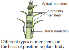
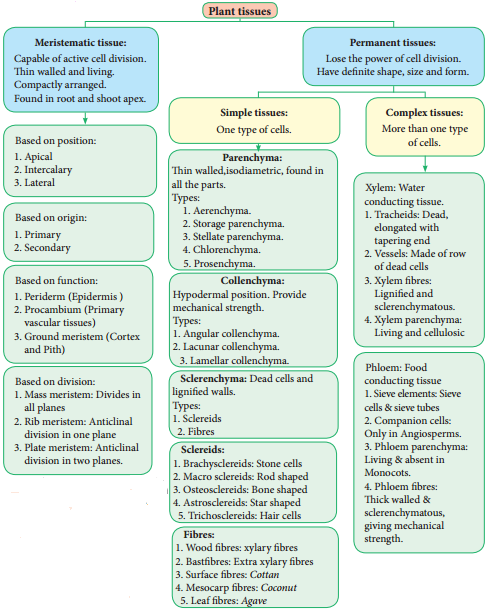
The Permanent tissues develop from apical meristem. They lose the power of cell division either permanently or temporarily. They are classified into two types:

  1. Simple permanent tissues.
  2. Complex permanent tissues.

Simple Permanent Tissues

Simple tissues are composed of one type of cells only. The cells are structurally and functionally similar. It is of three types.

  1. Parenchyma
  2. Collenchyma
  3. Sclerenchyma

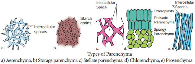
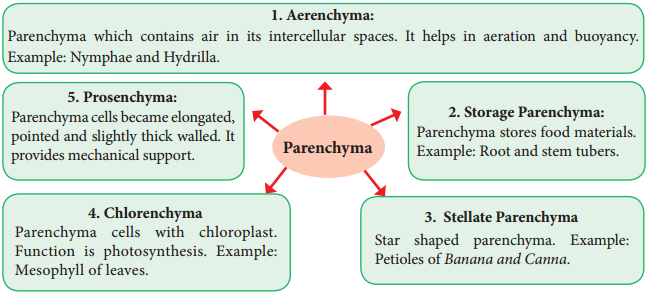
Parenchyma (Gk: Para-beside; enehein-to pour)

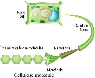
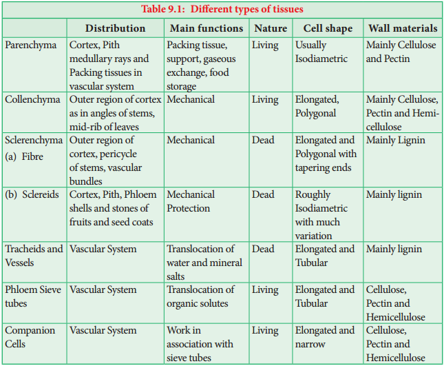
Parenchyma is generally present in all organs of the plant. It forms the ground tissue in a plant. Parenchyma is a living tissue and made up of thin walled cells. The cell wall is made up of cellulose. Parenchyma cells may be oval, polyhedral, cylindrical, irregular, elongated or armed. The tissue normally has prominent intercellular spaces and may store various types of materials like, water, air, ergastic substances.
Permanent Tissues img 1

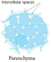
Occasionally Parenchyma cells which store resin, tannins, crystals of calcium carbonate, calcium oxalate are called idioblasts. Parenchyma is of different types and some of them are discussed as follows.
Permanent Tissues img 2
Permanent Tissues img 3

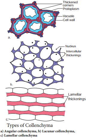
Collenchyma (Gk. Colla-glue; enchyma – an infusion)

Collenchyma is a simple, living mechanical tissue. Collenchyma generally occurs in hypodermis of dicot stem. It is absent in the roots and also occurs in petioles and pedicels. The cells are elongated and appear polygonal in cross section.

The cell wall is unevenly thickened. It contains more of hemicellulose and pectin besides cellulose. It provides mechanical support and elasticity to the growing parts of the plant. Collenchyma consists of narrow cells. It has only a few small chloroplast or none. Tannin maybe present in collenchyma. Based on pattern of pectinisation of the cell wall, there are three types of collenchyma.
Permanent Tissues img 4
Permanent Tissues img 5

Sclerenchyma (Gk. Sclerous – hard: enchyma-an infusion)

The sclerenchyma is dead cell and lacks protoplasm. The cells are long or short, narrow thick walled and lignified secondary walls.
The cell walls of these cells are uniformly and strongly thickened. sclerenchymatous cells are of two types:

  1. Sclereids
  2. Fibres

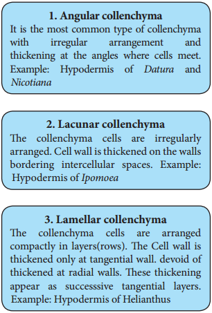
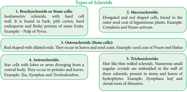
Sclereids (Stone Cells)

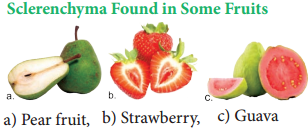
Sclereids are dead cells, usually these are isodiametric but some are elongated too. The cell wall is very thick due to lignification. Lumen is very much reduced. The pits may simple or branched. Sclereids are mechanical in function. They give hard texture to the seed coats, endosperms etc., Sclereids are classified into the following types.
Permanent Tissues img 6
Permanent Tissues img 7
Permanent Tissues img 8

Fibres

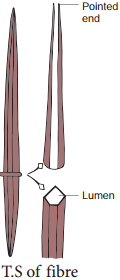
Fibres are very much elongated sclerenchyma cells with pointed tips. Fibres are dead cells and have lignified walls with narrow lumen. They have simple pits. They provide mechanical strength and protect them from the strong wind. It is also called supporting tissues. Fibres have a great commercial value in cottage and textile industries.

Fibres are of five types

1. Wood Fibres or Xylary Fibres

These fibres are associated with the secondary xylem tissue. They are also called xylary fibres. These fibres are derived from the vascular cambium. These are of two types.

  • Libriform Fibres
  • Fibre Tracheids

2. Bastfibres or Extra Xylary Fibres

These fibres are present in the phloem. Natural Bast fibres are strong and cellulosic. Fibres obtaining from the phloem or outer bark of jute, kenaf, flax and hemp plants. The so called pericyclic fibres are actually phloem fibres.
Permanent Tissues img 9

3. Surface Fibres

These fibres are produced from the surface of the plant organs. Cotton and silk cotton are the examples. They occur in the testa of seeds.

4. Mesocarp Fibres

Fibres obtained from the mesocarp of drupes like coconut.

5. Leaf Fibres

Fibres obtained from the leaf of Musa, Agave and Sensciveria.

Fibres in Our Daily Life

Economically fibres may be grouped as follows:-

1. Textile Fibres:

Fibres utilized for the manufacture of fabrics, netting and cordage etc.

Surface Fibres:
Example: Cotton

a. Surface Fibres:
Example: Cotton

b. Soft Fibres:
Example: Flax, Jute and Ramie

c. Hard Fibres:
Example: Sisal, Coconut, Pineapple, Abaca etc.

2. Brush Fibre:

Fibres utilized for the manufacture of brushes and brooms.

3. Rough Weaving Fibres:

Fibres utilized in making baskets, chairs, mats etc.

4. Paper Making Fibres:

Wood fibres utilized for paper making.

5. Filling Fibres:

Fibres used for stuffing cushions, mattresses, pillows, furniture etc. Example: Bombax and Silk cotton.

Complex Tissues

A complex tissue is a tissue with several types of cells but all of them function together as a single unit. It is of two types – xylem and phloem.

Xylem or Hadrome

The xylem is the principal water conducting tissue in a vascular plant. The term xylem was introduced by Nageli (1858) and is derived from the Gk. Xylos – wood. The xylem which is derived from Procambium is called primary xylem and the xylem which is derived from vascular cambium is called secondary xylem. Early formed primary xylem elements are called protoxylem, whereas the later formed primary xylem elements are called metaxylem.

Protoxylem lies towards the periphery and metaxylem that lies towards the centre is called Exarch. It is common in roots. Protoxylem lies towards the centre and meta xylem towards the periphery this condition is called Endarch. It is seen in stems. Protoxylem is located in the centre surrounded by the metaxylem is called Centrarch. In this type only one vascular strand is developed. Example: Selaginella sp.

Protoxylem is located in the centre surrounded by the metaxylem is called Mesarch. In this type several vascular strands are developed. Example: Ophioglossum sp

Xylem Consists of Four Types of Cells

  1. Tracheids
  2. Vessels or Trachea
  3. Xylem Parenchyma
  4. Xylem Fibres

1. Tracheids

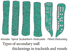
Tracheids are dead, lignified and elongated cells with tapering ends. Its lumen is broader than that of fibres. In cross section, the tracheids are polygonal. There are different types of cell wall thickenings due to the deposition of secondary wall substances.

They are annular (ring like), spiral (spring like), scalariform (ladder like) reticulate (net like) and pitted (uniformly thick except at pits). Tracheids are imperforated cells with bordered pits on their side walls. Only through this conduction takes place in Gymnosperms. They are arranged one above the other. Tracheids are chief water conducting elements in Gymnosperms and Pteridophytes. They also offer mechanical support to the plants.
Permanent Tissues img 10

2. Vessels or Trachea

Vessels are elongated tube like structure. They are dead cells formed from a row of vessel elements placed end to end. They are perforated at the end walls. Their lumen is wider than Tracheids. Due to the dissolution of entire cell wall, a single pore is formed at the perforation plate. It is called simple perforation plate, Example: Mangifera. If the perforation plate has many pores, it is called multiple perforation plate. Example Liriodendron.

The secondary wall thickening of vessels are annular, spiral, scalariform, reticulate, or pitted as in tracheids, Vessels are chief water conducting elements in Angiosperms and absent in Pteridophytes and Gymnosperms. In Gnetum of Gymnosperm, vessels occur. The main function is conduction of water, minerals and also offers mechanical strength.

3. Xylem Fibre

The fibres of sclerenchyma associated with the xylem are known as xylem fibres. Xylem fibres are dead cells and have lignified walls with narrow lumen. They cannot conduct water but being stronger provide mechanical strength. They are present in both primary and secondary xylem. Xylem fibres are also called libriform fibres.

The fibres are abundantly found in many plants. They occur in patches, in continuous bands and sometimes singly among other cells. Between fibres and normal tracheids, there are many transitional forms which are neither typical fibres nor typical tracheids. The transitional types are designated as fibretracheids. The pits of fibre-tracheids are smaller than those of vessels and typical tracheids.

4. Xylem Parernchyma

The parenchyma cells associated with the xylem are known as xylem parenchyma. These are the only living cells in xylem tissue. The cell wall is thin and made up of cellulose. Parenchyma arranged longitudinally along the long axis is called axial parenchyma Ray parenchyma is arranged in radial rows. Secondary xylem consists of both axial and ray parenchyma, Parenchyma stores food materials and also helps in conduction
of water.

Phloem to Leptome

Phloem is the food conducting complex tissues of vascular plants. The term phloem was coined by C. Nageli (1858). The Phloem which is derived from procambium is called primary phloem and the phloem which is derived from vascular cambium is called secondary phloem. Early formed primary phloem elements are called protophloem whereas the later formed primary phloem elements are called metaphloem. Protophloem is short lived. It gets crushed by the developing metaphloem.

Phloem Consists of Four Types of Cells

  1. Sieve elements
  2. Companion cells
  3. Phloem Parenchyma
  4. Phloem Fibres

1. Sieve Elements

Sieve elements are the conducting elements of the phloem. They are of two types, namely sieve cells and sieve tubes.

Sieve Cells

These are primitive type of conducting elements found in Pteridophytes and Gymnosperms. Sieve cells have sieve areas on their lateral walls only. They are not associated with companion cells.

Sieve Tubes

Sieve tubes are long tube like conducting elements in the phloem. These are formed from a series of cells called sieve tube elements. The sieve tube elements are arranged one above the other and form vertical sieve tube. The end wall contains a number of pores and it looks like a sieve.

So it is called as sieve plate. The sieve elements show nacreous thickenings on their lateral walls. They may possess simple or compound sieve plates. The function of sieve tubes are believed to be controlled by campanion cells.

In mature sieve tube, nucleus is absent. It contains a lining layer of cytoplasm. A special protein (P. Protein = Phloem Protein) called slime body is seen in it. In mature sieve tubes, the pores in the sieve plate are blocked by a substance called callose (callose plug). The conduction of food material takes place through cytoplasmic strands. Sieve tubes occur only in Angiosperms.
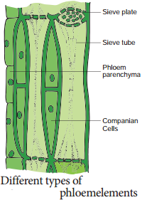
Permanent Tissues img 11

Companion Cells

The thin walled, elongated, specialized parenchyma cells, which are associated with the sieve elements, are called companion cells. These cells are living and they have cytoplasm and a prominent nucleus. They are connected to the sieve tubes through pits found in the lateral walls. Through these pits cytoplasmic connections are maintained between these elements. These cells are helpful in maintaining the pressure gradient in the sieve tubes.

Usually the nuclei of the companion cells serve for the nuclei of sieve tubes as they lack them. The companion cells are present only in Angiosperms and absent in Gymnosperms and Pteridophytes. They assist the sieve tubes in the conduction of food materials.

Phloem Parenchyma

The parenchyma cells associated with the phloem are called phloem parenchyma. These are living cells. They store starch and fats. They also contain resins and tannins in some plants. Primary phloem consists of axial parenchyma and secondary phloem consists of both axial and ray parenchyma. They are present in Pteridophytes, Gymnosperms and Dicots.

Phloem Fibres (or) Bast Fibres

The fibres of sclerenchyma associated with phloem are called phloem fibres or bast fibres. They are narrow, vertically elongated cells with very thick walls and a small lumen. Among the four phloem elements, phloem fibres are the only dead tissue. These are the strengthening as well as supporting cells.

Concept Map

Permanent Tissues img 12
Permanent Tissues img 13

Difference Between Meristematic Tissue and Permanent Tissue

Meristematic Tissue

Permanent Tissue

1. Cells Divide Repeatedly 1. Do Not Divide
2.  Cells are undifferentiated 2. Cells are fully differentiated
3. Cells are small and Isodiametric 3. Cells are variable in shape and size
4. Intercellular spaces are absent 4. Intercellular spaces are present
5. Vacuoles are absent 5. Vacuoles are present
6. Cell walls are thin 6. Cell walls are maybe thick or thin
7. Inorganic inclusions are absent 7. Inorganic inclusions are present


Difference Between Collenchyma and Schlerenchyma

Collenchyma

Schlerenchyma

1. Living Cells 1. Dead Cells
2. Contains Protoplasm 2. Do not have protoplasm
3. Cell walls are cellulosic 3. Cells walls are lignified
4. Thickening of cell wall is not uniform 4. Thickening of cell wall is uniform
5. Keeps the plant body soft 5. Keeps plant body stiff and hard
6. Sometimes it has chloroplast 6. Do not have chloroplast

Difference Between Fibre and Sclereids

Fibre

Sclereids

1. Long cells 1. Short cells
2. Narrow, Elongated pointed ends 2. Usually short and broad
3. Occurs in bundles 3. Occurs individually or in small groups
4. Commonly unbranched 4. Maybe branched
5. Derived directly from meristematic tissue 5. Develops from secondary sclerosis of parenchyma cells

Difference Between Tracheids and Fibres

Tracheids

Fibres

1. Not much elongated 1. Very long cells
2. Possess oblique end walls 2. Possess tapering end walls
3. Cell walls are not as thick as Fibres 3. Cell wall are thick and lignified
4. Possess various types of thickenings 4. Possess only pitted thickenings
5. Responsible for the conduction and also
mechanical support
5. Provide only mechanical support

Difference Between Sieve Cells and Sieve Tubes

Sieve Cells

Sieve Tubes

1. Have no companion cells 1. Have companion cells
2. The sieve areas do not form sieve plates 2. The sieve areas are confied to sieve plates
3. The sieve areas are not well differentiated 3. The sieve areas are well differentiated
4. They are elongated cells and are quite
long with tapering end walls
4. They consist of vertical cells placed one above the other forming long tubes connected at the walls by sieve pores
5. The sieve are smaller and numerous 5. The sieve pores are longer and fewer
6. Found in Pteridophytes and
Gymnosperms
6. Found in Angiosperms

Meristematic Tissue Definition, Characteristics and Classification

Learninsta presents the core concepts of Biology with high-quality research papers and topical review articles.

Meristematic Tissue Definition, Characteristics and Classification

Characteristics and Classification

The characters of meristematic tissues: (Gr. Meristos-Divisible)
Meristematic Tissue img 1

The term meristem was coined by C. Nageli 1858.

  • The meristematic cells are isodiametric and they may be, oval, spherical or polygonal in shape.
  • They generally have dense cytoplasm with prominent nucleus.
  • Generally the vacuoles are either small or absent.
  • Their cell wall is thin, elastic and made up of cellulose.
  • These are most actively dividing cells.
  • Meristematic cells are self-perpetuating.

Classification of Meristem

Meristem has been classified into several types on the basis of position, origin, function and division.

Theories of Meristem Organization and Function

Many anatomists illustrated the root and shoot apical meristems on the basis of number and arrangement and accordingly proposed the following theories – An extract of which is discussed below.

Shoot Apical Meristem
Apical Cell Theory

Apical cell theory is proposed by Hofmeister (1852) and supported by Nageli (1859). A single apical cell is the structural and functional unit.
Meristematic Tissue img 2

This apical cell governs the growth and development of whole plant body. It is applicable in Algae, Bryophytes and in some Pteridophytes.

Histogen Theory

Histogen theory is proposed by Hanstein (1868) and supported by Strassburgur. The shoot apex comprises three distinct zones.

1. Dermatogen:
It is the outermost layer. It gives rise to epidermis.

2. Periblem:
It is middle layer. That gives rise to cortex.

3. Plerome:
It is innermost layer. Which gives rise to stele

Tunica Corpus Theory

Tunica corpus theory is proposed by

A. Schmidt (1924).
Two zones of tissues are found in apical meris tem.

1. The tunica:
It is the peripheral zone of shoot apex, that forms epidermis.

2. The corpus:
It is the inner zone of shoot apex,that forms cortex and stele of shoot.

Root Apical Meristem

Root apex is present opposite to the shoot apex. The roots contain root cap at their apices and the apical meristem is present below the root cap. The different theories proposed to explain root apical meristem organization are given below.

Apical Cell Theory

Apical cell theory is proposed by Nageli. The single apical cell or apical initial composes the root meristem. The apical initial is tetrahedral in shape and produces root cap from one side. The remaining three sides produce epidermis, cortex and vascular tissues. It is found in vascular cryptogams.

Histogen Theory

Histogen theory is proposed by Hanstein (1868) and supported by Strassburgur. The histogen theory as appilied to the root apical meristem speaks of four histogen in the meristem. They are respectively,
Meristematic Tissue img 3

(i) Dermatogen:
It is the outermost layer. It gives rise to root epidermis.

(ii) Periblem:
It is the middle layer. It gives rise to cortex.

(iii) Plerome:
It is innermost layer. It gives rise to stele.

(iv) Calyptrogen:
It gives rise to root cap.

Korper Kappe Theory

Korper Kappe theory is proposed by Schuepp. There are two zones in root apex – Korper and Kappe

  1. Korper zone forms the body.
  2. Kappe zone forms the cap. This theory is equivalent to tunica corpus theory of shoot apex.
  3. The two divisions are distinguished by the type of T (also called Y) divisions.
  4. Korper is characterised by inverted T divisions and kappe by straight T divisions.

Quiescent Centre Concept

Quiescent centre concept was proposed by Clowes (1961) to explain root apical meristem activity. This centre is located between root cap and differentiating cells of the roots. The apparently inactive region of cells in root promeristem is called quiescent centre. It is the site of hormone synthesis and also the ultimate source of all meristematic cells of the meristem.

Nucleic Acids Definition, Structure, Features and its Types

Learninsta presents the core concepts of Biology with high-quality research papers and topical review articles.

Nucleic Acids Definition, Structure, Features and its Types

As we know DNA and RNA are the two kinds of nucleic acids. These were originally isolated from cell nucleus. They are present in all known cells and viruses with special coded genetic programme with detailed and specific instructions for each organism heredity.

DNA and RNA are polymers of monomers called nucleotides, each of which is composed of a nitrogen base, a pentose sugar and a phosphate. A purine or a pyrimidine and a ribose or deoxyribose sugar is called nucleoside. A nitrogenous base is linked to pentose sugar through n-glycosidic linkage and forms a nucleoside.

When a phosphate group is attached to a nucleoside it is called a nucleotide. The nitrogen base is a heterocyclic compound that can be either a purine (two rings) or a pyrimidine (one ring). There are 2 types of purines – adenine (A) and guanine (G) and 3 types of pyrimidines – cytosine (C), thymine (T) and uracil (U) (Figure 8.20 and 21).
Nucleic Acids img 1
Nucleic Acids img 2

A characteristic feature that differentiates DNA from RNA is that DNA contains nitrogen bases such as Adenine, guanine, thymine (5-methyl uracil) and cytosine and the RNA contains nitrogen bases such as adenine, guanine, cytosine and uracil instead of thymine.

The nitrogen base is covalently bonded to the sugar ribose in RNA and to deoxyribose (ribose with one oxygen removed from C2) in DNA. Phosphate group is a derivative of (PO43-) phosphoric acid, and forms phosphodiester linkages with sugar molecule (Figure 8.22).
Nucleic Acids img 3

Formation of Dinucleotide and Polynucleotide

Two nucleotides join to form dinucleotide that are linked through 3′- 5′ phosphodiester linkage by condensation between phosphate groups of one with sugar of other. This is repeated many times to makepolynucleotide.
Nucleic Acids img 4

Structure of DNA

Watson and Crick shared the Nobel Prize in 1962 for their discovery, along with Maurice Wilkins, who had produced the crystallographic data supporting the model.

Rosalind Franklin (1920 – 1958) had earlier produced the first clear crystallographic evidence for a helical structure. James Watson and Francis Crick of Cavendish laboratory in Cambridge built a scale model of double helical structure of DNA which is the most prevalent form of DNA, the B-DNA. This is the secondary structure of DNA.
Nucleic Acids img 5

As proposed by James Watson and Francis Crick, DNA consists of right handed double helix with 2 helical polynucleotide chains that are coiled around a common axis to form right handed B form of DNA. The coils are held together by hydrogen bonds which occur between complementary pairs of nitrogenous bases. The sugar is called 2′- deoxyribose because there is no hydroxyl at position 2′. Adenine and thiamine base pairs has two hydrogen bonds while guanine and cytosine base pairs have three hydrogen bonds.

As published by Erwin Chargaff in 1949, a purine pairs with pyrimidine and vice versa. Adenine (A) always pairs with Thymine (T) by double bond and Guanine (G) always pairs with Cytosine (C) by triple bond.

Features of DNA

If one strand runs in the 5′- 3′ direction, the other runs in 3′ – 5′ direction and thus are antiparallel (they run in opposite direction). The 5′ end has the phosphate group and 3’ end has the OH group.

The angle at which the two sugars protrude from the base pairs is about 120°, for the narrow angle and 240° for the wide angle. The narrow angle between the sugars generates a minor groove and the large angle on the other edge generates major groove.

Each base is 0.34 nm apart and a complete turn of the helix comprises 3.4 nm or 10 base pairs per turn in the predominant B form of DNA.

DNA helical structure has a diameter of 20A° and a pitch of about 34 A°. X-ray crystal study of DNA takes a stack of about 10 bp to go completely around the helix (360°).

Thermodynamic stability of the helix and specificity of base pairing includes:-

  1. The hydrogen bonds between the complementary bases of the double helix
  2. Atacking interaction between bases tend to stack about each other perpendicular to the direction of helical axis.
  3. Electron cloud interactions (π – π) between the bases in the helical stacks contribute to the stability of the double helix.

The phosphodiester linkages gives an inherent polarity to the DNA helix. They form strong covalent bonds, gives the strength and stability to the polynucleotide chain.
Nucleic Acids img 6

Plectonemic Coiling:

The two strands of the DNA are wrapped around each other in a helix, making it impossible to simply move them apart without breaking the entire structure. Whereas in paranemic coiling the two strands simply lie alongside one another, making them easier to pull apart.

Based on the helix and the distance between each turns, the DNA is of three forms – A DNA, B DNA and Z DNA (Figure 8.27).
Nucleic Acids img 7

Ribonucleic Acid (RNA)

Ribonucleic acid (RNA) is a polymeric molecule essential in various biological roles in coding, decoding, regulation and expression of genes. RNA is single stranded and is unstable when compared to DNA.
Nucleic Acids img 8

Types of RNA

mRNA (messenger RNA):
Single stranded, carries a copy of instructions for assembling amino acids into proteins. It is very unstable and comprises 5% of total RNA polymer. Prokaryotic mRNA (Polycistronic) carry coding sequences for many polypeptides. Eukaryotic mRNA (Monocistronic) contains information for only one polypeptide.

tRNA (transfer RNA):
Translates the code from mRNA and transfers amino acids to the ribosome to build proteins. It is highly folded into an elaborate 3D structure and comprises about 15% of total RNA. It is also called as soluble RNA.

rRNA (ribosomal RNA):
Single stranded, metabolically stable, make up the two subunits of ribosomes. It constitutes 80% of the total RNA. It is a polymer with varied length from 120-3000 nucleotides and gives ribosomes their shape. Genes for rRNA are highly conserved and employed for phylogenetic studies (Figure 8.28).
Nucleic Acids img 9

Enzymes Definition and its Types

Learninsta presents the core concepts of Biology with high-quality research papers and topical review articles.

Enzymes Definition and its Types

Enzymes are globular proteins that catalyse the many thousands of metabolic reactions taking place within cells and organism. The molecules involved in such reactions are metabolites. Metabolism consists of chains and cycles of enzyme-catalysed reactions, such as respiration, photosynthesis, protein synthesis and other pathways. These reactions are classified as:-

Anabolic (Building up of Organic Molecules):
Synthesis of proteins from amino acids and synthesis of polysaccharides from simple sugars are examples of anabolic reactions.

Catabolic (Breaking Down of larger Molecules):
Digestion of complex foods and the breaking down of sugar in respiration are examples of catabolic reactions (Figure 8.16).
Enzymes img 1

Enzymes can be extracellular enzyme as secreted and work externally exported from cells. Eg. digestive enzymes; or intracellular enzymes that remain within cells and work there. These are found inside organelles or within cells. Eg. insulin.

Properties of Enzyme

  • All are globular proteins.
  • They act as catalysts and effective even in small quantity.
  • They remain unchanged at the end of the reaction.
  • They are highly specific.
  • They have an active site where the reaction takes place.
  • Enzymes lower activation energy of the reaction they catalyse.

As molecules react, they become unstable, high energy intermediates. But they are in this transition state only momentarily. Energy is required to raise molecules to this transition state and this minimum energy needed is called the activation energy. This could be explained schematically by ‘boulder on hillside’ model of activation energy (Figure 8.17).
Enzymes img 2

Lock and Key Mechanism of Enzyme

In a enzyme catalysed reaction, the starting substance is the substrate. It is converted to the product. The substrate binds to the specially formed pocket in the enzyme – the active site, this is called lock and key mechanism of enzyme action.

As the enzyme and substrate form a ES complex, the substrate is raised in energy to a transition state and then breaks down into products plus unchanged enzyme (Figure 8.18).
Enzymes img 3

Enzyme Cofactors

Many enzymes require non-protein components called cofactors for their efficient activity. Cofactors may vary from simple inorganic ions to complex organic molecules. They are of three types: inorganic ions, prosthetic groups and coenzymes (Figure 8.19).
Enzymes img 4

Holoenzyme:
Active enzyme with its non protein component.

Apoenzyme:
The inactive enzyme without its non protein component.

Inorganic Ions

Help to increase the rate of reaction catalysed by enzymes. Example: Salivary amylase activity is increased in the presence of chloride ions.

Prosthetic Groups

Are organic molecules that assist in catalytic function of an enzyme. Flavin adenine dinucleotide (FAD) contains riboflavin (vit B2), the function of which is to accept hydrogen. ‘Haem’ is an iron-containing prosthetic group with an iron atom at its centre.

Coenzymes are Organic Compounds

Which act as cofactors but do not remain attached to the enzyme. The essential chemical components of many coenzymes are vitamins. Eg. NAD, NADP, Coenzyme A, ATP.

Classification of Enzymes
Enzymes are classified into six groups based on their mode of action.
Enzymes img 5

Uses of Enzymes

Enzyme

Source

Application

Bacterial protease Bacillus Biological detergents
Bacterial glucose isomerase Bacillus Fructose syrup manufacture
Fungal lactase Kluyvero-myces Breaking down of lactose to glucose and galactose
Amylases Aspergillus Removal of starch in woven cloth production

Proteins Definition and its Various Types

Learninsta presents the core concepts of Biology with high-quality research papers and topical review articles.

Proteins Definition and its Various Types

Proteins are the most diverse of all macromolecule. Proteins make up 2/3 of total dry mass of a cell. The term protein was coined by Gerardus Johannes Mulder and is derived form a greek word proteos which means of the first rank.

Amino acids are building blocks of proteins. There are about 20 different amino acids exist naturally. All amino acids have a basic skeleton consisting of a carbon (a-carbon) linked to a basic amino group.
Proteins img 1

(NH2), an acidic carboxylic group (COOH) and a hydrogen atom (H) and side chain or variable R group. The amino acid is both an acid and a base and hence is called amphoteric. A zwitterion also called as dipolar ion, is a molecule with two or more functional groups, of which at least one has a positive and other has a negative electrical charge and the net charge of the entire molecule is zero. The pH at which this happens is known as the isoelectric point (Figure 8.10).
Proteins img 2

Classification of Amino Acids

Based on the R group amino acids are classified as acidic, basic, polar, non-polar. The amino group of one amino acid reacts with carboxyl group of other amino acid, forming a peptide bond. Two amino acids can react together with the loss of water to form a dipeptide. Long strings of amino acids linked by peptide bonds are called polypeptides. In 1953, Fred Sanger first sequenced the Insulin protein (Figure 8.11 a and b).
Proteins img 3
Proteins img 4

Structure of Protein

Protein are synthesised on the ribosome as a linear sequence of amino acids which are held together by peptide bonds. After synthesis, the protein attains conformational change into a specific 3D form for proper functioning. According to the mode of folding, four levels of protein organisation have been recognised namely primary, secondary, tertiary and quaternary (Figure 8.12).
Proteins img 5

The Primary Structure:
Is linear arrangement of amino acids in a polypeptide chain.

Secondary Structure:
Arises when various functional groups are exposed on outer surface of the molecular interaction by forming hydrogen bonds. This causes the aminoacid chain to twist into coiled configuration called α-helix or to fold into a flat β-pleated sheets.

Tertiary Protein Structure:
Arises when the secondary level proteins fold into globular structure called domains.

Quaternary Protein Structure:
May be assumed by some complex proteins in which more than one polypeptide forms a large multiunit protein. The individual polypeptide chains of the protein are called subunits and the active protein itself is called a multimer.

For example:
Enzymes serve as catalyst for chemical reactions in cell and are non-specific. Antibodies are complex glycoproteins with specific regions of attachment for various organisms.

Protein Denaturation

Denaturation is the loss of 3D structure of protein. Exposure to heat causes atoms to vibrate violently, and this disrupts the hydrogen and ionic bonds. Under these conditions, protein molecules become elongated, disorganised strands. Agents such as soap, detergents, acid, alcohol and some disinfectants disrupt the interchain bond and cause the molecule to be non-functional (Figure 8.13).
Proteins img 6

Protein Bonding

There are four types of chemical bonds
Proteins img 7

Hydrogen Bond

It is formed between some hydrogen atoms of oxygen and nitrogen in polypeptide chain. The hydrogen atoms have a small positive charge and oxygen and nitrogen have small negative charge. Opposite charges attract to form hydrogen bonds. Though these bonds are weak, large number of them maintains the molecule in 3D shape.

Ionic Bond

It is formed between any charged groups that are not joined together by peptide bond. It is stronger than hydrogen bond and can be broken by changes in pH and temperature.

Disulfide Bond

Some amino acids like cysteine and methionine have sulphur. These form disulphide bridge between sulphur atoms and amino acids.

Hydrophobic Bond

This bond helps some protein to maintain structure. When globular proteins are in solution, their hydrophobic groups point inwards away from water.

Test for Proteins

The biuret test is used as an indicator for presence of protein as it gives a purple colour in the presence of peptide bonds (-C-N-). To protein solution, an equal quantity of sodium hydroxide solution is added and mixed. Then a few drops of 0.5% copper (II) sulphate is added with gentle mixing. A distinct purple colour develops without heating (Figure 8.15 a and b).
Proteins img 8
Proteins img 9

Lipids and its Various Types

Learninsta presents the core concepts of Biology with high-quality research papers and topical review articles.

Lipids and its Various Types

The term lipid is derived from greek word lipos, meaning fat. These substances are not soluble in polar solvent such as water but soluble in non-polar solvents such as benzene, ether, chloroform. This is because they contain long hydrocarbon chains that are non-polar and thus are hydrophobic. The main groups of compounds classified as lipids are triglycerides, phospholipids, steroids and waxes.

Triglycerides

Triglycerides are composed of single molecule of glycerol bound to 3 fatty acids. These include fats and oils. Fatty acids are long chain hydrocarbons with a carboxyl group at one end which binds to one of the hydroxyl groups of glycerol, thus forming an ester bond. Fatty acids are structural unit of lipids and are carboxylic acid of long chain hydrocarbons. The hydrocarbon can vary in length from 4 – 24 carbons and the fat may be saturated or unsaturated.

In saturated fatty acids the hydrocarbon chain is single bonded (Eg. palmitic acid, stearic acid) and in unsaturated fatty acids (Eg. oleic acid, linoleic acid) the hydrocarbon chain is double bonded (one/two/three). In general solid fats are saturated and oils are unsaturated, in which most are globules.

Lipids are molecules that contain hydrocarbons and make up the building blocks of the structure and function of living cells. Examples of lipids include fats, oils, waxes, certain vitamins (such as A, D, E and K), hormones and most of the cell membrane that is not made up of protein.

The Four Main Groups of Lipids Include:

  • Fatty acids (saturated and unsaturated)
  • Glycerides (glycerol-containing lipids)
  • Nonglyceride lipids (sphingolipids, steroids, waxes)
  • Complex lipids (lipoproteins, glycolipids)

A lipid is any of various organic compounds that are insoluble in water. They include fats, waxes, oils, hormones, and certain components of membranes and function as energy-storage molecules and chemical messengers.

Fats and lipids are an essential component of the homeostatic function of the human body. Lipids contribute to some of the body’s most vital processes. Lipids are fatty, waxy, or oily compounds that are soluble in organic solvents and insoluble in polar solvents such as water.

Examples of lipids include fats, oils, waxes, certain vitamins (such as A, D, E and K), hormones and most of the cell membrane that is not made up of protein. Lipids are not soluble in water as they are non-polar, but are thus soluble in non-polar solvents such as chloroform.

The main difference between lipids and fats is that lipids are a broad group of biomolecules whereas fats are a type of lipids. Fat is stored in the adipose tissue and under the skin of animals. It is mainly used as an energy-storage molecule in the body. Most steroids in the body serve as hormones.

Lipids are an important part of the body, along with proteins, sugars, and minerals. They can be found in many parts of a human: cell membranes, cholesterol, blood cells, and in the brain, to name a few ways the body uses them.

Within the body, lipids function as an energy reserve, regulate hormones, transmit nerve impulses, cushion vital organs, and transport fat-soluble nutrients. Fat in food serves as an energy source with high caloric density, adds texture and taste, and contributes to satiety.

Most people have high levels of fat in their blood because they eat too much high-fat food. Some people have high fat levels because they have an inherited disorder. High lipid levels may also be caused by medical conditions such as diabetes, hypothyroidism, alcoholism, kidney disease, liver disease and stress.

Lipids play diverse roles in the normal functioning of the body: they serve as the structural building material of all membranes of cells and organelles. they provide energy for living organisms – providing more than twice the energy content compared with carbohydrates and proteins on a weight basis.

The body uses three main nutrients to function – carbohydrate, protein, and fat. These nutrients are digested into simpler compounds. Carbohydrates are used for energy (glucose). Fats are used for energy after they are broken into fatty acids.
Lipids img 1

Carbohydrates and its Types

Learninsta presents the core concepts of Biology with high-quality research papers and topical review articles.

Carbohydrates and its Types

Carbohydrates are organic compounds made of carbon and water. Thus one molecule of water combines with a carbon atom to form CH2O and is repeated several (n) times to form (CH2O)n where n is an integer ranging from 3-7. These are also called as saccharides. The common term sugar refers to a simple carbohydrate such as a monosaccharide or disaccharide that tastes sweet are soluble in water (Figure 8.7).
Carbohydrates and its Types img 1

Monosaccharides – The Simple Sugars

Monosaccharides are relatively small molecules constituting single sugar unit. Glucose has a chemical formula of C6H12O6. It is a six carbon molecule and hence is called as hexose.

All monosaccharides contain one or two functional groups. Some are aldehydes, Eg: glucose and are referred as aldoses; other are ketones, Eg: Fructose and are referred as Ketoses.
Carbohydrates and its Types img 2

Disaccharides

Disaccharides are formed when two monosaccharides join together. An example is sucrose. Sucrose is formed from a molecule of α-glucose and a molecule of fructose. This is a condensation reaction releasing water. The bond formed between the glucose and fructose molecule by removal of water is called glycosidic bond. This is another example of strong, covalent bond.
Carbohydrates and its Types img 3

In the reverse process, a disaccharide is digested to the component monosaccharide in a hydrolysis reaction. This reaction involves addition of a water (hydro) molecule and splitting (lysis) of the glycosidic bond.

Polysaccharides

These are made of hundreds of monosaccharide units. Polysaccharides also called “Glycans”. Long chain of branched or unbranched monosaccharides are held together by glycosidic bonds. Polysaccharide is an example of giant molecule, a macromolecule and consists of only one type of monomer. Polysaccharides are insoluble in water and are sweetless. Cellulose is an example built from repeated units of glucose monomer (Figure 8.6).
Carbohydrates and its Types img 4

Depending on the function, polysaccharides are of two types – storage polysaccharide and structural polysaccharide.

Starch

Starch is a storage polysaccharide made up of repeated units of amylose and amylopectin. Starch grains are made up of successive layers of amylose and amylopectin, which can be seen as growth rings. Amylose is a linear, unbranched polymer which makes up 80% of starch. Amylopectin is a polymer with some 1, 6 linkages that gives it a branched structure.

Test for Starch

We test the presence of starch by adding a solution of iodine in potassium iodide. Iodine molecules fit nearly into the starch helix, producing a blue-black colour.
Carbohydrates and its Types img 5

  • Test on potato
  • Test on starch at varied concentrations
  • Starch – iodine reaction

Celluloses

Cellulose is a structural polysaccharide made up of thousands of glucose units. In this case, β-glucose units are held together by 1, 4 glycosidic linkage, forming long unbranched chains. Cellulose fibres are straight and uncoiled. It has many industrial uses which include cellulose fibres as cotton, nitrocellulose for explosives, cellulose acetate for fibres of multiple uses and cellophane for packing (Figure 8.7).
Carbohydrates and its Types img 6

Chitin

Chitin is a homo polysaccharide with amino acids added to form mucopolysaccharide. The basic unit is a nitrogen containing glucose derivative known as N-acetyl glucosamine. It forms the exoskeleton of insects and other arthropods. It is also present in the cell walls of fungi (Figure 8.8).
Carbohydrates and its Types img 7

Test for Reducing Sugars

Aldoses and ketoses are reducing sugars. This means that, when heated with an alkaline solution of copper (II) sulphate (a blue solution called benedict’s solution), the aldehyde or ketone group reduces Cu2+ ions to Cu+ ions forming brick red precipitate of copper(I) oxide.

In the process, the aldehyde or ketone group is oxidised to a carboxyl group (-COOH). This reaction is used as test for reducing sugar and is known as Benedict’s test. The results of benedict’s test depends on concentration of the sugar. If there is no reducing sugar it remains blue.

  • Sucrose is not a reducing sugar
  • The greater the concentration of reducing sugar, the more is the precipitate formed and greater is the colour change.

Other Sugar Compounds

Other Polysaccharides

Structure

Functions

Inulin Polymer of fructose It is not metabolised in the human body and is readily filtered through the kidney
Hyaluronic acid Heteroplayer of d glucuronic acid and D-N acetyl glucosamine It accounts for the toughness and flexibility of cartilage and tenson
Agar Mucopolysaccharide from red algae Used as solidifying agent in culture medium in laboratory
Heparin Glycosamino glycan contains variably sulphated disaccharide unit present in liver Used as an anticoagulant
Chondroitin sulphate Sulphated glycosaminoglycan composed of altering sugars (N-acetylglucosamine and glucuronic acid) Distery supplement for treatment of osteoarthritis
Keratan sulphate Sulphated glycosaminoglycan and is a structural carbohydrate Acts as cushion to absorb mechanical shock

Carbohydrates and its Types img 8

Primary and Secondary Metabolites

Learninsta presents the core concepts of Biology with high-quality research papers and topical review articles.

Primary and Secondary Metabolites

Most plants, fungi and other microbes synthesizes a number of organic compounds called as metabolites which are intermediates and products of metabolism. The term metabolite is usually restricted to small molecules. It can be catergorized into two types namely primary and secondary metabolites based on their role in metabolic process (Figure 8.4).
Primary and Secondary Metabolites img 1

Primary Metabolites

Are those that are required for the basic metabolic processes like photosynthesis, respiration, protein and lipid metabolism of living organisms.

Secondary Metabolites

Does not show any direct function in growth and development of organisms.
Primary and Secondary Metabolites img 2

Organic Molecules

Organic molecules may be small and simple. These simple molecules assemble and form large and complex molecules called macromolecules. These include four main classes – carbohydrates, lipids, proteins and nucleic acids. All macromolecules except lipids are formed by the process of polymerisation, a process in which repeating subunits termed monomers are bound into chains of different lengths. These chains of monomers are called polymers.

A primary metabolite is a kind of metabolite that is directly involved in normal growth, development, and reproduction. A secondary metabolite is typically present in a taxonomically restricted set of organisms or cells (plants, fungi, bacteria, etc).

The main difference between primary metabolites and secondary metabolites is that primary metabolites are directly involved in primary growth development and reproduction whereas secondary metabolites are indirectly involved in metabolisms while playing important ecological functions in the body.

Some common examples of primary metabolites include: ethanol, lactic acid, and certain amino acids. In higher plants such compounds are often concentrated in seeds and vegetative storage organs and are needed for physiological development because of their role in basic cell metabolism.

Examples of primary metabolites include proteins, enzymes, carbohydrates, lipids, vitamins, ethanol, lactic acid, butanol, etc. Some examples of secondary metabolites include steroids, essential oils, phenolics, alkaloids, pigments, antibiotics, etc.

Examples of secondary metabolites include antibiotics, pigments and scents. Secondary metabolites are produced by many microbes, plants, fungi and animals, usually living in crowded habitats, where chemical defense represents a better option than physical escape.

Metabolites are intermediate end products of metabolism. Primary metabolites are essential for the proper growth of microorganisms. Secondary metabolites are formed near the stationary phase of growth and are not involved in growth, reproduction and development.

The antibiotics are defined as “the complex chemical substances, the secondary metabolites which are produced by microorganisms and act against other microorganisms”. Those microorganisms which have capacity to produce more antibiotics can survive for longer time than the others producing antibiotics in less amount.

Definition:
A primary pollutant is an air pollutant emitted directly from a source. A secondary pollutant is not directly emitted as such, but forms when other pollutants (primary pollutants) react in the atmosphere.

Secondary metabolites are compounds that are not required for the growth or reproduction of an organism but are produced to confer a selective advantage to the organism. For example, they may inhibit the growth of organisms with which they compete and, as such, they often inhibit biologically important processes.

Water Importance and its Properties

Learninsta presents the core concepts of Biology with high-quality research papers and topical review articles.

Water Importance and its Properties

Water is the most abundant component in living organisms. Life on earth is inevitably linked to water. Water makes up 70% of human cell and upto 95% of mass of a plant cell (Figure 8.2).
Water img 1

Chemistry of Water

Water is a tiny polar molecule that can readily pass through membranes. Two electronegative atoms of oxygen share a hydrogen bonds of two water molecule. Thus, they can stick together by cohesion and results in lattice formation (Figure 8.3).
Water img 2

Properties of Water

  • Adhesion and Cohesion Property
  • High Latent Heat of Vaporisation
  • High Melting and Boiling Point
  • Universal Solvent
  • Specific Heat Capacity

Water is very important to the human body. Every one of your cells, organs and tissues use water to help with temperature regulation, keeping hydrated and maintaining bodily functions. In addition, water acts as a lubricant and cushions your joints. Drinking water is great for your overall health.

Our bodies use water in all the cells, organs, and tissues, to help regulate body temperature and maintain other bodily functions. Because our bodies lose water through breathing, sweating, and digestion, it’s crucial to rehydrate and replace water by drinking fluids and eating foods that contain water.

Your body uses water to sweat, urinate, and have bowel movements. Sweat regulates body temperature when you’re exercising or in warm temperatures. You need water to replenish the lost fluid from sweat. You also need enough water in your system to have healthy stool and avoid constipation.

Uses of Water

  • For Drinking
  • For Cleaning Dishes
  • For Cooking
  • For Watering Plants
  • For Washing Clothes
  • For Bathing
  • For Generation of Hydroelectricity
  • For Washing Car

It is said that too much consumption of water can lead to fluid overload in the body and imbalance in the body. Excess water can lead to lower sodium levels in the body, which may further lead to nausea, vomiting, cramps, fatigue, etal. This condition is known as hyponatremia.

It’s important to drink enough water during the day, however, it can be disruptive if you drink directly before bed. Avoid drinking water or any other fluids at least two hours before sleeping to prevent waking up at night.

Water helps your kidneys remove waste from your blood. If you don’t get enough water, that waste – along with acids – can build up. That can lead to your kidneys getting clogged up with proteins called myoglobin. Dehydration can also lead to kidney stones and urinary tract infections.

Eliminating food and water intake for a significant period of time is also known as starvation. Your body can be subject to starvation after a day or two without food or water. At that time, the body starts functioning differently to reduce the amount of energy it burns. Eventually, starvation leads to death.

Cell Division and its Difference Phases

Learninsta presents the core concepts of Biology with high-quality research papers and topical review articles.

Cell Division and its Difference Phases

Amitosis (Direct Cell Division)

Amitosis is also called direct or incipient cell division. Here there is no spindle formation and chromatin material does not condense. It consist of two steps: (Figure 7.2).
Cell Division img 1

Karyokinesis:

  • Involves division of nucleus.
  • Nucleus develops a constriction at the center and becomes dumbell shaped.
  • Constriction deepens and divides the nucleus into two.

Cytokinesis:

  • Involves division of cytoplasm.
  • Plasma membrane develops a constriction along nuclear constriction.
  • It deepens centripetally and finally divides the cell into two cells.

Example:
Cells of mammalian cartilage, macronucleus of Paramecium and old degenerating cells of higher plants.

Drawbacks of Amitosis

  • Causes unequal distribution of chromosomes.
  • Can lead to abnormalities in metabolism and reproduction.

Mitosis

Mitosis occurs in shoot and root tips and other meristematic tissues of plants associated with growth. The number of chromosomes in the parent and the daughter (Progeny) cells remain the same so it is also called as equational division.

Closed and Open Mitosis

In closed mitosis, the nuclear envelope remains intact and chromosomes migrate to opposite poles of a spindle within the nucleus (Figure 7.3). Example: Many single celled eukaryotes including yeast and slime molds. In open mitosis, the nuclear envelope breaks down and then reforms around the 2 sets of separated chromosome. Example: Most plants and animals.
Cell Division img 2

Mitosis is divided into four stages prophase, metaphase, anaphase and telophase (Figure 7.6).
Cell Division img 3

Prophase

Prophase is the longest phase in mitosis. Chromosomes become visible as long thin thread like structure, condenses to form compact mitotic chromosomes. In plant cells initiation of spindle fires takes place, nucleolus disappears. Nuclear envelope breaks down. Golgi apparatus and endoplasmic reticulum disappear.

In animal cell the centrioles extend a radial array of microtubules (Figure 7.4) and reach the poles of the cell. This arrangement of microtubules is called an aster. Plant cells do not form asters.
Cell Division img 4

Metaphase

Chromosomes (two sister chromatids) are attached to the spindle fires by kinetochore of the centromere. The spindle fires are made up of tubulin. The alignment of chromosome into compact group at the equator of the cell is known as metaphase plate.

This is the stage where the chromosomal morphology can be easily studied. Kinetochore is a DNA-Protein complex present at the centromere where microtubules are attached. It is a trilaminar disc like plate.

Anaphase

Each chromosome splits simultaneously and two daughter chromatids begin to migrate towards two opposite poles of a cell. Each centromere splits longitudinally into two, freeing the two sister chromatids from each other. When sister chromatids separate the actual partitioning of the replicated genome is complete.

APC (Anaphase Promoting Complex) is a cluster of proteins that induces the breaking down of cohesion proteins which leads to the separation of chromatids during mitosis (Figure 7.5). This it helps in the transition of metaphase to anaphase.
Cell Division img 5

Telophase

Two sets of daughter chromosomes reach opposite poles of the cell and mitotic spindle disappears. Division of genetic material is completed during karyokinesis, followed by cytokinesis (division of cytoplasm). Nucleolus and nuclear membranes reforms. Nuclear membrane form around each set of chromosomes. Now the chromosomes decondense.

In plants, phragmoplast are formed between the daughter cells. Cell plate is formed between the two daughter cells, reconstruction of cell wall takes place. Finally cells are separated by the distribution of organelles, macromolecules into two newly formed daughter cells.

Cytokinesis

Cytokinesis in Animal Cells It is a contractile process. The ring consists of a bundle of microfilaments assembled from actin and myosin. This firil generates a contractile force, that draws the ring inward forming a cleavage furrow in the cell. This it divides the cell into two.

Cytokinesis in Plant Cell

Division of the cytoplasm often starts during telophase. In plants, cell plate grows from centre towards lateral walls. Phragmoplast contains microtubules, actin filaments and vesicles from golgi apparatus and ER. Microtubule of the pharagmoplast move to the equator, fuse to form a new plasma membrane and the materials which are placed there becomes new cell wall.

The first stage of cell wall construction is a line dividing the newly forming cells called a cell plate. The cell plate eventually stretches right across the cell forming the middle lamella. Cellulose builds up on each side of the middle lamella to form the cell walls of two new plant cells.

Significance of Mitosis

Exact copy of the parent cell is produced by mitosis (genetically identical).

1. Genetic stability:
Daughter cells are genetically identical to parent cells.

2. Growth:
As multicellular organisms grow, the number of cells making up their tissue increases. The new cells must be identical to the existing ones.

3. Repair of Tissues:
Damaged cells must be replaced by identical new cells by mitosis.

4. Asexual Reproduction:
Asexual reproduction results in offspring that are identical to the parent. Example Yeast and Amoeba.

5. Flowering Plants:
In flowering plants, structure such as bulbs, corms, tubers, rhizomes and runners are produced by mitotic division. When they separate from the parent, they form a new individual.

The production of large numbers of offsprings in a short period of time, is possible only by mitosis. In genetic engineering and biotechnology, tissues are grown by mitosis (i.e. in tissue culture).

6. Regeneration:
Arms of star fish.

Meiosis

In Greek meioum means to reduce. Meiosis is unique because of synapsis, homologous recombination and reduction division. Meiosis takes place in the reproductive organs. It results in the formation of gametes with half the normal chromosome number.

Haploid sperms are made in testes; haploid eggs are made in ovaries of animals. In flowering plants meiosis occurs during microsporogenesis in anthers and megasporogenesis in ovule. In contrast to mitosis, meiosis produces cells that are not genetically identical. So meiosis has a key role in producing new genetic types which results in genetic variation.

Stages in Meiosis

Meiosis can be studied under two divisions i.e., meiosis I and meiosis II. As with mitosis, the cell is said to be in interphase when it is not dividing.

Meiosis I:
Reduction Division

Prophase I:
Prophase I is of longer duration and it is divided into 5 substages – Leptotene, Zygotene, Pachytene, Diplotene and Diakinesis (Figure 7.7).
Cell Division img 6

Leptotene:
Chromosomes are visible under light microscope. Condensation of chromosomes takes place. Paired sister chromatids begin to condense.

Zygotene:
Pairing of homologous chromosomes takes place and it is known as synapsis. Chromosome synapsis is made by the formation of synaptonemal complex. The complex formed by the homologous chromosomes are called as bivalent (tetrads).

Pachytene:
At this stage bivalent chromosomes are clearly visible as tetrads. Bivalent of meiosis I consists of 4 chromatids and 2 centromeres. Synapsis is completed and recombination nodules appear at a site where crossing over takes place between non-sister chromatids of homologous chromosome. Recombination of homologous chromosomes is completed by the end of the stage but the chromosomes are linked at the sites of crossing over. This is mediated by the enzyme recombinase.

Diplotene:
Synaptonemal complex disassembled and dissolves. The homologous chromosomes remain attached at one or more points where crossing over has taken place. These points of attachment where ‘X’ shaped structures occur at the sites of crossing over is called Chiasmata.

Chiasmata are chromatin structures at sites where recombination has been taken place. They are specialised chromosomal structures that hold the homologous chromosomes together.

Sister chromatids remain closely associated whereas the homologous chromosomes tend to separate from each other but are held together by chiasmata. This substage may last for days or years depending on the sex and organism.

Diakinesis:
Terminalisation of chiasmata, homologous chromosomes become short and condensed. Nucleolus and nuclear envelope disappears. Spindle fires assemble.

Metaphase I

Spindle fires are attached to the centromeres of the two homologous chromosomes. Bivalent (pairs of homologous chromosomes) aligned at the equator of the cell known as metaphase plate. The random distribution of homologous chromosomes in a cell in Metaphase I is called independent assortment.

Anaphase I

Homologous chromosomes are separated from each other by shortening of spindle fiers. Each homologous chromosomes with its two chromatids and undivided centromere move towards the opposite poles of the cells. The actual reduction in the number of chromosomes takes place at this stage. Homologous chromosomes which move to the opposite poles are either paternal or maternal in origin. Sister chromatids remain attached with their centromeres.

Telophase I

Haploid set of chromosomes are present at each pole. The formation of two daughter cells, each with haploid number of chromosomes takes place. Nuclei reassembled. Nuclear envelope forms around the chromosome and the chromosomes becomes uncoiled. Nucleolus reappears.

In plants after karyokinesis, cytokinesis takes place by which two daughter cells are formed by the cell plate between 2 groups of chromosomes known as dyad of cells (haploid). The stage between the two meiotic divisions is called interkinesis which is short-lived.

Meiosis II:
Equational Division

This division is otherwise called mitotic meiosis because it resembles mitosis. Since it includes all the stages of mitotic divisions.

Prophase II

The chromosome with 2 chromatids becomes short, condensed, thick and becomes visible. New spindle develops at right angles to the cell axis. Nuclear membrane and nucleolus disappear.

Metaphase II

Chromosome arranged at the equatorial plane of the spindle. Microtubules of spindle gets attached to the centromere of sister chromatids.

Anaphase II

Sister chromatids separate. The daughter chromosomes move to the opposite poles due to shortening of spindle fires. Centromere of each chromosome split, allowing to move towards opposite poles of the cells holding the sister chromatids.

Telophase II

Four groups of chromosomes are organised into four haploid nuclei. The spindle disappears. Nuclear envelope, nucleolus reappear. After karyokinesis, cytokinesis follows and four haploid daughter cells are formed, called tetrads.

Signifiance of Meiosis

  • This maintains a definite constant number of chromosomes in organisms.
  • Crossing over takes place and exchange of genetic material leads to variations among species.
  • These variations are the raw materials to evolution.
  • Meiosis leads to genetic variability by partitioning different combinations of genes into gametes through independent assortment.
  • Adaptation of organisms to various environmental stress.

Cell Division img 7

Difference Between Mitosis in Plants and Animals

Plants

Animals

Centrioles are absent Centrioles are present
Asters are not formed Asters are formed
Cell division involves the formation of a cell plate Cell division involves furrowing and cleavage of cytoplasm
Occurs mainly at meristem Occurs in tissues throughout the body


Mitosis

Meiosis

One division Two divisions
Number of chromosome remain the same Number of chromosomes is halved
Homologous chromosomes line up
separately on the metaphase plate
Homologous chromosomes line up in pairs at the
metaphase plate
Homologous chromosome do not pair up Homologous chromosome pairup to form bivalent
Chiasmata do not form and crossing over
never occurs
Chiasmata form and crossingover occurs
Daughter cells are genetically identical Daughter cells are genetically diffrent from parent cell
Two daughter cells are formed Four daughter cells are formed

Cell Cycle Definition and its Types

Learninsta presents the core concepts of Biology with high-quality research papers and topical review articles.

Cell Cycle Definition and its Types

Definition:
A series of events leading to the formation of new cell is known as cell cycle. The series of events include several phases.

History of a Cell

Year

Scientist  

Events

1665 Robert Hooke Coined word “Cell”
1670-74 Anthony van Laeeuwenhoek First living cells observed in microscope – Structure of bacteria
1831-33 Robert Brown Presence of nucleus in cells of orchid roots
1839 Jan Evangelista Purkyne (J.E. Purkinje) Coined “Protoplasm”
1838-39 Schleiden & Schwann Cell theory
1858 Rudolph Ludwig Carl Virchow Cell theory ‘omnis cellula e cellula’
1873 Anton Schneider Described chromosomes (Nuclear filaments) for the first time
1882 Walther Flemming Coined the word mitosis; chromosome behaviour
1883 Edouard Van Beneden Cell division in round worm
1888 Theodor Boveri Centrosome; Chromosome Theory

Duration of Cell Cycle

Different kinds of cells have varied duration for cell cycle phases. Eukaryotic cell divides every 24 hours. The cell cycle is divided into mitosis and interphase. In a cell cycle 95% is spent for interphase whereas the mitosis and cytokinesis last only for an hour.

Cell Cycle of a Proliferating Human Cell

Interphase
The different phases of cell cycle are as follows (Figure 7.1).

Phase

Time Duration (in hrs)

G1 11
S 8
G2 4
M 1

Longest part of the cell cycle, but it is of extremely variable length. At first glance the nucleus appears to be resting but this is not the case at all. The chromosomes previously visible as thread like structure, have dispersed. Now they are actively involved in protein synthesis, at least for most of the interphase.

G1 Phase

The first gap phase – 2C amount of DNA in cells of G1. Cells become metabolically active and grows by producing proteins, lipids, carbohydrates and cell organelles including mitochondria and endoplasmic reticulum. Many checkpoints control the cell cycle.

The check point are also called as the restriction point. First check point at the end of G1, determines a cells fate whether it will continue in the cell cycle and divide or enter a stage called G0 a quiescent stage, probably as specified cell or die. Cells are arrested in G1 due to:

  • Nutrient deprivation
  • Lack of growth factors or density dependant inhibition
  • Undergo metabolic changes and enter into G0 state.

Biochemicals inside cell activates the cell division. The proteins called kinases and cyclins activate genes and their proteins to perform cell division. Cyclins act as major checkpoint which operates in G1 to determine whether or not a cell divides.

G0 Phase

Some cells exit G1 and enters a quiescent stage called G0, where the cell remains metabolically active without proliferation. Cells can exist for long periods in G0 phase. In G0 cells cease growth with reduced rate of RNA and protein synthesis.

The G0 phase is not permanent. Mature neuron and skeletal muscle cell remain permanently in G0. Many
cells in animals remains in G0 unless called on to proliferate by appropriate growth factors or other extracellular signals. G0 cells are not dormant. S phase – Synthesis phase – cells with intermediate amounts of DNA.

Growth of the cell continues as replication of DNA occur, protein molecules called histones are synthesised and attach to the DNA. The centrioles duplicate in the cytoplasm. DNA content increases from 2C to 4C. G2 – The second Gap phase – 4C amount of DNA in cells of G2 and mitosis

Cell growth continues by protein and cell organelle synthesis, mitochondria and chloroplasts divide. DNA content remains as 4C. Tubulin is synthesised and microtubules are formed. Microtubles organise to form spindle fire. The spindle begins to form and nuclear division follows.

One of the proteins synthesized only in the G2 period is known as Maturation Promoting Factor (MPF). It brings about condensation of interphase chromosomes into the mitotic form. DNA damage checkpoints operates in G1S and G2 phases of the cell cycle.