Food Additives of Chemistry In Everday Life

Find free online Chemistry Topics covering a broad range of concepts from research institutes around the world.

Food Additives of Chemistry In Everday Life

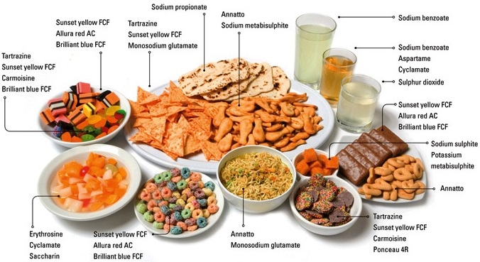
Have you ever noticed the ingredients that is printed on the cover of the packed food materials such as biscuits, chocolates etc… You might have noticed that emulsifiers such as 322, 472E, dough conditioners 223 etc… are used in the preparation, in addition to the main ingredients such as wheat flour, edible oil, sugar, milk solid etc… Do you think that these substances are necessary? Yes. These substances enhance the nutritive, sensory and practical value of the food.

They also increase the shelf life of food. The substances which are not naturally a part of the food and added to improve the quality of food are called food additives.

Important Categories of Food Additives

  • Aroma compounds
  • Food colours
  • Preservatives
  • Stabilizers
  • Artificial Sweeteners
  • Antioxidants
  • Buffering substances
  • Vitamins and minerals

Advantages of Food Additives:

  1. Uses of preservatives reduce the product spoilage and extend the shelf-life of food
  2. Addition of vitamins and minerals reduces the mall nutrient
  3. Flavouring agents enhance the aroma of the food
  4. Antioxidants prevent the formation of potentially toxic oxidation products of lipids and other food constituents

Preservatives:

Preservatives are capable of inhibiting, retarding or arresting the process of fermentation, acidification or other decomposition of food by growth of microorganisms. Organic acids such as benzoic acid, sorbic acid and their salts are potent inhibitors of a number of fungi, yeast and bacteria. Alkyl esters of hydroxy benzoic acid are very effective in less acidic conditions. Acetic acid is used mainly as a preservative for the preparation of pickles and for preserved vegetables.

Sodium metasulphite is used as preservatives for fresh vegetables and fruits. Sucrose esters with palmitic and steric acid are used as emulsifiers. In addition that some organic acids and their salts are used as preservatives. In addition to chemical treatment, physical methods such as heat treatment (pasteurisation and sterilisations), cold treatment (chilling and freezing) drying (dehydration) and irradiation are used to preserve food.

Antioxidants:

Antioxidants are substances which retard the oxidative deteriorations of food. Food containing fats and oils is easily oxidised and turn rancid. To prevent the oxidation of the fats and oils, chemical BHT (butylhydroxy toluene), BHA(Butylated hydroxy anisole) are added as food additives.

They are generally called antioxidants. These materials readily undergo oxidation by reacting with free radicals generated by the oxidation of oils, thereby stop the chain reaction of oxidation of food. Sulphur dioxide and sulphites are also used as food additives. They act as anti-microbial agents, antioxidants and enzyme inhibitors.

Sugar Substituents:

These compounds that are used like sugars (glucose, sucrose) for sweetening, but are metabolised without the influence of insulin are called sugar substituents. Eg. Sorbitol, Xylitol, Mannitol.

Artificial Sweetening Agents:

Synthetic compounds which imprint a sweet sensation and possess no or negligible nutritional value are called artificial sweeteners. Eg. Saccharin, Aspartame, sucralose, alitame etc…

Food Additives of Chemistry In Everday Life img 1

Chemistry In Everday Life of Drug and its Types

Find free online Chemistry Topics covering a broad range of concepts from research institutes around the world.

Chemistry In Everday Life of Drug and its Types

The word drug is derived from the French word “drogue” meaning “dry herb”. A drug is a substance that is used to modify or explore physiological systems or pathological states for the benefit of the recipient. It is used for the purpose of diagnosis, prevention, cure/relief of a disease.

The drug which interacts with macromolecular targets such as proteins to produce a therapeutic and useful biological response is called medicine. The specific treatment of a disease using medicine is known as chemotherapy. An ideal drug is the one which is nontoxic, bio-compatible and bio-degradable, and it should not have any side effects.

Generally, most of the drug molecules that are used now a days have the above properties at lower concentrations. However, at higher concentrations, they have side effects and become toxic. The medicinal value of a drug is measured in terms of its therapeutic index, which is defined as the ratio between the maximum tolerated dose of a drug (above which it become toxic) and the minimum curative dose (below which the drug is ineffctive). Higher the value of therapeutic index, safer is the drug.

Classification of Drugs:

Drugs are classified based on their properties such as chemical structure, pharmacological effect, target system, site of action etc. We will discuss some general classifications here.

Classification Based on the Chemical Structure:

In this classification, drugs with a common chemical skeleton are classified into a single group. For example, ampicillin, amoxicillin, methicillin etc.. all have similar structure and are classified into a single group called penicillin.

Similarly, we have other group of drugs such as opiates, steroids, catecholamines etc. Compounds having similar chemical structure are expected to have similar chemical properties. However, their biological actions are not always similar. For example, all drugs belonging to penicillin group have same biological action, while groups such as barbiturates, steroids etc.. have different biological action.

Penicillins

Chemistry In Everday Life of Drug and its Types img 1

Classification Based on Pharmacological Effect:

In this classification, the drugs are grouped based on their biological effect that they produce on the recipient. For example, the medicines that have the ability to kill the pathogenic bacteria are grouped as antibiotics.

This kind of grouping will provide the full range of drugs that can be used for a particular condition (disease). The physician has to carefully choose a suitable medicine from the available drugs based on the clinical condition of the recipient.

Examples:

Antibiotic drugs: amoxicillin, ampicillin,cefiime, cefpodoxime, erythromycin, tetracycline etc..
Antihypertensive drugs: propranolol, atenolol, metoprolol succinate, amlodipine etc…

Classification Based on the Target System (Drug Action):

In this classifiation, the drugs are grouped based on the biological system/process, that they target in the recipient. This classification is more specific than the pharmacological classification. For example, the antibiotics streptomycin and erythromycin inhibit the protein synthesis (target process) in bacteria and are classified in a same group. However, their mode of action is different. Streptomycin inhibits the initiation of protein synthesis, while erythromycin prevents the incorporation of new amino acids to the protein.

Classification Based on the Site of Action (Molecular Target):

The drug molecule interacts with biomolecules such as enzymes, receptors etc,, which are referred as drug targets. We can classify the drug based on the drug target with which it binds. This classification is highly specific compared to the others. These compounds often have a common mechanism of action, as the target is the same.

Drug-Target Interaction:

The biochemical processes such as metabolism (which is responsible for breaking down the food molecules and harvest energy in the form of ATP and biosynthesis of necessary biomolecules from the available precursor molecules using many enzymes), cell-signaling (senses any change in the environment using the receptor molecules and send signals to various processes to elicit an appropriate response) etc… are essential for the normal functioning of our body.

These routine processes may be disturbed by any external factors such as microorganism, chemicals etc.. or by a disorder in the system itself. Under such conditions we may have to take medicines to restore the normal functioning of the body.

These drug molecules interact with biomolecules such as proteins, lipids, etc.. that are responsible for different functions of the body. For example, proteins which act as biological catalysts are called enzymes and those which are important for communication systems are called receptors. The drug interacts with these molecules and modify the normal biochemical reactions either by modifying the enzyme activity or by stimulating/suppressing certain receptors.

Enzymes as Drug Targets:

In all living systems, the biochemical reactions are catalysed by enzymes. Hence, these enzyme actions are highly essential for the normal functioning of the system. If their normal enzyme activity is inhibited, then the system will be affected. T is principle is usually applied to kill many pathogens.

We have already learnt that in enzyme catalysed reactions, the substrate molecule binds to the active site of the enzyme by means of the weak interaction such as hydrogen bonding, van der Waals force etc… between the amino acids present in the active site and the substrate. When a drug molecule that has a similar geometry (shape) as the substrate is administered, it can also bind to the enzyme and inhibit its activity.In other words, the drug acts as an inhibitor to the enzyme catalyst.

These type of inhibitors are of en called competitive inhibitors. For example the antibiotic sulphanilamide, which is structurally similar to p-aminobenzoic acid (PABA) inhibits the bacterial growth. Many bacteria need PABA in order to produce an important coenzyme, folic acid.

When the antibiotic sulphanilamide is administered, it acts as a competitive inhibitor to the enzyme dihydropteroate synthase (DHPS) in the biosynthetic pathway of converting PABA into folic acid in the bacteria. It leads to the folic acid deficiency which retards the growth of the bacteria and can eventually kill them.

Chemistry In Everday Life of Drug and its Types img 2

In certain enzymes, the inhibitor molecule binds to a different binding site, which is commonly referred to as allosteric site, and causes a change in its active site geometry (shape). As a result, the substrate cannot bind to the enzyme. This type of inhibitors are called allosteric inhibitors.

Receptor as Drug Targets:

Many drugs exert their physiological effects by binding to a specifi molecule called a receptor whose role is to trigger a response in a cell. Most of the receptors are integrated with the cell membranes in such a way that their active site is exposed to outside region of the cell membrane.

The chemical messengers, the compounds that carry messages to cells, bind to the active site of these receptors. This brings about the transfer of message into the cell. These receptors show high selectivity for one chemical messenger over the others.

If we want to block a message, a drug that binds to the receptor site should inhibit its natural function. Such drugs are called antagonists. In contrast, there are drugs which mimic the natural messenger by switching on the receptor. These type of drugs are called agonists and are used when there is lack of chemical messenger.

Chemistry In Everday Life of Drug and its Types img 3

For example, when adenosine binds to the adenosine receptors, it induces sleepiness. On the other hand, the antagonist drug caffine binds to the adenosine receptor and makes it inactive. This results in the reduced sleepiness (wakefulness). The agonist drug, morphine, which is used as a pain killer, binds to the opioid receptors and activates them. This supress the neuro transmitters that causes pain.

Chemistry In Everday Life of Drug and its Types img 4

Thrapeutic Action of Different Classes of Drugs:

The developments in the field of biology allowed us to understand various biological process and their mechanism in detail. This enabled to develop new safer efficient drugs. For example, to treat acidity, we have been using weak bases such as aluminium and magnesium hydroxides. But these can make the stomach alkaline and trigger the production of much acid. Moreover, This treatment only relives the symptoms and does not control the cause.

Detailed studies reveal that histamines stimulate the secretion of HCl by activating the receptor in the stomach wall. This findings lead to the design of new drugs such as cimetidine, ranitidine etc.. which binds the receptor and inactivate them. These drugs are structurally similar to histamine. In this section, we shall discuss the therapeutic action of a few important classes of drugs.

Chemistry In Everday Life of Drug and its Types img 5

Chemistry In Everday Life of Drug and its Types img 6

Chemistry In Everday Life of Drug and its Types img 7

Chemistry In Everday Life of Drug and its Types img 8

Chemistry In Everday Life of Drug and its Types img 9

Biomolecules of Hormones

Find free online Chemistry Topics covering a broad range of concepts from research institutes around the world.

Biomolecules of Hormones

Hormone is an organic substance (e.g. a peptide or a steroid) that is secreted by one tissue. it limits the blood stream and induces a physiological response (e.g. growth and metabolism) in other tissues. It is an intercellular signalling molecule.

Virtually every process in a complex organism is regulated by one or more hormones: maintenance of blood pressure, blood volume and electrolyte balance, embryogenesis, hunger, eating behaviour, digestion – to name but a few.

Endocrine glands, which are special groups of cells, make hormones. The major endocrine glands are the pituitary, pineal, thymus, thyroid, adrenal glands, and pancreas. In addition, men produce hormones in their testes and women produce them in their ovary.

Chemically, hormones may be classified as either protein (e.g. insulin, epinephrine) or steroids (e.g. estrogen, androgen). Hormones are classified according to the distance over which they act as, endocrine, paracrine and autocrine hormones.

Endocrine hormones act on cells distant from the site of their release. Example: insulin and epinephrine are synthesized and released in the bloodstream by specialized ductless endocrine glands.

Paracrine hormones (alternatively, local mediators) act only on cells close to the cell that released them. For example, interleukin-1 (IL-1).

Autocrine hormones act on the same cell that released them. For example, protein growth factor interleukin-2 (IL-2).

Biomolecules of Hormones img 1

Only those cells with a specific receptor for a given hormone will respond to its presence even though nearly all cells in the body may be exposed to the hormone. Hormonal messages are therefore quite specifically addressed.

Most commonly, hormones are categorized into four structural groups, with members of each group having many properties in common:

  • Peptides and proteins
  • Steroids
  • Amino acid derivatives
  • Fatty acid derivatives – Eicosanoids

Some hormones that are products of endocrine glands are proteins or peptides, others are steroids. (The origin of hormones, their physiological role, and their mode of action are dealt with in the article hormone). None of the hormones has any enzymatic activity.

Some examples of protein hormones include growth hormone, which is produced by the pituitary gland, and follicle-stimulating hormone (FSH), which has an attached carbohydrate group and is thus classified as a glycoprotein.

Chemically, hormones may be classified as either proteins or steroids. All of the hormones in the human body, except the sex hormones and those from the adrenal cortex, are proteins or protein derivatives.

Biomolecules of Hormones img 2

Nucleic Acids Types and its Functions

Find free online Chemistry Topics covering a broad range of concepts from research institutes around the world.

Nucleic Acids Types and its Functions

The inherent characteristics of each and every species are transmitted from one generation to the next. It has been observed that the particles in nucleus of the cell are responsible for the transmission of these characteristics.

They are called chromosomes and are made up of proteins and another type of biomolecules called nucleic acids. There are mainly two types nucleic acids, the deoxyribonucleic acid (DNA) and ribonucleic acid (RNA). They are the molecular repositories that carry genetic information in every organism.

Composition and Structure of Nucleic Acids

Nucleic acids are biopolymers of nucleotides. Controlled hydrolysis of DNA and RNA yields three components namely a nitrogenous base, a pentose sugar and phosphate group.

Nitrogen Base

These are nitrogen containing organic compounds which are derivatives of two parent compounds, pyrimidine and purine. Both DNA and RNA have two major purine bases, adenine (A) and guanine (G). In both DNA and RNA, one of the pyrimidines is cytosine (C), but the second pyrimidine is thymine (T) in DNA and uracil (U) in RNA.

Nucleic Acids Types and its Functions img 1

Pentose Sugar:

Nucleic acids have two types of pentoses. The recurring deoxyribonucleotide units of DNA contain 2’-deoxy-D-ribose and the ribonucleotide units of RNA contain D-ribose. In nucleotides, both types of pentoses are in their β-furanose (closed five membered rings) form.

Nucleic Acids Types and its Functions img 2

Phosphate Group

Phosphoric acid forms phospho diester bond between nucleotides. Based on the number of phosphate group present in the nucleotides, they are classified into mono nucleotide, dinucleotide and trinucleotide.

Nucleosides and Nucleotides:

The molecule without the phosphate group is called a nucleoside. A nucleotide is derived from a nucleoside by the addition of a molecule of phosphoric acid. Phosphorylation occurs generally in the 5’ OH group of the sugar. Nucleotides are linked in DNA and RNA by phospho diester bond between 5’ OH group of one nucleotide and 3’ OH group on another nucleotide.

Sugar + Base → Nucleoside
Nucleoside + Phosphate → Nucleotide
nNucleotide → Polynucleotide (Nucleic Acid)

Double Strand Helix Structure of DNA

In early 1950s, Rosalind Franklin and Maurice Wilkins used X-ray diffraction to unravel the structure of DNA. The DNA fibers produced a characteristic diffraction pattern. The central X shaped pattern indicates a helix, whereas the heavy black arcs at the top and bottom of the diffraction pattern reveal the spacing of the stacked bases.

Nucleic Acids Types and its Functions img 3

The structure elucidation of DNA by Watson and Crick in 1953 was a momentous event in science. They postulated a 3-dimensional model of DNA structure which consisted of two antiparallel helical DNA chains wound around the same axis to form a right-handed double helix.

The hydrophilic backbones of alternating deoxyribose and phosphate groups are on the outside of the double helix, facing the surrounding water. The purine and pyrimidine bases of both strands are stacked inside the double helix, with their hydrophobic and ring structures very close together and perpendicular to the long axis, thereby reducing the repulsions between the charged phosphate groups. The offet pairing of the two strands creates a major groove and minor groove on the surface of the duplex.

Nucleic Acids Types and its Functions img 4

Nucleic Acids Types and its Functions img 5

The model revealed that, there are 10.5 base pairs (36 Å) per turn of the helix and 3.4 Å between the stacked bases. They also found that each base is hydrogen bonded to a base in opposite strand to form a planar base pair.

Two hydrogen bonds are formed between adenine and thymine and three hydrogen bonds are formed between guanine and cytosine. Other pairing tends to destabilize the double helical structure. This specific association of the two chains of the double helix is known as complementary base pairing. The DNA double helix or duplex is held together by two forces,

  • Hydrogen bonding between complementary base pairs
  • Base-stacking interactions

The complementary between the DNA strands is attributable to the hydrogen bonding between base pairs but the base stacking interactions are largely non-specific, make the major contribution to the stability of the double helix.

Types of RNA Molecules

Ribonucleic acids are similar to DNA. Cells contain up to eight times high quantity of RNA than DNA. RNA is found in large amount in the cytoplasm and a lesser amount in the nucleus. In the cytoplasm it is mainly found in ribosomes and in the nucleus, it is found in nucleolus.

RNA molecules are classified according to their structure and function into three major types

  • Ribosomal RNA (rRNA)
  • Messenger RNA (mRNA)
  • Transfer RNA (tRNA)

rRNA

rRNA is mainly found in cytoplasm and in ribosomes, which contain 60% RNA and 40% protein. Ribosomes are the sites at which protein synthesis takes place.

tRNA

tRNA molecules have lowest molecular weight of all nucleic acids. They consist of 73 – 94 nucleotides in a single chain. The function of tRNA is to carry amino acids to the sites of protein synthesis on ribosomes.

mRNA

mRNA is present in small quantity and very short lived. They are single stranded, and their synthesis takes place on DNA. The synthesis of mRNA from DNA strand is called transcription. mRNA carries genetic information from DNA to the ribosomes for protein synthesis. This process is known as translation.

Difference between DNA and RNA

DNA

RNA

It is mainly present in nucleus, mitochondria and chloroplast It is mainly present in cytoplasm, nucleolus and ribosomes
It contains deoxyribose sugar It contains ribose sugar
Base pair A = T. G = C Base pair A = U. C = G
Double standard molecules Single standard molecules
It’s life time is high It is Short lived
It is stable and hot hydrolysed easily by alkalis It is unstable and hydrolysed easily by alkalis
It can replicate itself It cannot replicate itself. It is formed from DNA.

More to Know

DNA finger printing Traditionally, one of the most accurate methods for placing an individual at the scene of a crime has been a fingerprint. With the advent of recombinant DNA technology, a more powerful tool is now available: DNA fingerprinting is (also called DNA typing or DNA profiling).

It was first invented by Professor Sir Alec Jeffrey sin 1984. The DNA finger print is unique for every person and can be extracted from traces of samples from blood, saliva, hair etc… By using this method we can detect the individual specific variation in human DNA.

In this method, the extracted DNA is cut at specific points along the strand with restriction of enzymes resulting in the formation of DNA fragments of varying lengths which were analysed by technique called gel electrophoresis. This method separates the fragments based on their size.

The gel containing the DNA fragments is then transferred to a nylon sheet using a technique called blotting. Then, the fragments will undergo autoradiography in which they were exposed to DNA probes (pieces of synthetic DNA that were made radioactive and that bound to the fragments).

A piece of X-ray film was then exposed to the fragments, and a dark mark was produced at any point where a radioactive probe had become attached. The resultant pattern of marks could then be compared with other samples. DNA fingerprinting is based on slight sequence differences (usually single base-pair changes) between individuals. These methods are proving decisive in court cases worldwide.

Nucleic Acids Types and its Functions img 6

Biological Functions of Nucleic Acids

In addition to their roles as the subunits of nucleic acids, nucleotides have a variety of other functions in every cell such as,

(i) Energy Carriers (ATP)

Nucleic Acids Types and its Functions img 7

(ii) Components of enzyme cofactors (Example: Coenzyme A, NAD+, FAD)

Nucleic Acids Types and its Functions img 8

(iii) Chemical messengers (Example: Cyclic AMP, cAMP)

Nucleic Acids Types and its Functions img 9

Biomolecules of Vitamins and Their Functions

Find free online Chemistry Topics covering a broad range of concepts from research institutes around the world.

Biomolecules of Vitamins and Their Functions

Vitamins are small organic compounds that cannot be synthesised by our body but are essential for certain functions. Hence, they must be obtained through diet. The requirements of these compounds are not high, but their deficiency or excess can cause diseases. Each vitamin has a specific function in the living system, mostly as co enzymes.

They are not served as energy sources like carbohydrates, lipids, etc. The name ‘Vitamin’ is derived from ‘vital amines’, referring to the vitamins earlier identified amino compounds. Vitamins are essential for the normal growth and maintainance of our health.

Classification of Vitamins

Vitamins are classified into two groups based on their solubility either in water or in fat.

Fat Soluble Vitamins:

These vitamins absorbed best when taken with fatty food and are stored in fatty tissues and livers. These vitamins do not dissolve in water. Hence they are called fat soluble vitamins. Vitamin A, D, E & K are fat-soluble vitamins.

Water Soluble Vitamins:

Vitamins B (B1, B2, B3, B5, B6, B7, B9 and B12) and C are readily soluble in water. On the contrary to fat soluble vitamins, these can’t be stored. The excess vitamins present will be excreted through urine and are not stored in our body. Hence, these two vitamins should be supplied regularly to our body. The missing numbers in B vitamins are once considered as vitamins but no longer considered as such, and the numbers that were assigned to them now form the gaps.

Table 14.2: Vitamins, their Sources, Functions and their Deficiency Disease

Biomolecules of Vitamins and Their Functions img 1
Biomolecules of Vitamins and Their Functions img 1a

A diverse range of biomolecules exist,

Including:

Small Molecules:

Lipids, fatty acids, glycolipids, sterols, monosaccharides. Vitamins.

Biological Function of Vitamins

Once growth and development are completed, vitamins remain essential nutrients for the healthy maintenance of the cells, tissues, and organs that make up a multicellular organism; they also enable a multicellular life form to efficiently use chemical energy provided by food it eats, and to help process
the proteins, All of the biomolecules that make up our cells are made up of strings of monomers.

For example, proteins are made up of strings of amino acids and nucleic acids are strings of nucleotides. The term for a long string of monomers is a polymer. The biomolecules, proteins, carbohydrates and nucleic acids are all polymers.

Vitamins and minerals are considered essential nutrients-because acting in concert, they perform hundreds of roles in the body. They help shore up bones, heal wounds, and bolster your immune system. They also convert food into energy, and repair cellular damage.

Vitamin, any of several organic substances that are necessary in small quantities for normal health and growth in higher forms of animal life. Vitamins are distinct in several ways from other biologically important compounds such as proteins, carbohydrates, and lipids.

Biomolecules of Lipids:

Find free online Chemistry Topics covering a broad range of concepts from research institutes around the world.

Biomolecules of Lipids:

Lipids are organic molecules that are soluble in organic solvents such as chloroform and methanol and insoluble in water. The word lipid is derived from the Greek work ‘lipos’ meaning fat. They are the principal components of cell membranes. In addition, they also act as energy source for living systems. Fat provide 2-3 fold higher energy compared to carbohydrates/proteins.

Classification of Lipids:

Based on their structures lipids can be classified as simple lipids, compound lipids and derived lipids. Simple lipids can be further classified into fats, which are esters of long chain fatty acids with glycerol (triglycerides) and waxes which are the esters of fatty acids with long chain monohydric alcohols (Bees wax).

Compound lipids are the esters of simple fatty acid with glycerol which contain additional groups. Based on the groups attached, they are further classified into phospholipids, glycolipids and lipoproteins. Phospholipids contain a phospho-ester linkage while the glycolipids contain a sugar molecule attached. The lipoproteins are complexes of lipid with proteins.

Biological Importance of Lipids

  1. Lipids are the integral component of cell membrane. They are necessary of structural integrity of the cell.
  2. The main function of triglycerides in animals is as an energy reserve. They yield more energy than carbohydrates and proteins.
  3. They act as protective coating in aquatic organisms.
  4. Lipids of connective tissue give protection to internal organs.
  5. Lipids help in the absorption and transport of fat soluble vitamins.
  6. They are essential for activation of enzymes such as lipases.
  7. Lipids act as emulsifier in fat metabolism.

They include fats, waxes, oils, hormones, and certain components of membranes and function as energy-storage molecules and chemical messengers. Together with proteins and carbohydrates, lipids are one of the principal structural components of living cells.

Biological substances that are insoluble in water are classified as lipids. This characteristic physical property of lipids makes them very different from other biomolecules like carbohydrates, proteins, and nucleic acids. Some lipids are used to store energy.

The four main groups of lipids include:

  • Fatty acids (saturated and unsaturated)
  • Glycerides (glycerol-containing lipids)
  • Nonglyceride lipids (sphingolipids, steroids, waxes)
  • Complex lipids (lipoproteins, glycolipids)

Carbohydrates, nucleic acids, and proteins are often found as long polymers in nature. Lipids are not usually polymers and are smaller than the other three, so they are not considered macromolecules by some sources 1, 2 start superscript, 1, comma, 2, end superscript.

Fats and Oils. A fat molecule consists of two main components-glycerol and fatty acids. Glycerol is an organic compound (alcohol) with three carbons, five hydrogens, and three hydroxyl (OH) groups.

A lipid is any of various organic compounds that are insoluble in water. They include fats, waxes, oils, hormones, and certain components of membranes and function as energy-storage molecules and chemical messengers.

Biomolecules of Lipids img 1

Biomolecules of Proteins

Find free online Chemistry Topics covering a broad range of concepts from research institutes around the world.

Biomolecules of Proteins

Proteins are most abundant biomolecules in all living organisms. The term protein is derived from Greek word ‘Proteious’ meaning primary or holding first place. They are main functional units for the living things. They are involved in every function of the cell including respiration. Proteins are polymers of α-amino acids.

Amino Acids

Amino acids are compounds which contain an amino group and a carboxylic acid group. The protein molecules are made up a-amino acids which can be represented by the following general formula.

Biomolecules of Proteins img 1

There are 20 α-amino acids commonly found in the protein molecules. Each amino acid is given a trivial name, a three letter code and a one letter code. In writing the amino acid sequence of a protein, generally either one letter or three letter codes are used.

Classification of α-amino acids

The amino acids are classified based on the nature of their R groups commonly known as side chain. They can be classified as acidic, basic and neutral amino acids. They can also be classified as polar and non-polar (hydrophobic) amino acids.

Biomolecules of Proteins img 2

Amino acids can also be classified as essential and non-essential amino acids based on the ability to be synthesise by the human. The amino acids that can be synthesised by us are called non-essential amino acids (Gly, Ala, Glu, Asp, Gln, Asn, Ser, Cys, Tyr & Pro) and those needs to be obtained through diet are called essential amino acids (Phe, Val, Th, Trp, Ile, Met, His, Arg, Leu and Lys). These ten essential amino acids can be memorised by mnemonic called PVT TIM HALL.

Although the vast majority of plant and animal proteins are formed by these 20 α-amino acids, many other amino acids are also found in the cells. These amino acids are called as non-protein amino acids. Example: ornithine and citrulline (components of urea cycle where ammonia is converted into urea)

Properties of Amino Acid

Amino acids are colourless, water soluble crystalline solids. Since they have both carboxyl group and amino group their properties differ from regular amines and carboxylic acids. The carboxyl group can lose a proton and become negatively charged or the amino group can accept a proton to become positively charged depending upon the pH of the solution. At a specific pH the net charge of an amino acid is neutral and this pH is called isoelectric point. At a pH above the isoelectric point the amino acid will be negatively charged and positively charged at pH values below the isoelectric point.

In aqueous solution the proton from carboxyl group can be transferred to the amino group of an amino acid leaving these groups with opposite charges. Despite having both positive and negative charges this molecule is neutral and has amphoteric behaviour. These ions are called zwitter ions.

Biomolecules of Proteins img 3

Except glycine all other amino acids have at least one chiral carbon atom and hence are optically active. They exist in two forms namely D and L amino acids. However, L-amino acids are used predominantly by the living organism for synthesising proteins. Presence of D-amino acids has been observed rarely in certain organisms.

Peptide Bond Formation

The amino acids are linked covalently by peptide bonds. The carboxyl group of the first amino acid react with the amino group of the second amino acid to give an amide linkage between these amino acids. This amide linkage is called peptide bond.

The resulting compound is called a dipeptide. Addition an another amino acid to this dipeptide a second peptide bond results in tripeptide. This we can generate tetra peptide, penta peptide etc… When you have more number of amino acids linked this way you get a polypeptide. If the number of amino acids are less it is called as a polypeptide, if it has large number of amino acids (and preferably has a function) then it is called a protein.

Biomolecules of Proteins img 4

The amino end of the peptide is known as N-terminal or amino terminal while the carboxy end is called C-terminal or carboxy terminal. In general protein sequences are written from N-Terminal to C-Terminal. The atoms other than the side chains (R-groups) are called main chain or the back bone of the polypeptide.

Classification of Proteins

Proteins are classified based on their structure (overall shape) into two major types. They are fibrous proteins and globular proteins.

Fibrous Proteins

Fibrous proteins are linear molecules similar to fires. These are generally insoluble in water and are held together by disulphide bridges and weak intermolecular hydrogen bonds. The proteins are often used as structural proteins. Example: Keratin, Collagen etc…

Globular Proteins

Globular proteins have an overall spherical shape. The polypeptide chain is folded into a spherical shape. These proteins are usually soluble in water and have many functions including catalysis (enzyme). Example: myoglobin, insuline

Biomolecules of Proteins img 5

Structure of Proteins

Proteins are polymers of amino acids. Their three dimensional structure depends mainly on the sequence of amino acids (residues). The protein structure can be described at four hierarchal levels called primary, secondary, tertiary and quaternary structures as shown in the figure 14.16

1. Primary Structure of Proteins:

Proteins are polypeptide chains, made up of amino acids are connected through peptide bonds. The relative arrangement of the amino acids in the polypeptide chain is called the primary structure of the protein. Knowledge of this is essential as even small changes have potential to alter the overall structure and function of a protein.

Biomolecules of Proteins img 6

2. Secondary Structure of Proteins:

The amino acids in the polypeptide chain forms highly regular shapes (sub-structures) through the hydrogen bond between the carbonyl oxygen image 7 and the neighbouring amine hydrogen (-NH) of the main chain. α-Helix and β-strands or sheets are two most common sub-structures formed by proteins.

α-Helix

In the α-helix sub-structure, the amino acids are arranged in a right handed helical (spiral) structure and are stabilised by the hydrogen bond between the carbonyl oxygen of one amino acid (nth residue) with amino hydrogen of the fifth residue (n+4th residue). The side chains of the residues protrude outside of the helix. Each turn of an α-helix contains about 3.6 residues and is about 5.4 Å long. The amino acid proline produces a kink in the helical structure and often called as a helix breaker due to its rigid cyclic structure.

β-Strand

β-Strands are extended peptide chain rather than coiled. The hydrogen bonds occur between main chain carbonyl group one such strand and the amino group of the adjacent strand resulting in the formation of a sheet like structure. This arrangement is called β-sheets.

Biomolecules of Proteins img 7

3. Tertiary Structure:

The secondary structure elements (α-helix & β-sheets) further folds to form the three dimensional arrangement. This structure is called tertiary structure of the polypeptide (protein). Teritary structure of proteins are stabilised by the interactions between the side chains of the amino acids. These interactions include the disulphide bridges between cysteine residues, electrostatic, hydrophobic, hydrogen bonds and van der Waals interactions.

4. Quaternary Structure

Some proteins are made up of more than one polypeptide chains. For example, the oxygen transporting protein, haemoglobin contains four polypeptide chains while DNA polymerase enzyme that make copies of DNA, has ten polypeptide chains. In these proteins the individual polypeptide chains (subunits) interacts with each other to form the multimeric structure which are known as quaternary structure. The interactions that stabilises the tertiary structures also stabilises the quaternary structures.

Biomolecules of Proteins img 8

Denaturation of Proteins

Each protein has a unique three-dimensional structure formed by interactions such as disulphide bond, hydrogen bond, hydrophobic and electrostatic interactions. These interactions can be disturbed when the protein is exposed to a higher temperature, by adding certain chemicals such as urea, alteration of pH and ionic strength etc., It leads to the loss of the three-dimensional structure partially or completely. The process of a losing its higher order structure without losing the primary structure, is called denaturation. When a protein denatures, its biological function is also lost.

Since the primary structure is intact, this process can be reversed in certain proteins. This can happen spontaneously upon restoring the original conditions or with the help of special enzymes called cheperons (proteins that help proteins to fold correctly).

Example: Coagulation of egg white by action of heat.

Biomolecules of Proteins img 9

Importance of Proteins

Proteins are the functional units of living things and play vital role in all biological processes

  1. All biochemical reactions occur in the living systems are catalysed by the catalytic proteins called enzymes.
  2. Proteins such as keratin, collagen act as structural back bones.
  3. Proteins are used for transporting molecules (Haemoglobin), organelles (Kinesins) in the cell and control the movement of molecules in and out of the cells (Transporters).
  4. Antibodies help the body to fifth various diseases.
  5. Proteins are used as messengers to coordinate many functions. Insulin and glucagon control the glucose level in the blood.
  6. Proteins act as receptors that detect presence of certain signal molecules and activate the proper response.
  7. Proteins are also used to store metals such as iron (Ferritin) etc.

Enzymes:

There are many biochemical reactions that occur in our living cells. Digestion of food and harvesting the energy from them, and synthesis of necessary molecules required for various cellular functions are examples for such reactions. All these reactions are catalysed by special proteins called enzymes. These biocatalysts accelerate the reaction rate in the orders of 105 and also make them highly specific.

The high specif city is followed allowing many reactio ns to occur within the cell. For example, the Carbonic anhydrase enzyme catalyses the interconversion of carbonic acid to water and carbon dioxide. Sucrase enzyme catalyses the hydrolysis of sucrose to fructose and glucose. Lactase enzyme hydrolyses the lactose into its constituent monosaccharides, glucose and galactose.

Mechanism of Enzyme Action:

Enzymes are biocatalysts that catalyse a specific biochemical reaction. They generally activate the reaction by reducing the activation energy by stabilising the transition state. In a typical reaction enzyme (E) binds with the substrate (S) molecule reversibly to produce an enzyme-substrate complex (ES). During this stage the substrate is converted into product and the enzyme becomes free, and ready to bind to another substrate molecule. More detailed mechanism is discussed in the unit XI surface chemistry.

Biomolecules of Proteins img 10

Biomolecules of Carbohydrates:

Find free online Chemistry Topics covering a broad range of concepts from research institutes around the world.

Biomolecules of Carbohydrates:

Carbohydrates are the most abundant organic compounds in every living organism. They are also known as saccharides (derived from Greek word ‘sakcharon’ which means sugar) as many of them are sweet. They are considered as hydrates of carbon, containing hydrogen and oxygen in the same ratio as in water. Chemically, they are defined as polyhydroxy aldehydes or ketones with a general formula Cn(H2O)n. Some common examples are glucose (monosaccharide), sucrose (disaccharide) and starch (polysaccharide).

Biomolecules of Carbohydrates img 1

Carbohydrates are synthesised by green leaves during photo synthesis, a complex process in which sun light provides the energy to convert carbon dioxide and water into glucose and oxygen. Glucose is then converted into other carbohydrates and is consumed by animals.

Biomolecules of Carbohydrates img 2

Configuration of Carbohydrates:

Almost all carbohydrates are optically active as they have one or more chiral carbons. The number of optical isomers depends on the number of chiral carbons (2n isomers, where n is the total number of chiral carbons). We have already learnt in XI standard to represent an organic compound using Fischer projection formula. Fischer has devised a projection formula to relate the structure of a carbohydrate to one of the two enantiomeric forms of glyceraldehyde (Figure 14.2).

Biomolecules of Carbohydrates img 3

Based on these structures, carbohydrates are named as D or L. The carbohydrates are usually named with two prefixes namely D or L and followed by sign either (+) or (-). Carbohydrates are assigned the notation (D/L) by comparing the confiuration of the carbon that is attached to – CH2OH group with that of glyceraldehyde. For example D-glucose is so named because the H and OH on C5 carbon are in the same configuration as the H and OH on C2 carbon in D-Glyceraldehyde.

There + and – sign indicates the dextro rotatory and levo rotatory respectively. Dextro rotatory compounds rotate the plane of plane polarised light in clockwise direction while the levo rotatory compounds rotate in anticlockwise direction. The D or L isomers can either be dextro or levo rotatory compounds. Dextro rotatory compounds are represented as D-(+) or L-(+) and the levo rotatory compounds as D-(-) or L-(-)

Classification of Carbohydrates:

Carbohydrates can be classified into three major groups based on their product of hydrolysis, namely monosaccharides, oligosaccharides and polysaccharides.

Monosaccharides:

Monosaccharides are carbohydrates that cannot be hydrolysed further and are also called simple sugars. Monosaccharides have general formula Cn(H2O)n. While there are many monosaccharides known only about 20 of them occur in nature. Some common examples are glucose, fructose, ribose, erythrose.

Monosaccharides are further classified based on the functional group present (aldoses or ketoses) and the number of carbon present in the chain (trioses, tetroses, pentoses, hexoses etc…). If the carbonyl group is an aldehyde, the sugar is an aldose. If the carbonyl group is a ketone, the sugar is a ketose. The most common monosaccharides have three to eight carbon atoms.

Table 14.1 Different Types of Monosaccharides:

No.of carbon atoms in the chain

Functional group present Type of sugar

Example

3 Aldehyde Aldotriose Glyceraldehyde
3 Ketone Ketotriose Dihydroxy acetone
4 Aldehyde Aldotetrose Erythrose
4 Ketone Ketotetrose Erythrulose
5 Aldehyde Aldopentose Ribose
5 Ketone Ketopentose Ribulose
6 Aldehyde Aldohexose Glucose
6 Ketone Ketohexose Fructose

Glucose

Glucose is a simple sugar which serves as a major energy source for us. It is the most important and most abundant sugar. It is present in honey, sweet fruits such as grapes and mangoes etc… Human blood contains about 100 mg/dL of glucose, hence it is also known as blood sugar. In the combined form it is present in sucrose, starch, cellulose etc.,

Biomolecules of Carbohydrates img 4

Preparation of Glucose

1. When sucrose (cane sugar) is boiled with dilute H2SO4 in alcoholic solution, it undergoes hydrolysis and
give glucose and fructose.

Biomolecules of Carbohydrates img 5

2. Glucose is produced commercially by the hydrolysis of starch with dilute HCl at high temperature under pressure.

Biomolecules of Carbohydrates img 6

Structure of Glucose

Glucose is an aldohexose. It is optically active with four asymmetric carbons. Its solution is dextrorotatory and hence it is also called as dextrose. The proposed structure of glucose is shown in the figure 14.4 which was derived based on the following evidences.

Biomolecules of Carbohydrates img 7

1. Elemental analysis and molecular weight determination show that the molecular formula of glucose is C6H12O6.

2. On reduction with concentrated HI and red phosphorus at 373K, glucose gives a mixture of n hexane and 2-iodohexane indicating that the six carbon atoms are bonded linearly.

Biomolecules of Carbohydrates img 8

3. Glucose reacts with hydroxylamine to form oxime and with HCN to form cyanohydrin. These reactions indicate the presence of carbonyl group in glucose.

Biomolecules of Carbohydrates img 9

4. Glucose gets oxidized to gluconic acid with mild oxidizing agents like bromine water suggesting that the carbonyl group is an aldehyde group and it occupies one end of the carbon chain. When oxidised using strong oxidising agent such as conc. nitric acid gives glucaric acid (saccharic acid) suggesting the other end is occupied by a primary alcohol group.

Biomolecules of Carbohydrates img 10

5. Glucose is oxidised to gluconic acid with ammonical silver nitrate (Tollen’s reagent) and alkaline copper sulphate (Fehling’s solution). Tollen’s reagent is reduced to metallic silver and Fehling’s solution to cuprous oxide which appears as red precipitate. These reactions further confirm the presence of an aldehyde group.

Biomolecules of Carbohydrates img 11

6. Glucose forms penta acetate with acetic anhydride suggesting the presence of fie alcohol groups.

Biomolecules of Carbohydrates img 12

7. Glucose is a stable compound and does not undergo dehydration easily. It indicates that not more than one hydroxyl group is bonded to a single carbon atom. Thus the five the hydroxyl groups are attached to five different carbon atoms and the sixth carbon is an aldehyde group.

8. The exact spacial arrangement of -OH groups was given by Emil Fischer as shown in Figure 14.4. The glucose is referred to as D(+) glucose as it has D configuration and is dextrorotatory.

Biomolecules of Carbohydrates img 13

Cyclic Structure of Glucose

Fischer identified that the open chain penta hydroxyl aldehyde structure of glucose, that he proposed, did not completely explain its chemical behaviour. Unlike simple aldehydes, glucose did not form crystalline bisulphite compound with sodium bisulphite. Glucose does not give Schif ’s test and the penta acetate derivative of glucose was not oxidized by Tollen’s reagent or Fehling’s solution. Thus behaviour could not be explained by the open chain structure.

Biomolecules of Carbohydrates img 14

In addition, glucose is found to crystallise in two different forms depending upon the crystallisation conditions with different melting points (419 and 423 K). In order to explain these it was proposed that one of the hydroxyl group reacts with the aldehyde group to form a cyclic structure (hemiacetal form) as shown in figure 14.5. This also results in the conversion of the achiral aldehyde carbon into a chiral one leading to the possibility of two isomers.

These two isomers differ only in the configuration of C1 carbon. These isomers are called anomers. The two anomeric forms of glucose are called α and β-forms. This cyclic structure of glucose is similar to pyran, a cyclic compound with 5 carbon and one oxygen atom, and hence is called pyranose form.

The specific rotation of pure α- and β-(D) glucose are 112º & 18.7º respectively. However, when a pure form of any one of these sugars is dissolved in water, slow interconversion of α-D glucose and β-D glucose via open chain form occurs until equilibrium is established giving a constant specific rotation +53º. This phenomenon
is called mutarotation.

Epimers and Epimerisation:

Sugar differing in configuration at an asymmetric centre is known as epimers. The process by which one epimer is converted into other is called epimerisation and it requires the enzymes epimerase. Galactose is converted to glucose by this manner in our body.

Biomolecules of Carbohydrates img 15

Fructose

Fructose is another commonly known monosaccharide having the same molecular formula as glucose. It is levorotatory and a ketohexose. It is present abundantly in fruits andhence it is also called as fruit sugar.

Preparation

1. From Sucrose

Fructose is obtained from sucrose by heating with dilute H2SO4 or with the enzyme invertase

Biomolecules of Carbohydrates img 16

Fructose is separated by crystallisation. The mixture having equal amount of glucose and fructose is termed as invert sugar.

2. From Inulin

Fructose is prepared commercially by hydrolysis of Inulin (a polysaccharide) in an acidic medium.

Biomolecules of Carbohydrates img 17

Structure of Fructose:

Fructose is the sweetest of all known sugars. It is readily soluble in water. Fresh solution of fructose has a specific rotation -133° which changes to -92° at equilibrium due to mutarotation. Similar to glucose the structure of fructose is deduced from the following facts.

1. Elemental analysis and molecular weight determination of fructose show that it has the molecular formula C6H12O6

2. Fructose on reduction with HI and red phosphorus gives a mixture of n – hexane (major product) and 2 – iodohexane (minor product). This reaction indicates that the six carbon atoms in fructose are in a straight chain.

Biomolecules of Carbohydrates img 18

3. Fructose reacts with NH2OH and HCN. It shows the presence of a carbonyl groups in the fructose.

4. Fructose reacts with acetic anhydride in the presence of pyridine to form penta acetate. This reaction indicates the presence of five hydroxyl groups in a fructose molecule.

5. Fructose is not oxidized by bromine water. This rules out the possibility of absence of an aldehyde (-CHO) group.

6. Partial reduction of fructose with sodium amalgam and water produces mixtures of sorbitol and mannitol which are epimers at the second carbon. New asymmetric carbon is formed at C-2. This confirms the presence of a keto group.

Biomolecules of Carbohydrates img 19

On oxidation with nitric acid, it gives glycollic acid and tartaric acids which contain smaller number of carbon atoms than in fructose.

Biomolecules of Carbohydrates img 20

This shows that a keto group is present in C-2. It also shows that 1° alcoholic groups are present at C-1 and C-6. Based on these evidences, the following structure is proposed for fructose (Figure 14-7)

Biomolecules of Carbohydrates img 21

Cyclic Structure of Fructose

Like glucose, fructose also forms cyclic structure. Unlike glucose it forms a five membered ring similar to furan. Hence it is called furanose form. When fructose is a component of a saccharide as in sucrose, it usually occurs in furanose form.

Biomolecules of Carbohydrates img 22

Disaccharides

Disaccharides are sugars that yield two molecules of monosaccharides on hydrolysis. This reaction is usually catalysed by dilute acid or enzyme. Disaccharides have general formula Cn(H2O)n-1. In disaccharides two monosaccharide’s are linked by oxide linkage called ‘glycosidic linkage’, which is formed by the reaction of the anomeric carbon of one monosaccharide reacts with a hydroxyl group of another monosaccharide.

Example: Sucrose, Lactose, Maltose

Sucrose: Sucrose, commonly known as table sugar is the most abundant disaccharide. It is obtained mainly from the juice of sugar cane and sugar beets. Insects such as honey bees have the enzyme called invertases that catalyzes the hydrolysis of sucrose to a glucose and fructose mixture.

Biomolecules of Carbohydrates img 23

Honey in fact, is primarily a mixture of glucose, fructose and sucrose. On hydrolysis sucrose yields equal amount of glucose and fructose units.

Biomolecules of Carbohydrates img 24

Sucrose (+66.6°) and glucose (+52.5°) are dextrorotatory compounds while fructose is levo rotatory (-92.4°). During hydrolysis of sucrose the optical rotation of the reaction mixture changes from dextro to levo. Hence, sucrose is also called as invert sugar.

Structure:

In sucrose, C1 of α-D-glucose is joined to C2 of β-D-fructose. The glycosidic bond thus formed is called α-1,2 glycosidic bond. Since, both the carbonyl carbons (reducing groups) are involved in the glycosidic bonding, sucrose is a non-reducing sugar.

Lactose:

Lactose is a disaccharide found in milk of mammals and hence it is referred to as milk sugar. On hydrolysis, it yields galactose and glucose. Here, the β-D-galactose and β-D-glucose are linked by β-1,4 glycosidic bond as shown in the figure 14.10. The aldehyde carbon is not involved in the glycosidic bond hence, it retains its reducing property and is called a reducing sugar.

Biomolecules of Carbohydrates img 25

Maltose:

Maltose derives its name from malt from which it is extracted. It is commonly called as malt sugar. Malt from sprouting barley is the major source of maltose. Maltose is produced during digestion of starch by the enzyme α-amylase.

Biomolecules of Carbohydrates img 27

Maltose consists two molecules of α-D-glucose units linked by an α-1, 4 glycosidic bond between anomeric carbon of one unit and C-4 of the other unit. Since one of the glucose has the carbonyl group intact, it also acts as a reducing sugar.

Polysaccharides:

Polysaccharides consist of large number of monosaccharide units bonded together by glycosidic bonds and are the most common form of carbohydrates. Since, they do not have sweet taste polysaccharides are called as non-sugars. They form linear and branched chain molecules.

Polysaccharides are classified into two types, namely, homopolysaccharides and heteropolysaccharides depending upon the constituent monosaccharides. Homopolysaccharides are composed of only one type of monosaccharides while the heteropolysaccharides are composed of more than one. Example: starch, cellulose and glycogen (homopolysaccharides); hyaluronic acid and heparin (heteropolysaccharides).

STARCH

Starch is used for energy storage in plants. Potatoes, corn, wheat and rice are the rich sources of starch. It is a polymer of glucose in which glucose molecules are lined by α(1, 4) glycosidic bonds. Starch can be separated into two fractions namely, water soluble amylose and water insoluble amylopectin. Starch contains about 20 % of amylose and about 80% of amylocpectin.

Amylose is composed of unbranched chains upto 4000 α-D-glucose molecules joined by α(1, 4)glycosidic bonds. Amylopetin contains chains upto 10000 α-D-glucose molecules linked by α(1, 4)glycosidic bonds. In addition, there is a branching from linear chain. At branch points, new chains of 24 to 30 glucose molecules are linked by α(1, 6)glycosidic bonds. With iodine solution amylose gives blue colour while amylopectin gives a purple colour.

Biomolecules of Carbohydrates img 28

Cellulose

Cellulose is the major constituent of plant cell walls. Cotton is almost pure cellulose. On hydrolysis cellulose yields D-glucose molecules. Cellulose is a straight chain polysaccharide. The glucose molecules are linked by β(1, 4)glycosidic bond.

Biomolecules of Carbohydrates img 29

Cellulose is used extensively in the manufacturing paper, cellulose fires, rayon explosive, (Gun cotton – Nitrated ester of cellulose) and so on. Human cannot use cellulose as food because our digestive systems do not contain the necessary enzymes (glycosidases or cellulases) that can hydrolyse the cellulose.

Glycogen:

Glycogen is the storage polysaccharide of animals. It is present in the liver and muscles of animals. Glycogen is also called as animal starch. On hydrolysis it gives glucose molecules. Structurally, glycogen resembles amylopectin with more branching. In glycogen the branching occurs every 8-14 glucose units opposed to 24-30 units in amylopectin. The excessive glucose in the body is stored in the form of glycogen.

Importance of Carbohydrates

  1. Carbohydrates, widely distributed in plants and animals, act mainly as energy sources and structural polymers.
  2. Carbohydrate is stored in the body as glycogen and in plant as starch.
  3. Carbohydrates such as cellulose which is the primary components of plant cell wall, is used to make paper, furniture (wood) and cloths (cotton)
  4. Simple sugar glucose serves as an instant source of energy.
  5. Ribose sugars are one of the components of nucleic acids.
  6. Modified carbohydrates such as hyaluronate (glycosaminoglycans) act as shock absorber and lubricant.

Cyanides and Isocyanides

Find free online Chemistry Topics covering a broad range of concepts from research institutes around the world.

Cyanides and Isocyanides

These are the derivatives of hydrocyanic acid (HCN), and is known to exist in two tautomeric forms

Cyanides and Isocyanides img 1

Two types of alkyl derivatives can be obtained. Those derived by replacement of H – atom of hydrogen cyanide by the alkyl groups are known as alkyl cyanides (R-C≡N) and those obtained by the replacement of H – atom of hydrogen isocyanide are known as alkyl isocyanides

Cyanides and Isocyanides img 2

In IUPAC system, alkyl cyanides are named as “alkanenitriles” whereas aryl cyanides as “arenecarbonitrile”.

Table: Nomenclature of Cyanides

Cyanides and Isocyanides img 3

Methods of Preparation of Cyanides

1. From Alkyl Halides

When alkyl halides are treated in the solution NaCN (or) KCN, alkyl cyanides are obtained. In this reaction a new carbon – carbon bond is formed.

Example

Cyanides and Isocyanides img 4

Aryl cyanide cannot be prepared in this method because of their less reactivity towards nucleophilic substitution. Aryl cyanides are prepared using Sandmeyers reactions.

2. By Dehydration of Primary Amides and Aldoximes with P2O5

Cyanides and Isocyanides img 5

3. By dehydration of ammonium carboxylates with P2O5

Cyanides and Isocyanides img 6

This method suitable for large scale preparation of alkyl cyanides.

4. From Grignard Reagent

Methyl magnesium bromide on treatment with cyanogen chloride (Cl – CN) forms ethanenitrile.

Cyanides and Isocyanides img 7

Properties of Cyanides

Physical Properties

The lower members (up to C14) are colourless liquids with a strong characteristic sweet smell. The higher members are crystalline solids, They are moderately soluble in water but freely souble in organic solvents. They are poisonous. They have higher boiling points than analogous acetylenes due to their high dipole moments.

Chemical Properties

1. Hydrolysis

On boiling with alkali (or) a dilute mineral acid, the cyanides are hydrolysed to give carboxylic acids.

For Example

Cyanides and Isocyanides img 8

2. Reduction

On reduction with LiAlH4 2(or) Ni/H2, alkyl cyanides yields primary amines.

Cyanides and Isocyanides img 9

3. Condensation Reaction

(a) Thorpe Nitrile Condensation

Self condensation of two molecules of alkyl nitrile (containing α-H atom) in the presence of sodium to form iminonitrile.

Cyanides and Isocyanides img 10

(b) The nitriles containing α – hydrogen also undergo condensation with esters in the presence of sodamide in ether to form ketonitriles. This reaction is known as “Levine and Hauser” acetylation.

This reaction involves replacement of ethoxy (OC2H5) group by methylnitrile (- CH2CN) group and
is called as cyanomethylation reaction.

Cyanides and Isocyanides img 11

Alkyl Isocyanides (Carbylamines)

Nomenclature of Isocyanides

They are commonly named as Alkyl isocyanides. The IUPAC system names them as alkylcarbylamines

Table: Nomenclature of Alkylisocyanides

Cyanides and Isocyanides img a

Methods of Preparation of Isocyanides

1. From Primary Amines (Carbylamines Reaction)

Both aromatic as well as aliphatic amines on treatment with CHCl3 in the presence of KOH give carbylamines.

Cyanides and Isocyanides img 12

2. From Alkyl Halides

Ethyl bromide on heating with ethanolic solution of AgCN give ethyl isocyanide as major product and ethyl cyanide as minor product.

Cyanides and Isocyanides img 13

3. From N – alkyl formamide. By reaction with POCl3 in pyridine.

Cyanides and Isocyanides img 14

Properties of Isocyanides

Physical Properties

  • They are colourless, highly unpleasant smelling volatile liquids and are much more poisonous than the cyanides.
  • They are only slightly soluble in water but are soluble in organic solvents.
  • They are relatively less polar than alkyl cyanides. Ths, their melting point and boiling point are lower than cyanides.

Chemical Properties

1. Hydrolysis:

Alkyl isocyanides are not hydrolysed by alkalies. However they are hydrolysed with dilute mineral acids to give primary amines and formic acids.

Cyanides and Isocyanides img 15

2. Reduction:

When reduced catalytically (or) by nascent hydrogen, they give secondary amines.

Cyanides and Isocyanides img 16

3. Isomerisation:

When Alkyl isocyanides and heated at 250°C, they change into the more stable, isomeric cyanides

Cyanides and Isocyanides img 17

4. Addition Reaction:

Alkyl isocyanides add on halogen, sulphur, and oxygen to form the corresponding addition compounds.

Cyanides and Isocyanides img 18

Uses of Organic Nitrogen Compounds

Nitroalkanes

  1. Nitromethane is used as a fuel for cars
  2. Chloropicrin (CCl3NO2) is used as an insecticide
  3. Nitroethane is used as a fuel additive and precursor to explosive and they are good solvents for polymers, cellulose ester, synthetic rubber and dyes etc.,
  4. 4% solution of ethylnitrite in alcohol is known as sweet spirit of nitre and is used as diuretic.

Nitrobenzene

  1. Nitrobenzene is used to produce lubricating oils in motors and machinery.
  2. It is used in the manufacture of dyes, drugs, pesticides, synthetic rubber, aniline and explosives like TNT, TNB.

Cyanides and Isocyanides

1. Alkyl cyanides are important intermediates in the organic synthesis of larger number of compounds like acids, amides, esters, amines etc.

2. Nitriles are used in textile industry in the manufacture of nitrile rubber and also as a solvent particularly in perfume industry.

Organic Compounds of Diazonium Salts

Find free online Chemistry Topics covering a broad range of concepts from research institutes around the world.

Organic Compounds of Diazonium Salts

We have just learnt that aromatic amines on treatment with (NaNO2+HCl) gives diazonium salts. They are stable only for a short time and hence are used immediately after preparation.

Example

Diazonium Salts img 1

Resonance Structure

The stability of arene diazonium salt is due to the dispersal of the positive charge over the benzene ring.

Diazonium Salts img 2

Method of Preparation of Diazonium Salts

We have already learnt that benzene diazonium chloride is prepared by the reaction of aniline with nitrous acid (Which is produced by the reaction of NaNO2 and HCl) at 273 – 278K

Physical Properties

  • Benzene diazonium chloride is a colourless, crystalline solid.
  • These are readily soluble in water and stable in cold water. However it reacts with warm water.
  • Their aqueous solutions are neutral to litmus and conduct electricity due to the presence to ions.
  • Benzenediazonium tetrafloro borate is soluble in water and stable at room temperature.

Chemical Reactions

Benzene diazoniumchloride gives two types of chemical reactions

A. Replacement reactions involving loss of nitrogen

In these reactions diazonium group is replaced by nucleophiles such as X, CN, H, OH
etc.,

B. Reactions involving retention of diazogroup. Coupling reaction.

A. Replacement Reactions Involving loss of Nitrogen

1. Replacement by Hydrogen

Benzene diazonium chloride on reduction with mild reducing agents like hypophosphrous acid (phosphinic acid) or ethanol in the presence of cuprous ion gives benzene. This reaction proceeds through a free-radical chain mechanism.

Diazonium Salts img 3

2. Replacement by Chlorine, Bromine, Cyanide group

(a) Sandmeyer Reaction

On mixing freshly prepared solution of benzene diazonium chloride with cuprous halides (chlorides and bromides), aryl halides are obtained. This reaction is called Sandmeyer reaction. When diazonium salts are treated with cuprous cyanide, cyanobenzene is obtained.

Diazonium Salts img 4

(b) Gattermann Reaction

Conversion of benzene diazonium chloride into chloro/bromo arenes can also be effected using hydrochloric/hydrobromic acid and copper powder. This reaction is called Gattermann reaction.

Diazonium Salts img 5

The yield in Sandmeyer reaction is found to be better than the Gattermann reaction.

3. Replacement by Iodine

Aqueous solution of benzene diazonium chloride is warmed with KI to form iodobenzene

Diazonium Salts img 6

4. Replacement of Flourine (Baltz – schiemann reaction)

When benzene diazonium chloride is treated with floroboric acid, benezene diazonium tetra flouroborate is precipitated which on heating decomposes to give flourobenzene.

Diazonium Salts img 7

5. Replacement by Hydroxyl Group

Benzene diazonium chloride solution is added slowly to a large volume of boiling water to get phenol.

Diazonium Salts img 8

6. Replacement by Nitrogroup

When diazonium flouroborate is heated with aqueous sodium nitrite solution in the presence of copper, the diazonium group is replaced by – NO2 group.

Diazonium Salts img 9

7. Replacement by Aryl Group (Gomberg Reaction)

Benzene diazonium chloride reacts with benzene in the presence of sodium hydroxide to give biphenyl. This reaction in known as the Gomberg reaction.

Diazonium Salts img 10

8. Replacement by Carboxylic Acid Group

When diazonium flouroborate is heated with acetic acid, benzoic acid is obtained. This reaction is used to convert the of aliphatic carboxylic acid into aromatic carboxylic acid.

Diazonium Salts img 11

B. Reactions Involving Retention of Diazo Group

9. Reduction to Hydrazines

Certain reducing agents like SnCl2/HCl ; Zn dust/CH3COOH, sodium hydrosulphite, sodium sulphite etc. reduce benzene diazonium chloride to phenyl hydrazine.

Diazonium Salts img 12

10. Coupling Reactions

Benzene diazonium chloride reacts with electron rich aromatic compounds like phenol, aniline to form brightly coloured azo compounds. Coupling generally occurs at the para position. If para position is occupied then coupling occurs at the ortho position. Coupling tendency is enhanced if an electron donating group is present at the para – position to – N2Cl group. This is an electrophilic substitution.

Diazonium Salts img 13

Aryl flourides and iodides cannot be prepared by direct halogenation and the cyano group cannot be introduced by nucleophilic substitution of chlorine in chlorbenzene. For introducing such a halide group, cyano group -OH, NO2 etc.. benzenediazonium chloride is a very good intermediate Diazo compounds obtained from the coupling reactions of diazonium salts are coloured and are used as dyes.

Amines – Classification

Find free online Chemistry Topics covering a broad range of concepts from research institutes around the world.

Amines – Classification

Amines – classification

Amines - Classification img 1

Nomenclature

(a) Common System:

In common system, an aliphatic amine is named by prefixing alkyl group to amine. The prefixes di-,tri-, and tetra-, are used to describe two, three (or) four same substituent’s.

(b) IUPAC System:

Amines - Classification img 2

Structure of Amines

Like, ammonia, nitrogen atom of amines is trivalent and carries a lone pair of electron and sp3 hybridised, out of the four sp3 hybridised orbitals of nitrogen, three sp3 orbitals overlap with orbitals of hydrogen (or) alkyl groups of carbon, the fourth sp3 orbital contains a lone pair of electron.

Hence, amines posses pyramidal geometry. Due to presence of lone pair of electron C-N-H (or) C-N-C bond angle is less than the normal tetrahedral bond angle 109.50. For example, the C-N-C bond angle of trimethylamine is 1080 which is lower than tetrahedral angle and higher than the H-N-H bond angle of 107˚. This increase is due to the repulsion between the bulky methyl groups.

Amines - Classification img 3

General Methods of Preparation Amines

Aliphatic and aromatic amines are prepared by the following methods.

1. From Nitro Compounds

Reduction of Nitro compounds using H2/Ni (or) Sn/HCl or Pd/H2 gives primary amines.

Amines - Classification img 4

2. From Nitriles

(a) Reduction of alkyl or aryl cyanides with H2/Ni (or) LiAlH4 (or) Na/C2H5OH gives primary amines. The reduction reaction in which Na/C2H5OH is used as a reducing agent is called mendius reaction.

Amines - Classification img 5

(b) Reduction of isocyanides with sodium amalgam / C2H2OH gives secondary amines

Amines - Classification img 6

3. From Amides

(a) Reduction of amides with LiAlH4 gives amines

Amines - Classification img 7

(b) Hoffann’s Degradation Reaction

When Amides are treated with bromine in the presence of aqueous or ethanolic solution of KOH, primary amines with one carbon atom less than the parent amides are obtained.

Example:

Amines - Classification img 8

4. From Alkyl Halides

(a) Gabriel Phthalimide Synthesis

Gabriel synthesis is used for the preparation of Aliphatic primary amines. Phthalimide on treatment with ethanolic KOH forms potassium salt of phthalimide which on heating with alkyl halide followed by alkaline hydrolysis gives primary amine. Aniline cannot be prepared by this method because the arylhalides do not undergo nucleophilic substitution with the anion formed by phthalimide.

Amines - Classification img 9

(b) Hoffann’s Ammonolysis

When Alkyl halides (or) benzylhalides are heated with alcoholic ammonia in a sealed tube, mixtures of 1°, 2° and 3° amines and quaternary ammonium salts are obtained.

Amines - Classification img 10

This is a nucleophilic substitution, the halide ion of alkyl halide is substituted by the – NH2 group. The product primary amine so formed can also has a tendency to act as a nucleophile and hence if excess alkyl halide is taken, further nucleophilic substitution takes place leading to the formation of quarternary ammonium salt. However, if the process is carried out with excess ammonia, primary amine is obtained as the major product.

The order of reactivity of alkylhalides with amines.

RI > RBr > RCl

(c) Alkyl halide can also be converted to primary amine by treating it with sodium azide (NaN3) followed by the reduction using lithium aluminium hydride.

Amines - Classification img 11

(d) Preparation of Aniline from Chlorobenzene

When chlorobenzene is heated with alcoholic ammonia, aniline is obtained.

Amines - Classification img 12

5. Ammonolysis of Hydroxyl Compounds

(a) when vapour of an alcohol and ammonia are passed over alumina, W2O5 (or) silica at 400°C, all types of
amines are formed. This method is called Sabatier – Mailhe method.

Amines - Classification img 13

(b) Phenol reacts with ammonia at 300°C in the presence of anhydrous ZnCl2 to give aniline

Amines - Classification img 14

Properties of Amines

1. Physical State and Smell

The lower aliphatic amines (C1 – C2) are colourless gases and have ammonia like smell and those with four or
more carbons are volatile liquids with fish like smell. Aniline and other arylamines are usually colourless but when exposed to air they become coloured due to oxidation.

2. Boiling Point

Due to the polar nature of primary and secondary amines, can form intermolecular hydrogen bonds using their lone pair of electorn on nitrogen atom. There is no such H-bonding in tertiary amines.

Amines - Classification img 15

The boiling point of various amines follows the order,

Amines - Classification img 16

Amines have lower boiling point than alcohols because nitrogen has lower electronegative value than oxygen and hence the N-H bond is less polar than -OH bond.

Table Boiling points of amines, alcohols and alkanes of comparable molecular weight.

Compound

Molecular Mass

Boiling Point (K)

CH3(CH3)2NH2 59 321
C2H5-NH-CH3 59 308
(CH3)3N 59 277
CH3CH(OH)CH3 60 355
CH3CH2CH2CH3 58 272.5

Solubility

Lower aliphatic amines are soluble in water, because they can form hydrogen bonds with water molecules. However, solubility decreases with increase in molecular mass of amines due to increase in size of the hydrophobic alkyl group. Amines are insoluble in water but readily soluble in organic solvents like benzene, ether etc.

Chemical Properties

The lone pair of electrons on nitrogen atom in amines makes them basic as well as nucleophilic. They react with acids to form salts and also react with electrophiles. They form salts with mineral acids

Example:

Amines - Classification img 17

Expression for Basic Strength of Amines

In the aqueous solutions, the following equilibrium exists and it lies far to the left hence amines are weak bases compared to NaOH.

Amines - Classification img 18

The basicity constant Kb gives a measure of the extent to which the amine accepts the hydrogen ion (H+) from water, we know that,

Larger the value of Kb or smaller the value of pKb, stronger is the base.

Table: pKb values of Amines in Aqueous solution. (pKb for NH3 is 4.74)

Amines - Classification img 19

Influence of Structure on Basic Character of Amines

The factors which increase the availability of electron pair on nitrogen for sharing with an acid will increase the basic character of an amine. When a +I group like an alkyl group is attached to the nitrogen increase the electron density on nitrogen which makes the electron pair readily available for protonation.

(a) Hence alkyl amines are stronger bases than ammonia.

Consider the reaction of an alkyl amine image 19 with a proton

Amines - Classification img 20

The electron – releasing alkyl group R pushes electron towards nitrogen in the amine Amines - Classification img 21 and provide unshared electron pair more available for sharing with proton. Therefore, the expected order of basicity of aliphatic amines (in gas phase) is

Amines - Classification img 22

The above order is not regular in their aqueous solution as evident by their pKb values given in the table.

To compare the basicity of amines, the inductive effect, solvation effect, steric hindrance, etc., should be taken into consideration.

Solvation Effect

In the aqueous solution, the substituted ammonium cations get stabilized not only by electron releasing (+I) effect of the alkyl group but also by solvation with water molecules. The greater the size of the ion, lesser will be the solvation.

The order of stability of the protonated amines is greater the size of the ion, lesser is the solvation and lesser is the stability. In case of secondary and tertiary amines, due to steric hindrance, the alkyl groups decrease the number of water molecules that can approach the protonated amine. Therefore the order of basicity is,

Amines - Classification img 23

Based on these effects we can conclude that the order of basic strength in case of alkyl substituted amines in aqueous solution is

Amines - Classification img 24

The resultant of +I effect, steric effect and hydration effect cause the 2° amine, more basic.

Basic Strength of Aniline

In aniline, the NH2 group is directly attached to the benzene ring. The lone pair of electron on nitrogen atom in aniline gets delocalised over the benzene ring and hence it is less available for protonation makes the, aromatic amines (aniline) less basic than NH3.

In case of substituted aniline, electron releasing groups like -CH3, -OCH3, -NH2 increase the basic strength and electron withdrawing group like -NO2,-X,-COOH decrease the basic strength.

Table pKb’s of substituted anilines (pKb value of aniline is 9.376)

Amines - Classification img 25

The relative basicity of amines follows the below mentioned order

Alkyl amines > Aralkyl amines > Ammonia > N – Aralkyl amines > Aryl amines

Chemical Properties of Amines

1. Alkylation

Amines reacts with alkyl halides to give successively 2° and 3° amines and quaternary ammonium salts.

Amines - Classification img 26

2. Acylation

Aliphatic/aromatic primary and secondary amines react with acetyl chloride (or) acetic anhydride in presence of pyridine to form N – alkyl acetamide.

Example

Amines - Classification img 27

3. Schotten – Baumann Reaction

Aniline reacts with benzoylchloride (C6H5COCl) in the presence of NaOH to give N – phenyl benzamide. This reaction is known as Schotten – Baumann reaction. The acylation and benzoylation are nucleophilic substitutions.

Amines - Classification img 28

4. Reaction With Nitrous Acid

Three classes of amines react differently with nitrous acid which is prepared in situ from a mixture of NaNO2 and HCl.

(a) Primary Amines

(i) Ethylamine reacts with nitrous acid to give ethyl diazonium chloride, which is unstable and it is converted to ethanol by liberating N2.

Amines - Classification img 29

(ii) Aniline reacts with nitrous acid at low temperature (273 – 278 K) to give benzene diazonium chloride which is stable for a short time and slowly decomposes even at low temperatures. This reaction is known as diazotization.

Amines - Classification img 30

(b) Secondary Amines

Alkyl and aryl secondary amines react with nitrous acid to give N – nitroso amine as yellow oily liquid which is insoluble in water.

Amines - Classification img 31

This reaction is known as Libermann’s nitroso test,

(c) Teritiary Amine

(i) Aliphatic tertiary amine reacts with nitrous acid to form trialkyl ammonium nitrite salt, which is soluble in water.

Amines - Classification img 32

(ii) Aromatic tertiary amine reacts with nitrous acid at 273K to give p – nitroso compound.

Amines - Classification img 33

5. Carbylamine Reaction

Aliphatic (or) aromatic primary amines react with chloroform and alcoholic KOH to give isocyanides (carbylamines), which has an unpleasant smell. This reaction is known as carbylamine test. This test used to identify the primary amines.

Amines - Classification img 34

6. Mustard Oil Reaction

(i) When primary amines are treated with carbon disulphide (CS2), N – alkyldithio carbomic acid is formed which on subsequent treatment with HgCl2, give an alkyl isothiocyanate.

Amines - Classification img 35

(ii) When aniline is treated with carbon disulphide, or heated together, S-diphenylthio urea is formed, which on boiling with strong HCl, phenyl isothiocyanate (phenyl mustard oil), is formed.

Amines - Classification img 36

These reactions are known as Hofmann – Mustard oil reaction. This test is used to identify the primary amines.

7. Electrophilic Substitution Reactions in Aniline

The image 36 group is a strong activating group. In aniline the NH2 is directly attached to the benzene ring, the lone pair of electrons on the nitrogen is in conjugation with benzene ring which increases the electron density at ortho and para position, thereby facilitating the electrophilic attack at ortho and para positions.

Amines - Classification img 37

(i) Bromination

Aniline reacts with Br2/H2O to give 2, 4, 6 – tribromo aniline a white precipitate.

Amines - Classification img 38

To get mono bromo compounds, – NH2 is first acylated to reduce its activity.

Amines - Classification img 39

When aniline is acylated, the lone pair of electron on nitrogen is delocalised by the neighbouring carbonyl group by resonance. Hence it is not easily available for conjugation with benzene ring.

Amines - Classification img 40

The acetylamino group is thus less activating than the amino group in electrophilic substitution reaction.

(ii) Nitration

Direct nitration of aniline gives o and p – nitro aniline along with dark coloured ‘tars’ due to oxidation. Moreover in a strong acid medium aniline is protonated to form anilinium ion which is m – directing and hence m – nitro aniline is also formed.

Amines - Classification img 41

To get para product, the – NH2 group is protected by acetylation with acetic anhydride. Then, the nitrated product is hydrolysed to form the product.

Amines - Classification img 42

(iii) Sulphonation

Aniline reacts with Conc. H2SO4 to form anilinium hydrogen sulphate which on heating with H2SO4
at 453 – 473K gives p – aminobenzene sulphonic acid, commonly known as sulphanilic acid, as the major product.

Amines - Classification img 43

(iv) Aniline

It does not under go Friedel – Crafts reaction (alkylation and acetylation) we know aniline is basic in nature and it donates its lone pair to the lewis acid AlCl3 to form an adduct which inhibits further the electrophilic substitution reaction.