Sericulture – Diseases and Pests of Silkworm

Learninsta presents the core concepts of Biology with high-quality research papers and topical review articles.

Sericulture – Diseases and Pests of Silkworm

Silk is Nature’s gift to mankind and a commercial fiber of animal origin other than wool. Being eco-friendly, biodegradable and self-sustaining material; silk has assumed special relevance in present age. Sericulture is an agro – based industry, the term which denotes commercial production of silk through silkworm rearing. Historical evidence reveals that sericulture was practiced in China long back and they preserved the secret for more than 3000 years and maintained monopoly in silk trade with the rest of the world.

According to Western historians, mulberry cultivation spread to India about 140BC from China through Tibet. The fabulous silk from China and India were carried to European countries. The 7000 mile lengthy road, historically called the “Silk road” passing through Baghdad, Tashkent, Damascus and Istanbul was used for silk transport. Today more than 29 countries in the world are practicing sericulture and producing different kinds of silk. India stands second in silk production next to China.

Production of silk from the silk worm, by rearing practices on a commercial scale is called sericulture. It is an agrobased industry comprising three main components:

  • Cultivation of food plants for the silkworms
  • Rearing of silkworms, and
  • Reeling and spinning of silk.

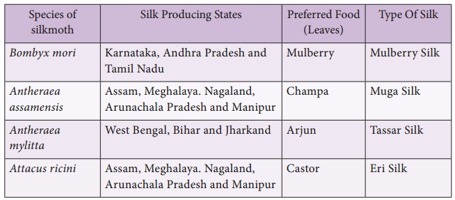
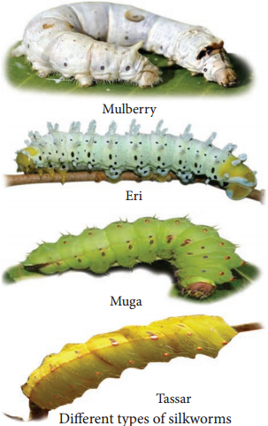
The first two are agricultural and the last one is an industrail component. Only few species of silkworms are used in the sericulture industry (Table 12.1 and Figure 12.2).
Table 12.1: Different Types of Silkworm
Sericulture img 1
Sericulture img 2

Life Cycle of Bombyx Mori

The adult of Bombyx mori is about 2.5 cm in length and pale creamy white in colour. Due to heavy body and feeble wings, flight is not possible by the female moth. This moth is unisexual in nature and does not feed during its very short life period of 2-3 days. Just after emergence, male moth copulates with female for about 2-3 hours and if not separated, they may die after few hours of copulating with female.

Just after copulation, female starts egg laying which is completed in 1-24 hours. A single female moth lays 400 to 500 eggs depending upon the climatic conditions. Two types of eggs are generally found namely diapause type and non-diapause type.

The diapause type is laid by silkworms inhabiting the temperate regions, whereas silkworms belonging to subtropical regions like India lay non-diapause type of eggs. The eggs after ten days of incubation hatch into larva called about 3 mm in length and is pale, yellowishwhite in colour. The caterpillars are provided with well developed mandibulate type of mouth-parts adapted to feed easily on the mulberry leaves.

After 1st, 2nd, 3rd and 4th moultings caterpillars get transformed into 2nd, 3rd, 4th and 5th instars respectively (Figure 12.3). It takes about 21 to 25 days after hatching. The fully grown caterpillar is 7.5 cm in length.
Sericulture img 3

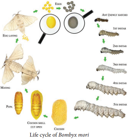
It develops salivary glands, stops feeding and undergoes pupation. The caterpillars stop feeding and move towards the corner among the leaves and secretes a sticky fluid through their silk gland. The secreted fluid comes out through spinneret (a narrow pore situated on the hypopharynx) and takes the form of long fie thread of silk which hardens on exposure to air and is wrapped around the body of caterpillar in the forms of a covering called as cocoon.

It is the white coloured bed of the pupa whose outer threads are irregular while the inner threads are regular. The length of continuous thread secreted by a caterpillar for the formation of cocoon is about 1000-1200 metres which requires 3 days to complete. The pupal period lasts for 10 to 12 days and the pupae cut through the cocoon and emerge into adult moth.

On the basis of the moults which they undergo during their larval life, B. mori is divided into three races – tri-moulters, tetramoulters and pentamoulters. Based on voltinism (the number of broods raised per year), three kinds of races are recognized in mulberry silkworm – univoltines (one brood only), bivoltines (two broods only) and multivoltines (more than two broods).

Cultivation of Food Plants for the Silkworms

The first component, is to grow the food plants for the silkworms. Mulberry leaves are widely used as food for silkworm Bombyx mori and the cultivation of mulberry is called as Moriculture. Presently improved mulberry varieties like Victory1, S36, G2 and G4 which can withstand various agro – climatic and soil conditions are used for planting.

The favourable season for cultivating of the mulberry plants is June, July, November and December. The mulberry crop production technology includes land preparation, preparation of cutting, planting techniques, maintenance of mulberry nursery, disease and pest management and uprooting for raising new mulberry gardens. Mulberry is also being grown as tree plant at an height of 123-152 cm with 20 x 20 cm or 25 x 25 cm spacing to harvest better silkworm cocoon crops.

Rearing of Silkworms

The second component is the rearing of silkworm. A typical rearing house (6m x 4m x 3.5m) is constructed on an elevated place under shade to accommodate 100 dfls (disease free layings). Space of 1m should be provided surrounding the rearing house. Sufficient windows and ventilators should be provided for free circulation of air inside the rearing house.

The windows and ventilators should be covered with nylon net to restrict the entry of uzi flies and other insects. Apart from the specified area of the rearing house; the following appliances such as hygrometer, power sprayers, rearing stands, foam pads, wax coated paraffin papers, nylon nets, baskets for keeping leaves, gunny bags, rotary or bamboo mountages and drier are needed for effective rearing of silkworms. The steps involved in rearing process of silkworm are disinfection of rearing house, incubation of eggs, brushing, young larval rearing and late age larval rearing.

The selected healthy silk moths are allowed to mate for 4 hours. Female moth is then kept in a dark plastic bed, it lays about 400 eggs in 24 hours; the female is taken out, crushed and examined for any disease, only certified disease-free eggs are reared for industrial purpose. The eggs are incubated in an incubator. The small larvae (caterpillars) hatch between 7-10 days.

These larvae are kept in trays inside a rearing house at a temperature of about 20°C – 25°C. These are first fed on chopped mulberry leaves. After 4-5 days fresh leaves are provided. As the larvae grow, they are transferred to fresh leaves on clean trays, when fully grown they spin cocoons. Their maturity is achieved in about 45 days. At this stage the salivary glands (silk glands) starts secreting silk to spin cocoons.

Post Cocoon Processing

The method of obtaining silk thread from the cocoon is known as post cocoon processing. This includes stifling and reeling. The process of killing the cocoons is called stifling. The process of removing the threads from the killed cocoon is called reeling. For reeling silk the cocoons are gathered about 8 – 10 days after spinning had begun. The cocoons are first treated by steam or dry heat to kill the insect inside.

This is necessary to prevent the destruction of the continuous fibre by the emergence of the month. The cocoons are then soaked in hot water (95° – 97°C) for 10-15 minutes to soften the gum that binds the silk threads together. This process is called cooking.

The “cooked” cocoons are kept in hot water and the loose ends of the thread are caught by hand. Threads from several cocoons are wound together on spinning wheels (Charakhas) to form the reels of raw silk. Only about onehalf of the silk of each cocoon is reelable, the remainder is used as a silk waste and formed into spun silk. Raw silk thus obtained is processed through several treatments to bring about the luster on the thread.

Uses of Silk

1. Silk fibers are utilized in preparing silk clothes. Silk fibers are now combined with other natural or synthetic fibers to manufacture clothes like Teri-Silk, Cot-Silk etc. Silk is dyed and printed to prepare ornamented fabrics. They are generally made from Eri-silk or spun silk.

2. Silk is used in industries and for military purposes.

3. It is used in the manufacture of fishing fibers, parachutes, cartridge bags, insulation coils for telephone, wireless receivers, tyres of racing cars, filter fibres, in medical dressings and as suture materials.

Diseases and Pests of Silkworm:

The profitable silk industry is threatened by various diseases caused by the virus, fungal, bacterial and protozoan infections but also by insect predators, birds and other higher animals. Ants, crows, kites, rats, feed upon silk worms thereby causing a great loss to silk industry.

Pebrine, is a dangerous disease to in silkworms and the causative organism is Nosema bombycis, a protozoan. This silkworm disease is transmitted through the egg of the mother silkworm and also through ingestion of contaminated food.

Flacherie generally occurs in the mature larvae and is caused mainly by bacteria like Streptococcus and Staphylococcus. Grasserie is a most dominant and serious viral disease. It is caused by Bombyx mori nuclear polyhedrosis virus (BmNPV) a Baculovirus, which belongs to sub group ‘A’ of the Baculoviridae. Among the fungal diseases, white muscardine is common. This disease is caused by fungus Beauveria bassiana.

Vermiculture Definition and its Advantages

Learninsta presents the core concepts of Biology with high-quality research papers and topical review articles.

Vermiculture Definition and its Advantages

Vermiculture is the process of using earthworms to decompose organic food waste, into a nutrient-rich material capable of supplying necessary nutrients which helps to sustain plant growth. The aim is to continually increase the number of worms to have a sustainable harvest.

The excess worms can either be used to expand a vermicomposting operation or sold to customers. Vermicompost is the primary goal of vermiculture. Technically the worm castings are pure worm waste and are fine and nutrient rich organic soil amendment.

Vermicompost on the other hand, is comprised of the castings, bits of bedding and other organic matter. Essentially, though the terms are used interchangeably, they are both worm manure and are valuable for improving soil health. Applications of earthworm in technology of composting and bioremediation of soils and other activities is called Vermitech (Sultan Ismail, 1992).

The disposal of solid wastes (biodegradable and non – biodegradable) remains a serious challenge in most of the countries. Earthworms play a vital role in maintaining soil fertility; hence these worms are called as “farmer’s friends”.

These are also called as “biological indicators of soil fertility”. The reason is that they support bacteria, fungi, protozoans and a host of other organisms which are essential for sustaining a healthy soil.

The breakdown of organic matter by the activity of the earthworms and its elimination from its body is called vermicast. It is a finely divided granular material and is noted for its porosity, aeration, drainage and moisture holding capacity and serves as rich organic manure. Earthworms are divided into two major groups. The first group, the humus formers, dwell on the surface and feed on organic matter.

They are generally darker in colour. These worms are used for vermicomposting. The second group, the humus feeders, are burrowing worms that are useful in making the soil porous, and mixing and distributing humus through out the soil.

There are different endemic (native) species of earthworms cultured in India for vermicomposting such as Periyonyx excavatus, Lampito mauritii, Octochaetona serrata. Some earthworm species have been introduced from other countries and called as exotic species Eg. Eisenia fetida, Eudrilus eugeniae.

Vermicomposting

Vermicompost is the compost produced by the action of earthworms in association with all other organisms in the compost unit. Vermicompost bed may be selected on upland or an elevated level as it prevents the stagnation of water. You may construct a cement pit of 3x2x1m size (LxWxD) over ground surface using bricks. The size of pit may vary as per availability of raw materials.

Cement pot or well rings are practically good. Provision should be made for excess water to drain. The vermibed should not be exposed to direct sunlight and hence shade may be provided (Figure. 12.1). The first layer of vermibed contains gravel at about 5 cm in height, followed by coarse sand to a thickness of 3.5 cm, which will facilitate the drainage of excess water.
Vermiculture img 1

Earthworms collected from native soil prefer a layer of local soil in their compost beds. If local soil earthworms are used, add a layer of native loamy soil for about 15 cm on top of the gravel sand layer and introduce earthworms into it.

For exotic species such as Eisenia fetida and Eudrilus eugeniae, the layer of soil is not needed. The unit can now be loaded with digested biomass or animal dung such as cow dung that has lost its heat.

The number of earthworms to be introduced in an unit depends on the size of the vermibed prepared. Earthworms such as Periyonyx, Excavatus, Eisenia fetida or Eudrilus eugeniae are introduced on the top. Jute bags or cardboards or broad leaves are used to cover the unit. As worms require moisture, water management is most important for the survival of the earthworms. Too little or too much of water is not good for the worms.

Earthworms release their castings on the surface. One can start harvesting this from the surface on noticing the castings on the surface. It may take several days for the entire biomass to be composted depending on the amount of biomass.

When all the compost is harvested, earthworms can be handpicked by creating small conical heaps of harvested compost and leaving in sunlight for a few hours. The earthworms then move down and settle at the bottom of the heap as a cluster.

Earthworms from the lower layers of the compost can be recovered and the worms can be transferred to new composting units. Vermiwash is a liquid collected after the passage of water through a column of vermibed. It is useful as a foliar spray to enhance plant growth and yield. It is obtained from the burrows or drilospheres formed by earthworms. Nutrients, plant growth promoter substances and some useful microorganisms are present in vermiwash.

Earthworms can be used for recycling of waste food, leaf, litter and biomass to prepare a good fertilizer in container known as wormery or wormbin. Earthworm Pests and Diseases Earthworms are subjected to attack by a variety of pests.

Most outbreaks are the result of poor bed management. Earthworm enemies include ants, springtails, centipedes, slugs, mites, certain beetle larvae, birds, rats, snakes, mice, toads, and other insects or animals which feed on worms.

The earthworm has a number of internal parasites including numerous protozoa, some nematodes, and the larvae of certain flies. Larger predators can be excluded from worm beds by proper construction of the bins, and by use of screens or gratings at the bottom and top of the beds.

Advantages of Using Vermicompost

People are aware about benefits of organic inputs in farming. Vermicompost is excellent organic manure for sustainable agropractices. So, marketing vermicompost is now a potential and flurishing industry. Retail marketing of vermicompost in urban areas is most promising.

Vermicompost is neatly packed in designed and printed packets for sale. People of different age groups are involved in the production and selling of vermicompost. Marketing of vermicompost can provide a supplementary income.

  1. Vermicompost is rich in essential plant nutrients.
  2. It improves soil structure texture, aeration, and water holding capacity and prevents soil erosion.
  3. Vermicompost is a rich in nutrients and an eco-friendly amendment to soil for farming and terrace gardening.
  4. It enhances seed germination and ensures good plant growth.

Scope of Zoology Science and its Importance

Learninsta presents the core concepts of Biology with high-quality research papers and topical review articles.

Scope of Zoology Science and its Importance

Studying Zoology can provide self employment opportunities and you can become an entrepreneur. Economic Zoology is a branch of science that deals with economically useful animals. It involves the study of application of animals for human welfare.

The need of Zoology is not just to improve our economic condition but also to provide food security and provide employment opportunities. Based on the economic importance, animals can be categorized as:

  • Animals for food and food products
  • Economically beneficial animals
  • Animals of aesthetic importance
  • Animals for scientific research

Zoology offers immense scope to a candidate who has done master’s degree in the subject. They can work as Animal Behaviourist, Conservationist, Wildlife Biologist, Zoo Curator, Wildlife Educator, Zoology faculty, Forensic experts, lab technicians, and Veterinarians.

Tools and Techniques in Biology:
Animal Physics

Structure and Function of Genes:
Animal Behaviour

Genetics Evolution:
Ecology

Cell and Molecular Biology:
Embryology

There are generally not a plethora of employment opportunities with a zoology degree, unless you have some connections and experience that have you ready to go. It is also not a lucrative field. However, if you have a passion to study animals or do related research, it’s a pathway.

B.Sc Zoology and B.Sc Botany are the two most popular B.Sc courses under Life Science. The scope of both B.Sc Zoology and B.Sc Botany is wide and can be a good option for candidates who want to go for research work in future that involves the study of living forms in the ecosystem.

Zoologists are also hired for zoos, wildlife services, botanical gardens, conservation organizations, national parks, nature reserves, universities, laboratories, aquariums, animal clinics, fisheries and aquaculture, museums, research, pharmaceutical Companies, veterinary hospitals, etc.

Chemistry has more scope than zoology and botany. If you want to pursue your career in teaching and in research then go for zoology and botany whereas in chemistry you have other scopes to work with Food manufacturing industries, chemical industries, pharmaceutical industries also teaching and research.

They are both “hard”. There is a lot of science and math in each. However, both botany and zoology are broad topics covering many sub-subjects. If you enter university to study one or the other of these, you will probably narrow your interests, and probably your career, by the time you graduate.

Designing and conducting research projects and studies of animals. Studying the characteristics of animals and their behaviors. Collecting and analyzing biological data and specimens. Writing papers, reports, and articles that explain research findings.
Scope of Zoology img 1

Mechanism of Hormone Action

Learninsta presents the core concepts of Biology with high-quality research papers and topical review articles.

Mechanism of Hormone Action

Hormones circulate in the blood but their concentration can increase or decrease based on the requirement of the body. This is controlled by feedback mechanisms. These mechanisms control the secretion of endocrine glands by stimulating the hypothalamus, pituitary or both, which inturn governs the secretion of a particular hormone.

In positive feedback, the secretion of the hormone increases where as in negative feedback further secretion of hormone slows down. Feedback mechanisms are the key factors for maintaining homeostasis in our body.

Hormones are classified into three major groups as peptide hormones, steroid hormones and amino acid derived hormones based on their chemical structure.

Peptide hormones cannot cross the phospolipid cell membrane and bind to the receptors on the exterior cell surface. They are are transported to the golgi, which is the site of modification. It acts as a first messenger in the cell. Hormones on binding to their receptors do not enter the target cell but generate the production of second messengers such as cyclic AMP (c AMP), which in turn regulates cellular metabolism.

This is catalyzed by the enzyme adenylate cyclase. The interaction between the hormone at the surface and the effect brought out by cAMP within the cell is known as signaling cascade. At each step there is a possibility of amplification. (Figure 11.17)
Mechanism of Hormone Action img 1

  • One hormone molecule may bind to multiple receptor molecules before it is degraded.
  • Each receptor may activate several adenylate cyclases each of which make much cAMP.
  • Thus there is more signal after each step.

The actions of cAMP are terminated by phosphodiesterases. The effect of peptide hormones like insulin, glucagon, somatotropin are usually short lived because they work through second messenger system.

Steroid hormones can easily cross the cell membrane, and bind to their receptors, which are intracellular or intranuclear. Upon binding to the receptors, they pair up with another receptor – hormone complex (dimerize). This dimer can then bind to DNA and alter its transcription. (Figure 11.18)
Mechanism of Hormone Action img 2

The effect of steroid hormones such as aldosterone, oestrogen, FSH are long lived, as they alter the amount of mRNA and protein in a cell. Amino acid derived hormones are derived from one or two aminoacid with a few additional modifications. Thyroid hormone is synthesised from tyrosine and includes the addition of several iodine atoms.

Epinephrine an amino acid derivative may function through second messenger system like peptide hormones or they may actually enter the cell and function like steroid hormones.

Hormones activate target cells by diffusing through the plasma membrane of the target cells (lipid-soluble hormones) to bind a receptor protein within the cytoplasm of the cell, or by binding a specific receptor protein in the cell membrane of the target cell (water-soluble proteins).

There are two modes of hormonal action. A: Activation of cell-surface receptors and coupled second-messenger systems, with a variety of intracellular consequences.

Hormone levels are primarily controlled through negative feedback, in which rising levels of a hormone inhibit its further release. The three mechanisms of hormonal release are humoral stimuli, hormonal stimuli, and neural stimuli.

This type of mechanism is shown by lipid soluble hormones such as fatty acids and steroids that can easily pass through the plasma membrane. They possess intracellular receptors. The hormones bind to the target receptor that activates the enzymatic activity of the cell to bring about biochemical changes.

Mechanism of hormone action is not the same in all categories of hormones is a proteinaceous hormone, has large molecular weight and is insoluble in lipids, therefore, it cannot enter the target cell. Thus, it binds with the membrane bound receptor present on ovarian cell membrane.

The action involves secretion of by the thyroid gland into the circulation, uptake of target tissues, activation or inactivation of by deiodinase enzymes, binding of to nuclear receptors that act as ligand-regulated transcription factors, and regulation of expression of target genes.

What is the most common mechanism of hormone control? With negative feedback, the most common mechanism of hormone control, some feature of hormone action directly or indirectly inhibits further hormone secretion so that the hormone level returns to an ideal level or set point.

Hormones bind to specific proteins (hormone receptors) in the target tissues and produce effect on them. There are two types of receptors: membrane bound receptors (hormone receptors present on the cell membrane of the target cell) and intracellular receptors (receptors present inside the target cell).

The mechanism by which peptide hormones act upon specific target tissues to evoke characteristic functional responses is believed to be initiated by interaction with a highly special- ized portion of the plasma membrane, the so called hormone receptor site.

Hypo and Hyper Activity of Endocrine Glands and Related Disorders

Learninsta presents the core concepts of Biology with high-quality research papers and topical review articles.

Hypo and Hyper Activity of Endocrine Glands and Related Disorders

The hyper secretion and hypo secretion of hormones leads to several disorders. Dwarfism is due to hyposecretion of growth hormone (GH) in children, skeletal growth and sexual maturity is arrested. They attain a maximum height of 4 feet only (Figure 11.8).
Hypo and Hyper Activity of Endocrine Glands and Related Disorders img 1

Gigantism is due to hypersecretion of growth hormone (GH) in children. Overgrowth of skeletal structure occurs (up to 8 feet) and the visceral growth is not appropriate with that of limbs. Figure 11.9.
Hypo and Hyper Activity of Endocrine Glands and Related Disorders img 2

Acromegaly is due to excessive secretion of growth hormone in adults. Over growth of hand bones, feet bones, jaw bones, malfunctioning of gonads, enlargement of viscera, tongue, lungs, heart, liver, spleen and endocrine gland like thyroid, adrenal etc., are the symptoms of acromegaly. (Figure 11.10)
Hypo and Hyper Activity of Endocrine Glands and Related Disorders img 3

In infants, hypothyroidism causes cretinism. A cretin shows retarded skeletal growth, absence of sexual maturity, retarded mental ability, thick wrinkled skin, protruded enlarged tongue, bloated face, thick and short limbs occurs. The other symptoms are low BMR, slow pulse rate, subnormal body temperature and elevated blood cholesterol levels. (Figure 11.11)
Hypo and Hyper Activity of Endocrine Glands and Related Disorders img 4

Hyposecretion of thyroid in adults causes myxodema. It is otherwise called Gull’s disease. This disease is characterised by decreased mental activity, memory loss, slowness of movement, speech, and general weakness of body, dry coarse skin, scarce hair, puffy appearance, disturbed sexual function, low BMR, poor appetite, and subnormal body temperature. (Figure 11.12)
Hypo and Hyper Activity of Endocrine Glands and Related Disorders img 5

Grave’s disease also called as thyrotoxicosis or exophthalmic goitre. This disease is caused due to hyper secretion of thyroid. It is characterised by enlargement of thyroid gland, increased BMR (50% – 100%), elevated respiratory and excretory rates, increased heart beat, high BP, increased body temperature, protrusion of eyeball and weakness of eye muscles and weight loss. (Figure 11.13)
Hypo and Hyper Activity of Endocrine Glands and Related Disorders img 6

Simple goitre is also known asEndemic goitre. It is caused due to hyposecretion of thyroxine. The symptoms includes enlargement of thyroid gland, fall in serum thyroxine level, increased TSH secretion. (Figure 11.14)
Hypo and Hyper Activity of Endocrine Glands and Related Disorders img 7

Tetany is caused due to the hyposecretion of parathyroid hormone (PTH). Due to hyposecretion of PTH serum calcium level decreases (Hypocalcemia), as a result serum phosphate level increases. Calcium and phosphate excretion level decreases. Generalized convulsion, locking of jaws increased heart beat rate, increased body temperature, muscular spasm are the major symptoms of tetany.

Hyperparathyroidism is caused due to excess PTH in blood. Demineralisation of bone, cyst formation, softening of bone, loss of muscle tone, general weakness, renal disorders are the symptoms of hyperparathyroidism.

Addison’s disease is caused due to hyposecretion of glucocorticoids and mineralocorticoids from the adrenal cortex. Muscular weakness, low BP., loss of appetite, vomiting, hyper pigmentation of skin, low metabolic rate, subnormal temperature, reduced blood volume, weight loss are the symptoms that occur in Addison’s disease (Figure 11.15). Reduced aldosterone secretion increases urinary excretion of NaCl and water and decreases potassium excretion leading to dehydration.
Hypo and Hyper Activity of Endocrine Glands and Related Disorders img 8

Cushing’s syndrome is caused due to excess secretion of cortisol. Obesity of the face and trunk, redness of face, hand, feet, thin skin, excessive hair growth, loss of minerals from bone (osteoporosis) systolic hypertension are features of Cushing’s syndrome. Suppression of sexual function like atrophy of gonads are the other symptoms of Cushing’s syndrome. (Figure 11.16)
Hypo and Hyper Activity of Endocrine Glands and Related Disorders img 9

Hypoglycaemia is due to increased secretion of insulin thereby blood glucose level decreases. In this disorder blood glucose level lowers than normal fasting index. Increased heartbeat, weakness, nervousness, headache, confusion, lack of co-ordination, slurred speech, serious brain defects like epilepsy and coma occurs.

Hyperglycaemia is otherwise known as Diabetes mellitus. It is caused due to reduced secretion of insulin. As the result, blood glucose level is elevated. Diabetes mellitus is of two types, Type I Diabetes and Type II Diabetes. Type I diabetes is also known Insulin dependent diabetes, caused by the lack of insulin secretion due to illness or viral infections.

Type II diabetes is also known as Non – Insulin dependent diabetes, caused due to reduced sensitivity to insulin, often called as insulin resistance. Symptoms of diabetes includes, polyurea (excessive urination), polyphagia (excessive intake of food), polydipsia (excessive consumption liquids due to thirst), ketosis (breakdown of fat into glucose results in accumulation of ketone bodies) in blood. Gluconeogenesis (Conversion of non – carbohydrate form like amino acids and fat into glucose) also occur in diabetes.

Diabates insipidus is caused due to hyposecretion of vasopressin (ADH) from neurohypophysis. The symptom includes frequent urination (polyurea) and excessive consumption of liquids due to thirst (polydipsia).

Human Endocrine System Various Types and its Function

Learninsta presents the core concepts of Biology with high-quality research papers and topical review articles.

Human Endocrine System Various Types and its Function

There are two glandular systems such as the exocrine glands and the endocrine glands. The exocrine glands secrete enzymes, saliva and sweat and have ducts that carry their substances to the membrane surfaces. Example: salivary gland and gastric gland. The endocrine glands, called ductless glands produce hormones and lack ducts; they release their hormone to the surrounding tissue fluid.

The hormones circulate around the body and eventually reach the target organs. Endocrine glands (Figure: 11. 1) include the pituitary, thyroid, parathyroid, pineal, adrenal, thymus and are also known as exclusive endocrine glands.

The hypothalamus along with its neural function also produces hormones and is considered as a neuro endocrine gland. In addition several organs such as pancreas, gastro intestinal tract epithelium, kidney, heart, gonads and placenta are also have endocrine tissues and are known as partial endocrine glands.
Human Endocrine System img 1
Table 11.1 Chemical Nature of Hormones
Human Endocrine System img 2

Hypothalamus

Hypothalamus is a small cone shaped structure that projects downward from the brain ending into the pituitary stalk. It interlinks both the nervous system and endocrine system. Though pituitary gland is known as master endocrine glands that controls the other endocrine glands, but it is, in turn controlled by the hypothalamus. Hypothalamus contains groups of neurosecretory cells. It produces neurotransmitters which regulate the secretions of the pituitary (Figure 11. 2). The hormones produced by the hypothalamus act either as a releasing hormone or as an inhibitory hormone.
Human Endocrine System img 3
Table 11.2 The major hypothalamic hormones and their functions
Human Endocrine System img 4

In the basal region of the brain, the hypothalamic hypophyseal portal blood vessel connects hypothalamus and anterior pituitary. It allows hypothalamic hormones to control anterior pituitary secretion. The posterior pituitary is connected with hypothalamus by a nerve bundle called hypothalamic hypophyseal axis. It produces nerve signal that control the posterior pituitary secretion. Hypothalamus maintains homeostasis, blood pressure, body temperature, cardio and fluid electrolyte balance of the body. As the part of limbic system it influences various emotional responses.

Pituitary Gland or Hypophysis

The pituitary gland (means to grow under) is ovoid in shape and is located in the sella turcica, a bony cavity of the sphenoid bone at the base of brain and connected to the hypothalamic region of the brain by a stalk called infundibulum. It is about one centimetre in diameter and 0.5 gm in weight. The pituitary consists of two lobes, anterior glandular adenohypophysis and posterior neural neurohypophysis.

The anterior lobe originates from the embryonic invagination of pharyngeal epithelium called Rathke’s pouch and the posterior lobe is originates from the base of the brain as an outgrowth of hypothalamus. Anatomically the adenohypophysis has three lobes or zones namely pars intermedia, pars distalis and pars tuberalis. The neurohypophysis is otherwise known as pars nervosa.

The anterior lobe of pituitary secretes six tropic hormones such as growth hormone (GH), thyroid stimulating hormone (TSH), adreno corticotropic hormone (ACTH), follicle stimulating hormone (FSH), luteinizing hormone (LH), luteotropic hormone (LTH) and melanocyte stimulating hormone (MSH) (in lower animals only). The posterior lobe of pituitary secretes the hormones namely vasopressin and oxytocin.

Hormones of Adenohypophysis

(i) Growth Hormone (GH):

It is also known as somatotropic hormone (STH) or Somatotropin. It is a peptide hormone. Growth hormone promotes growth of all the tissues and metabolic process of the body. It influences the metabolism of carbohydrates, proteins and lipids and increases the rate of protein biosynthesis in the cells.

It stimulates chondrogenesis (cartilage formation), osteogenesis (bone formation) and helps in the retention of minerals like nitrogen, potassium, phosphorus, sodium etc., in the body. GH increases the release of fatty acid from adipose tissue and decreases the rate of glucose utilization for energy by the cells. Thus it conserves glucose for glucose dependent tissues, such as the brain.

(ii) Throid Stimulating Hormone (TSH) or Thyrotropin:

TSH is a glycoprotein hormone, which stimulates the thyroid gland to secrete Tri-iodothyronine (T3) and thyroxine (T4). TSH secretion is regulated by negative feedback mechanism. It’s release from the anterior pituitary is induced by the thyrotropin releasing hormone (TRH). When thyroxine level in the blood increases, TRH acts on both the pituitary and hypothalamus to inhibit TSH secretion.

(iii) Adreno Cortico Tropic Hormone

(ACTH):

ACTH is a peptide hormone that stimulates the adrenal cortex to secrete glucocorticoids and mineralocorticoids. It stimulates melanin synthesis in melanocytes, induces the release of fatty acids from adipose tissues and stimulates insulin secretion. ACTH secretion is regulated by negative feedback mechanism.

(iv) Follicle Stimulating Hormone (FSH):

FSH is a glycoprotein hormone which regulates the functions of the gonads (ovary and testis). In males, FSH along with androgens acts on the germinal epithelium of seminiferous tubules and stimulates the production and release of sperms (spermatogenesis). In females, FSH acts on the ovaries and brings about the development and maturation of graffian follicles.

(v) Luteinizing Hormone (LH):

LH is a glycoprotein hormone which is also known as interstitial cell stimulating hormone (ICSH). In males, ICSH acts on the interstitial cells of testis to produce the male sex hormone, testosterone. In females, LH along with FSH matures the ovarian follicles.

LH independently induces ovulation, maintains the corpus luteum and promotes synthesis and release of ovarian hormones. FSH and LH are collectively referred as gonadotropins. FSH and LH are not produced during childhood. The secretion of FSH and LH starts only during pre pubertal period.

(vi) Luteotropic Hormone (LTH):

LTH is also called luteotropin or lactogenic hormone or prolactin or mammotropin. It is a protein hormone which stimulates milk secretion after the child birth in females. High prolactin secretion during lactation suppresses LH secretion and ovulation since it induces the corpus luteum hence named as luteo tropic hormone.

Hormones of Neurohypophysis

(i) Vasopressin or Antidiuretic Hormone (ADH) :

ADH is a peptide hormone which promotes reabsorption of water and electrolytes by distal tubules of nephron and thereby reduces loss of water through urine. Hence it is called as anti diuretic hormone. It also causes constriction of blood vessels when released in large amount and increases blood pressure. ADH deficiency causes Diabetes insipidus which induces the production of large amount of urine.

(ii) Oxytocin (Means Quick Birth):

It is a peptide hormone which stimulates vigorous contraction of the smooth muscles of uterus during child birth and ejection of milk from the mammary glands.

Pineal Gland

In human, the pineal gland or epiphysis cerebri or conarium is located behind the third ventricle of brain and is formed of parenchymal cells and interstitial cells. It secretes the hormone, melatonin, which plays a central role in the regulation of circadian rhythm of our body and maintains the normal sleep wake cycle. It also regulates the timing of sexual maturation of gonads. In addition melatonin also influences metabolism, pigmentation, menstrual cycle and defence mechanism of our body.

Thyroid Gland

The butterfly shaped thyroid gland is a bilobed gland located below the larynx on each side of upper trachea. It is the largest endocrine gland in the body. Its two lateral lobes are connected by a median tissue mass called isthmus.

Each lobe is made up of many lobules. The lobules consist of follicles called acini (acinus in singular). Each acinus is lined with glandular, cuboidal or squamous epithelial cells. The lumen of acinus is filled with colloid, a thick glycoprotein mixture consisting of thyroglobulin molecules.

Hormones of the thyroid gland are often called the major metabolic hormones. The follicular cells of thyroid gland secrete two hormones namely tri-iodothyronine (T3) and thyroxine or tetra – iodothyronine (T4). The parafollicular cells or ‘C’ cells of thyroid gland secrete a hormone called thyrocalcitonin. Iodine is essential for the normal synthesis of thyroid hormones.

Thyroid releasing hormone from the hypothalamus stimulates the adenohypophysis to secrete TSH, which inturn stimulates the thyroid gland to secrete the thyroid hormones. Thyroid hormones show a negative feedback effect on the hypothalamus and pituitary (Figure 11. 3).
Human Endocrine System img 5

Functions of Thyroxine or Tetraiodothyronine (T4):

Thyroxine regulates the basal metabolic rate (BMR) and body heat production. It stimulates protein synthesis and promotes growth. It is essential for the development of skeletal and nervous system. Thyroxine plays an important role in maintaining blood pressure. It reduces serum cholesterol levels, Optimum levels of thyroxine in blood is necessary for gonadial functions.

TCT is a polypeptide hormone, which regulates the blood calcium and phosphate levels. It reduces the blood calcium level and opposes the effects of parathyroid hormone.
Human Endocrine System img 6

Parathyroid Gland

In human, four tiny parathyroid glands are found in the posterior wall of the thyroid glands. This gland is composed of two types of cells, the chief cells and oxyphil cells. The chief cells secrete parathyroid hormone (PTH) and the functions of oxyphil cells are not known.

Parathyroid Hormone or Parathormone (PTH)

PTH is a hypercalcemic hormone. It is a peptide hormone involved in controlling the calcium and phosphate homeostasis. The secretion of PTH is controlled by calcium level in the blood. It increases the blood calcium level by stimulating osteoclasts to dissolve the bone matrix.

As a result calcium and phosphate are released into the blood. PTH enhances the reabsorption of calcium and excretion of phosphates by the renal tubules and promotes activation of vitamin D to increase calcium absorption by intestinal mucosal cells.

Thymus Gland

Thymus gland is partially an endocrine and partially a lymphoid organ. It is a bilobed structure located just above the heart and aorta, behind the sternum. It is covered by firous capsule and anatomically it is divisible into an outer cortex and an inner medulla.

It secretes four hormones such as thymulin, thymosin, thymopoietin and thymic humoral factor (THF). The primary function of thymus is the production of immuno competent ‘T’ lymphocytes which provides cell mediated immunity.
Human Endocrine System img 7

Adrenal Gland

A pair of adrenal glands are located at the anterior end of the kidneys, hence also called suprarenal glands. Anatomically the outer region is the cortex and the inner region is the medulla. Histologically the adrenal cortex has three distinct zones, zona glomerulosa, zona fasciculata and zona reticularis.

Zona glomerulosa an outer thin layer constitutes about 15% of adrenal cortex, and secretes mineralocorticoids. Zona fasciculata, the middle widest layer constitutes about 75% of adrenal cortex and secretes glucocorticoids such as cortisol, corticosterone and trace amounts of adrenal androgen and oestrogen. Zona reticularis, an inner zone of adrenal cortex constitute about 10% of adrenal cortex and secretes the adrenal androgen, trace amount of oestrogen and glucocorticoids.

Adrenal Medulla:

It is the central part of adrenal gland and is composed of ovoid and columnar cells, which are found around the network of blood capillaries. Adrenalin (epinephrine) and nor adrenalin (nor epinephrine) are the two hormones secreted by the adrenal medulla. Both adrenalin and nor adrenalin are catecholamines.

Function of Adrenal Hormones:

Glucocorticoids stimulate gluconeogensis, lipolysis and proteolysis (the life saving activity). Cortisol is a glucocorticoid involved in maintaining cardio vascular and kidney functions. It produces anti inflammatory reactions and suppresses the immune response.

It stimulates the RBC production. It is also known as stress combat hormone. Mineralocorticoids regulates water and electrolyte balance of our body. Aldosterone stimulates the reabsorption of sodium and water and eliminates potassium and phosphate ions through excretion, thus it helps in maintaining electrolytes, osmotic pressure and blood pressure. Adrenal androgen plays a role in hair growth in the axial region, pubis and face during puberty.

The adrenal medulla secretes the hormones adrenalin and noradrenalin and are referred as “3F hormone” (fight, flight and fright hormone). Adrenalin increases liver glycogen breakdown into glucose and increases the release of fatty acids from fat cells.

During emergency it increases heart beat rate and blood pressure. It stimulates the smooth muscles of cutaneous and visceral arteries to decrease blood flow. It increases blood flow to the skeletal muscles thereby increases the metabolic rate of skeletal muscles, cardiac muscles and nervous tissue.
Human Endocrine System img 8
Human Endocrine System img 9

Pancreas

Pancreas is a composite gland which performs both exocrine and endocrine functions. It is located just below the stomach as a leaf like structure. The pancreas is composed of two major tissues such the acini and islets of langerhans.

Acini secretes digestive enzymes and the islets of langerhans secretes hormones like insulin and glucagon. Human pancreas has one to two million islets of langerhans. In each islet about 60% cells are beta cells, 30% cells are alpha cells and 10% cells are delta cells. The alpha cells secrete glucagon, the beta cells secrete insulin and delta cells secrete somatostatin.

Insulin:

Insulin is a peptide hormone and plays an important role in glucose homeostasis. It’s main effect is to lower blood glucose levels by increasing the uptake of glucose into the body cells, especially muscle and fat cells. Insulin also inhibits the breakdown of glycogen to glucose, the conversion of amino acids or fats to glucose, so insulin is rightly called a hypoglycemic hormone.

Glucagon:

Glucagon is a polypeptide hormone. It is a potent hyperglycemic hormone that acts on the liver and promotes the breakdown of glycogen to glucose (Glygogenolysis), synthesis of glucose from lactic acid and from non-carbohydrate molecules (Gluconeogenesis).

Glucagon releases glucose from the liver cells, increasing the blood glucose levels. Since glucagon reduces the cellular uptake and utilisation of glucose it is called a hyperglycemic hormone. Prolonged hyperglycemia leads to the disorder called diabetes mellitus.

Gonads

Testis:

A pair of testis is present in the scrotal sac of males. The testis functions as a sex organ and also as an endocrine gland. The testis is composed of seminiferous tubules and interstitial cells or Leydig cells. The Leydig cells secrete several male sex hormones, collectively called androgens, mainly testosterone.

Functions of Testosterone:

Under the influence of FSH and LH, testosterone initiates maturation of male reproductive organs, and the appearance of secondary sexual characters, muscular growth, growth of facial and axillary hair, masculine voice and male sexual behaviour. It enhances the total bone matrix and plays a stimulating role in the process of spermatogenesis.

Ovary:

Females have a pair of ovaries located in the pelvic region of the abdomen. The ovary is composed of ovarian follicles and stromal tissues. It produces the eggs or ova. The ovaries secrete the steroid hormones oestrogen and progesterone. Oestrogen is responsible for the maturation of reproductive organs and the development of secondary sexual characters at puberty.

Along with progesterone, oestrogens promotes breast development and initiate the cyclic changes during menstrual cycle. Progesterone prepares the uterus for implantation of the fertilized ovum. It decreases the uterine contraction during pregnancy and stimulates the development of mammary glands and milk secretion. It is responsible for premenstrual changes in the uterus and is essential for the formation of placenta.

Hormones of Heart, Kidney and Gastro Intestinal Tract

Some tissues of the heart, kidney and gastro intestinal tract acts as partial endocrine glands. In the heart, cardiocytes on the atrial wall’s secretes an important peptide hormone called atrial natriuretic factor (ANF). When blood pressure is increased, ANF is secreted and causes dilation of the blood vessels to reduce the blood pressure.

In kidneys, hormones such as renin, erythropoietin and calcitriol are secreted. Renin is secreted by juxta glomerular cells (JGA), which increases blood pressure when angiotensin is formed in blood. Erythropoietin is also secreted by the JGA cells of the kidney and stimulates erythropoiesis (formation of RBC) in bone marrow. Calcitriol is sercreted by proximal tubules of nephron. It is an active form of vitamin D3 which promotes calcium and phosphorus absorption from intestine and accelerates bone formation.

Gastro Intestinal Tract Hormones

Group of specialized endocrine cells present in gastro-intestinal tract secretes hormones such as gastrin, cholecystokinin (CCK), secretin and gastric inhibitory peptides (GIP). Gastrin acts on the gastric glands and stimulates the secretion of HCl and pepsinogen.

Cholecystokinin (CCK) is secreted by duodenum in response to the presence of fat and acid in the diet. It acts on the gall bladder to release bile into duodenum and stimulates the secretion of pancreatic enzymes and its discharge. Secretin acts on acini cells of pancreas to secrete bicarbonate ions and water to neutralize the acidity. Gastric inhibitory peptide (GIP) inhibits gastric secretion and motility.

Endocrine Glands and Hormones

Learninsta presents the core concepts of Biology with high-quality research papers and topical review articles.

Endocrine Glands and Hormones

Physiological functions of our human body is regulated and coordinated by both neural and endocrine systems. The endocrine system influences the metabolic activities by means of hormones (hormone means to excite) which are chemical messengers released into the blood and circulated as chemical signals and acts specifically on certain organs or tissues called target organs or target tissues.

Hormones may speed up or slow down or alter the activity of the target organs. The hormones secreted do not remain permanently in the blood but are converted by the liver into inactive compounds and excreted by the kidneys.

Hormones are chemical messengers because they act as organic catalysts and coenzymes to perform specific functions in the target organs. The target organs contain receptor molecules either on the surface or within the cell.

Although different hormones come in contact with cells, only the cells that contain receptor molecules specific for the hormone are physiologically activated. A single hormone may have multiple effects on a single target tissue or on different target tissues.

Many hormones exhibit long term changes like growth, puberty and pregnancy. Hormones often influence many organs and organ systems at the same time. Serious deficiency or excess secretion of hormones leads to disorders. Hormones coordinate different physical, physiological, mental activities and maintain homeostasis. Hormones are composed of water soluble proteins or peptides or amines or fat soluble steroids.

While many parts of the body make hormones, the major glands that make up the endocrine system are the:

  • Hypothalamus
  • Pituitary
  • Thyroid
  • Parathyroids
  • Adrenals
  • Pineal body
  • The ovaries
  • The testes

Endocrine glands are ductless glands and release the substances that they make (hormones) directly into the bloodstream. These glands form part of the endocrine system and information on them is included in this website. There is another type of gland called an exocrine gland (e.g. sweat glands, lymph nodes).

The following are integral parts of the endocrine system:

Hypothalamus. The hypothalamus is located at the base of the brain, near the optic chiasm where the optic nerves behind each eye cross and meet.

  • Pineal body
  • Pituitary
  • Thyroid and parathyroid
  • Thymus
  • Adrenal gland
  • Pancreas
  • Ovary

The symptoms of an endocrine disorder vary widely and depend on the specific gland involved. However, most people with endocrine disease complain of fatigue and weakness. Blood and urine tests to check your hormone levels can help your doctors determine if you have an endocrine disorder.

When you are facing a diagnosis of a hormonal condition, like diabetes or thyroid disease, your doctor may suggest you see an endocrinologist. You may be wondering why you need to see a specialist instead of simply sticking with your primary doctor.

Endocrinologists are qualified to diagnose and treat conditions like diabetes, thyroid diseases, infertility, growth issues, metabolic disorders, osteoporosis, some cancers, and disorders in the hormone-producing adrenal glands and pituitary glands.

Beginning the examination with the hands establishes a personal connection with the patient and reinforces the trust between physician and patient that was initiated during the history. It opens the possibility for an equal exchange between doctor and patient as both observe and talk about the hands.
Endocrine Glands and Hormones img 1

Sensory Reception and Processing

Learninsta presents the core concepts of Biology with high-quality research papers and topical review articles.

Sensory Reception and Processing

Our senses make us aware of changes that occur in our surroundings and also within our body. Sensation [awareness of the stimulus] and perception [interpretation of the meaning of the stimulus] occur in the brain.

Receptors are Classified Based on Their Location:

1. Exteroceptors are located at or near the surface of the body. These are sensitive to external stimuli and receive sensory inputs for hearing, vision, touch, taste and smell.

2. Interoceptors are located in the visceral organs and blood vessels. They are sensitive to internal stimuli. Proprioceptors are also a kind of interoceptors. They provide information about position and movements of the body.

These are located in the skeletal muscles, tendons, joints, ligaments and in connective tissue coverings of bones and muscles. Receptors based on the type of stimulus are shown in Table 10.3.
Sensory Reception and Processing img 1

Photoreceptor – Eye

Eye is the organ of vision; located in the orbit of the skull and held in its position with the help of six extrinsic muscles. They are superior, inferior, lateral, median rectus muscles, superior oblique and inferior oblique muscles. These muscles aid in the movement of the eyes and they receive their nerve innervation from III, IV and VI cranial nerves.

Eyelids, eye lashes and eye brows are the accessory structures useful in protecting the eyes. The eye lids protect the eyes from excessive light and foreign objects and spread lubricating secretions over the eyeballs.

Eyelashes and the eyebrows help to protect the eyeballs from foreign objects, perspiration and also from the direct rays of sunlight. Sebaceous glands at the base of the eyelashes are called ciliary glands which secrete a lubricating fluid into the hair follicles.

Lacrymal glands, located in the upper lateral region of each orbit, secrete tears. Tears are secreted at the rate of 1mL/day and it contains salts, mucus and lysozyme enzyme to destroy bacteria. The conjunctiva is a thin, protective mucous membrane found lining the outer surface of the eyeball (Figure 10.13).
Sensory Reception and Processing img 2

The eye has two compartments, the anterior and posterior compartments. The anterior compartment has two chambers, first one lies between the cornea and iris and the second one lies between the iris and lens. These two chambers are filled with watery fluid called aqueous humor.

The posterior compartment lies between the lens and retina and it is filled with a jelly like fluid called vitreous humor that helps to retain the spherical nature of the eye. Eye lens is transparent and biconvex, made up of long columnar epithelial cells called lens fires. These cells are accumulated with the proteins called crystalline.

The Eye Ball

The eye ball is spherical in nature. The anterior one – sixth of the eyeball is exposed; the remaining region is fitted well into the orbit. The wall of the eye ball consists of three layers: firous Sclera, vascular Choroid and sensory Retina (Figure 10.14).
Sensory Reception and Processing img 3

The outer coat is composed of dense non-vascular connective tissue. It has two regions: the anterior cornea and the posterior sclera. Cornea is a non-vascular transparent coat formed of stratified squamous epithelium which helps the cornea to renew continuously as it is very vulnerable to damage from dust. Sclera forms the white of the eye and protects the eyeball.

Posteriorly the sclera is innervated by the optic nerve. At the junction of the sclera and the cornea, is a channel called ‘canal of schlemm’ which continuously drains out the excess of aqueous humor.

Choroid

Is highly vascularized pigmented layer that nourishes all the eye layers and its pigments absorb light to prevent internal reflection. Anteriorly the choroid thickens to form the ciliary body and iris. Iris is the coloured portion of the eye lying between the cornea and lens. The aperture at the centre of the iris is the pupil through which the light enters the inner chamber.

Iris is made of two types of muscles the dilator papillae (the radial muscle) and the sphincter papillae (the circular muscle). In the bright light, the circular muscle in the iris contract; so that the size of pupil decreases and less light enters the eye.

In dim light, the radial muscle in the iris contract; so that the pupil size increases and more light enters the eye. Smooth muscle present in the ciliary body is called the ciliary muscle which alters the convexity of the lens for near and far vision.

The ability of the eyes to focus objects at varying distances is called accommodation which is achieved by suspensory ligament, ciliary muscle and ciliary body. The suspensory ligament extends from the ciliary body and helps to hold the lens in its upright position. The ciliary body is provided with blood capillaries that secrete a watery fluid called aqueous humor that fills the anterior chamber.

Retina Forms the Inner Most Layer of the Eye and it Contains Two Regions:

A sheet of pigmented epithelium (non visual part) and neural visual regions. The neural retina layer contains three types of cells: photoreceptor cells – cones and rods (Figure 10.15 and Table 10.4), bipolar cells and ganglion cells.

The yellow flat spot at the centre of the posterior region of the retina is called macula lutea which is responsible for sharp detailed vision. A small depression present in the centre of the yellow spot is called fovea centralis which contains only cones.

The optic nerves and the retinal blood vessels enter the eye slightly below the posterior pole, which is devoid of photo receptors; hence this region is called blind spot.
Sensory Reception and Processing img 4
Differences between rod and cone cells
Sensory Reception and Processing img 5

Mechanism of Vision

When light enters the eyes, it gets refracted by the cornea, aqueous humor and lens and it is focused on the retina and excites the rod and cone cells. The photo pigment consists of Opsin, the protein part and Retinal, a derivative of vitamin A.

Light induces dissociation of retinal from opsin and causes the structural changes in opsin. This generates an action potential in the photoreceptor cells and is transmitted by the optic nerves to the visual cortex of the brain, via bipolar cells, ganglia and optic nerves, for the perception of vision.

Refractive Errors of Eye

Myopia (near sightedness):

The affected person can see the nearby objects but not the distant objects. This condition may result due to an elongated eyeball or thickened lens; so that the image of distant object is formed in front of the yellow spot. This error can be corrected using concave lens that diverge the entering light rays and focuses it on the retina.

Hypermetropia (Long Sightedness):

The affected person can see only the distant objects clearly but not the objects nearby. This condition results due to a shortened eyeball and thin lens; so the image of closest object is converged behind the retina. This defect can be overcome by using convex lens that converge the entering light rays on the retina.

Presbyopia:

Due to aging, the lens loses elasticity and the power of accommodation. Convex lenses are used to correct this defect.

Astigmatism

Is due to the rough (irregular) curvature of cornea or lens. Cylindrical glasses are used to correct this error (Figure 10.16).
Sensory Reception and Processing img 6

Cataract:

Due to the changes in nature of protein, the lens becomes opaque. It can be corrected by surgical procedures.

Phonoreceptor

The ear is the site of reception of two senses namely hearing and equilibrium. Anatomically, the ear is divided into three regions: the external ear, the middle ear and internal ear.

The external ear consists of pinna, external auditory meatus and ear drum. The pinna is flap of elastic cartilage covered by skin. It collects the sound waves. The external auditory meatus is a curved tube that extends up to the tympanic membrane [the ear drum]. The tympanic membrane is composed of connective tissues covered with skin outside and with mucus membrane inside.

There are very fine hairs and wax producing sebaceous glands called ceruminous glands in the external auditory meatus. The combination of hair and the ear wax [cerumen] helps in preventing dust and foreign particles from entering the ear.

The middle ear is a small air-filled cavity in the temporal bone. It is separated from the external ear by the eardrum and from the internal ear by a thin bony partition; the bony partition contains two small membrane covered openings called the oval window and the round window.

The Middle Ear Contains Three Ossicles:

Malleus [hammer bone], incus [anvil bone] and stapes [stirrup bone] which are attached to one another. The malleus is attached to the tympanic membrane and its head articulates with the incus which is the intermediate bone lying between the malleus and stapes.

The stapes is attached to the oval window in the inner ear. The ear ossicles transmit sound waves to the inner ear. A tube called Eustachian tube connects the middle ear cavity with the pharynx. This tube helps in equalizing the pressure of air on either sides of the ear drum.

Inner ear is the fluid filled cavity consisting of two parts, the bony labyrinth and the membranous labyrinths. The bony labyrinth consists of three areas: cochlea, vestibule and semicircular canals. The cochlea is a coiled portion consisting of 3 chambers namely: scala vestibuli and scala tympani – these two are filled with perilymph; and the scala media is filled with endolymph.

At the base of the cochlea, the scala vestibule ends at the ‘oval window’ whereas the scala tympani ends at the ‘round window’ of the middle ear. The chambers scala vestibuli and scala media are separated by a membrane called Reisner’s membrane whereas the scala media and scala tympani are separated by a membrane called Basilar membrane (Figure 10.17)
Sensory Reception and Processing img 7

Organ of Corti

The organ of Corti (Figure.10.18) is a sensory ridge located on the top of the Basilar membrane and it contains numerous hair cells that are arranged in four rows along the length of the basilar membrane. Protruding from the apical part of each hair cell is hair like structures known as stereocilia. During the conduction of sound wave, stereocilia makes a contact with the stiff gel membrane called tectorial membrane, a roof like structure overhanging the organ of corti throughout its length.

Mechanism of Hearing

Sound waves entering the external auditory meatus fall on the tympanic membrane. This causes the ear drum to vibrate, and these vibrations are transmitted to the oval window through the three auditory ossicles. Since the tympanic membrane is 17-20 times larger than the oval window, the pressure exerted on the oval window is about 20 times more than that on the tympanic membrane.

This increased pressure generates pressure waves in the fluid of perilymph. This pressure causes the round window to alternately bulge outward and inward meanwhile the basilar membrane along with the organ of Corti move up and down.

These movements of the hair alternately open and close the mechanically gated ion channels in the base of hair cells and the action potential is propagated to the brain as sound sensation through cochlear nerve.
Sensory Reception and Processing img 8

Defects of Ear

Deafness may be temporary or permanent. It can be further classified into conductive deafness and sensory-neural deafness. Possible causes for conductive deafness may be due to

  • The blockage of ear canal with earwax
  • Rupture of eardrum
  • Middle ear infection with fluid accumulation
  • Restriction of ossicular movement. In sensory-neural deafness, the defect may be in the organ of Corti or the auditory nerve or in the ascending auditory pathways or auditory cortex.

Organ of Equilibrium

Balance is part of a sense called proprioception, which is the ability to sense the position, orientation and movement of the body. The organ of balance is known as the vestibular system which is located in the inner ear next to the cochlea. The vestibular system is composed of a series of fluid filled sacs and tubules.

These sacs and tubules contain endolymph and are kept in the surrounding perilymph (Figure 10.19). These two fluids, perilymph and endolymph, respond to the mechanical forces, during changes occurring in body position and acceleration.
Sensory Reception and Processing img 8

The utricle and saccule are two membranous sacs, found nearest the cochlea and contain equilibrium receptor regions called maculae that are involved in detecting the linear movement of the head. The maculae contain the hair cells that act as mechanorecptors.

These hair cells are embeded in a gelatinous otolithic membrane that contains small calcareous particles called otoliths. This membrane adds weight to the top of the hair cells and increase the inertia.

The canals that lie posterior and lateral to the vestibule are semicircular canals; they are anterior, posterior and lateral canals oriented at right angles to each other. At one end of each semicircular canal, at its lower end has a swollen area called ampulla. Each ampulla has a sensory area known as crista ampullaris which is formed of sensory hair cells and supporting cells. The function of these canals is to detect rotational movement of the head.

Oldfactory Receptors

The receptors for taste and smell are the chemoreceptors. The smell receptors are excited by air borne chemicals that dissolve in fluids. The yellow coloured patches of oldfactory epithelium form the oldfactory organs that are located on the roof of the nasal cavity.

The oldfactory epithelium is covered by a thin coat of mucus layer below and oldfactory glands bounded connective tissues, above. It contains three types of cells: supporting cells, Basal cells and millions of pin shaped oldfactory receptor cells (which are unusual bipolar cells).

The oldfactory glands and the supporting cells secrete the mucus. The unmyelinated axons of the oldfactory receptor cells are gathered to form the filaments of oldfactory nerve [cranial nerve I] which synapse with cells of oldfactory bulb. The impulse, through the oldfactory nerves, is transmitted to the frontal lobe of the brain for identification of smell and the limbic system for the emotional responses to odour.

Gustatory Receptor:

The sense of taste is considered to be the most pleasurable of all senses. The tongue is provided with many small projections called papillae which give the tongue an abrasive feel. Taste buds are located mainly on the papillae which are scattered over the entire tongue surface.

Most taste buds are seen on the tongue (Figure 10.20) few are scattered on the sof palate, inner surface of the cheeks, pharynx and epiglottis of the larynx. Taste buds are flask-shaped and consist of 50 – 100 epithelial cells of two major types.
Sensory Reception and Processing img 10

Gustatory epithelial cells (taste cells) and Basal epithelial cells (Repairing cells) Long microvilli called gustatory hairs project from the tip of the gustatory cells and extends through a taste pore to the surface of the epithelium where they are bathed by saliva.

Gustatory hairs are the sensitive portion of the gustatory cells and they have sensory dendrites which send the signal to the brain. The basal cells that act as stem cells, divide and differentiate into new gustatory cells (Figure 10.20).
Sensory Reception and Processing img 11

Skin-Sense of Touch

Skin is the sensory organ of touch and is also the largest sense organ. This sensation comes from millions of microscopic sensory receptors located all over the skin and associated with the general sensations of contact, pressure, heat, cold and pain.

Some parts of the body, such as the finger tips have a large number of these receptors, making them more sensitive. Some of the sensory receptors present in the skin (Figure 10.21) are:

Tactile Merkel Disc

Is light touch receptor lying in the deeper layer of epidermis.

Hair Follicle Receptors

Are light touch receptors lying around the hair follicles.

Meissner’s Corpuscles

Are small light pressure receptors found just beneath the epidermis in the dermal papillae. They are numerous in hairless skin areas such as finger tips and soles of the feet.

Pacinian Corpuscles

Are the large egg shaped receptors found scattered deep in the dermis and monitoring vibration due to pressure. It allows to detect different textures, temperature, hardness and pain.

Ruffi Endings

Which lie in the dermis responds to continuous pressure.

Krause End Bulbs

Are thermoreceptors that sense temperature.

Reflex Action and Reflex Arc

Learninsta presents the core concepts of Biology with high-quality research papers and topical review articles.

Reflex Action and Reflex Arc

When dust falls in our eyes, the eyelids close immediately not waiting for our willingness; on touching a hot pan, the hand is withdrawn rapidly. Do you know how this happens?

The spinal cord remains as a connecting functional nervous structure in between the brain and effector organs. But sometimes when a very quick response is needed, the spinal cord can effect motor initiation as the brain and brings about an effect. This rapid action by spinal cord is called reflex action. It is a fast, involuntary, unplanned sequence of actions that occurs in response to a particular stimulus.

The nervous elements involved in carrying out the reflex action constitute a reflex arc or in other words the pathway followed by a nerve impulse to produce a reflex action is called a reflex arc (Figure 10.12).
Reflex Action and Reflex Arc img 1

Functional Components of a Reflex Arc

Sensory Receptor:
It is a sensory structure that responds to a specific stimulus.

Sensory Neuron:
This neuron takes the sensory impulse to the grey (afferent) matter of the spinal cord through the dorsal root of the spinal cord.

Interneurons:
One or two interneurons may serve to transmit the impulses from the sensory neuron to the motor neuron.

Motor Neuron:
It transmits impulse from CNS to the effector organ.

Effector Organs:
It may be a muscle or gland which responds to the impulse received. There are two types of reflexes. They are:-

(1) Unconditional Reflex:

Is an inborn reflex for an unconditioned stimulus. It does not need any past experience, knowledge or training to occur; Ex: blinking of an eye when a dust particle about to fall into it, sneezing and coughing due to foreign particle entering the nose or larynx.

(2) Conditioned Reflex:

Is a respone to a stimulus that has been acquired by learning. This does not naturally exists in animals. Only an experience makes it a part of the behaviour. Example: excitement of salivary gland on seeing and smelling a food. The conditioned reflex was first demonstrated by the Russian physiologist Pavlov in his classical conditioning experiment in a dog. The cerebral cortex controls the conditioned reflex.

Peripheral Neural System (PNS)

PNS consists of all nervous tissue outside the CNS. Components of PNS include nerves, ganglia, enteric plexuses and sensory receptors. A nerve is a chord like structure that encloses several neurons inside. Ganglia (singular-ganglion) are small masses of nervous tissue, consisting primarily of neuron cell bodies and are located outside the brain and spinal cord.

Enteric plexuses are extensive networks of neurons located in the walls of organs of the gastrointestinal tract. The neurons of these plexuses help in regulating the digestive system. The specialized structure that helps to respond to changes in the environment i.e. stimuli are called sensory receptor which triggers nerve impulses along the afferent fires to CNS. PNS comprises of cranial nerves arising from the brain and spinal nerves arising from the spinal cord.

(A) Cranial Nerves:

There are 12 pairs of cranial nerves, of which the first two pairs arise from the fore brain and the remaining 10 pairs from the mid brain. Other than the Vagus nerve, which extends into the abdomen, all cranial nerves serve the head and face.

(B) Spinal Nerves:

31 pairs of spinal nerves emerge out from the spinal cord through spaces called the intervertebral foramina found between the adjacent vertebrae. The spinal nerves are named according to the region of vertebral column from which they originate

  • Cervical nerves (8 pairs)
  • Thoracic nerves (12 pairs)
  • Lumbar nerves (5 pairs)
  • Sacral nerves (5 pairs)
  • Coccygeal nerves (1 pair)

Each spinal nerve is a mixed nerve containing both afferent (sensory) and efferent (motor) fibres. It originates as two roots:

  • A posterior dorsal root with a ganglion outside the spinal cord and
  • An anterior ventral root with no external ganglion.

Somatic Neural System (SNS)

The somatic neural system (SNS or voluntary neural system) is the part of the peripheral neural system associated with the voluntary control of body movements via skeletal muscles. The sensory and motor nerves that innervate striated muscles form the somatic neural system. Major functions of the somatic neural system include voluntary movement of the muscles and organs, and reflex movements.

Autonomic Neural System

The autonomic neural system is auto functioning and self governed. It is a part of peripheral neural system that innervates smooth muscles, glands and cardiac muscle. This system controls and coordinates the involuntary activities of various organs. ANS controlling centre is in the hypothalamus. Autonomic neural system comprises the following components:

Preganglionic Neuron

Whose cell body is in the brain or spinal cord; its myelinated axon exits the CNS as part of cranial or spinal nerve and ends in an autonomic ganglion.

Autonomic Ganglion

Consists of axon of preganglionic neuron and cell bodies of postganglionic neuron.

Postganglionic Neuron

Conveys nerve impulses from autonomic ganglia to visceral effector organs. The autonomic neural system consists of Sympathetic neural system and Parasympathetic neural system.

Central Neural System (CNS) | Brain | Spinal Cord

Learninsta presents the core concepts of Biology with high-quality research papers and topical review articles.

Central Neural System (CNS) | Brain | Spinal Cord

The CNS includes the brain and the spinal cord, which are protected by the bones of the skull and vertebral column. During its embryonic development, CNS develops from the ectoderm.

Brain

The brain acts as the command and control system. It is the site of information processing. It is located in the cranial cavity and is covered by three cranial meninges. The outer thick layer is Duramater which lines the inner surface of the cranial cavity; the median thin layer is Arachnoid mater which is separated from the duramater by a narrow subdural space.

The innermost layer is Piamater which is closely adhered to the brain but separated from the arachnoid mater by the subarachnoid space. The brain is divided into three major regions: Forebrain, Midbrain and Hindbrain.

Fore Brain

It Comprises the Following Regions:

Cerebrum and Diencephalon. Cerebrum is the ‘seat of intelligence’ and forms the major part of the brain. The cerebrum consists of an outer cortex, inner medulla and basal nuclei. The superficial region of the cerebrum is called cerebral cortex, which looks grey due to the presence of unmyelinated nerve cells. Cerebral cortex consists of neuronal cell body, dendrites, associated glial and blood vessels. The surface of the cerebrum shows many convolutions (folds) and grooves.

The folds are called gyri (singular gyrus); the shallow grooves between the gyri are called sulci (singular sulcus) and deep grooves are called fissures. These sulci and gyri increase the surface area of the cerebral cortex. Several sulci divide the cerebrum into eight lobes: a pair of frontals, parietals, temporals and occipital lobes (Figure 10.7 & Table 10.2).
Central Neural System (CNS) - Brain - Spinal Cord img 1
Central Neural System (CNS) - Brain - Spinal Cord img 2

A median longitudinal fissure divides the cerebrum longitudinally into two cerebral hemispheres (Figure 10.7). A transverse fissure separates the cerebral hemispheres from the cerebellum. The hemispheres are connected by a tract of nerve fires called corpus callosum. Cerebral cortex has three functional areas namely sensory areas occur in the parietal, temporal and occipital lobes of the cortex.
Central Neural System (CNS) - Brain - Spinal Cord img 1

They receive and interpret the sensory impulses. Motor area of the cortex which controls voluntary muscular movements lies in the posterior part of the frontal lobes. The areas other than sensory and motor areas are called Association areas that deal with integrative functions such as memory, communications, learning and reasoning. Inner to the cortex is medulla which is white in colour and acts as a nerve tract between the cortex and the diencephalon.

Diencephalon consists largely of following three paired structures. Epithalamus forms the roof of the diencephalon and it is a non-nervous tissue. The anterior part of epithalamus is vascular and folded to form the anterior choroid plexus. Just behind the choroid plexus, the epithalamus forms a short stalk which ends in a rounded body called pineal body which secretes the hormone, melatonin which regulates sleep and wake cycle.

Thalamus is composed of grey mater which serves as a relay centre for impulses between the spinal cord, brain stem and cerebrum. Within the thalamus, information is sorted and edited and plays a key role inlearning and memory. It is a major coordinating centre for sensory and motor signalling.
Central Neural System (CNS) - Brain - Spinal Cord img 3

Hypothalamus forms the floor of the diencephalon. The downward extension of the hypothalamus, the infundibulum connects the hypothalamus with the pituitary gland. The hypothalamus contains a pair of small rounded body called mammillary bodies that are involved in olfactory reflxes and emotional responses to odour.

Hypothalamus maintains homeostasis and has many centres which control the body temperature, urge for eating and drinking. It also contains a group of neurosecretory cells which secrete the hypothalamic hormones. Hypothalamus also acts as the satiety centre.

Limbic System

The inner part of the cerebral hemisphere constitutes the limbic system. The main components of limbic system are oldfactory bulbs, cingulate gyrus, mammillary body, amygdala, hippocampus and hypothalamus. The limbic system is called ‘emotional brain’ because it plays a primary role in the regulation of pleasure, pain, anger, fear, sexual feeling and affection. The hippocampus and amygdala also play a role in memory (Figure 10.9).
Central Neural System (CNS) - Brain - Spinal Cord img 4

Brain stem is the part of the brain between the spinal cord and the diencephalon. It consists of mid brain, pons varolii and medulla oblongata (Figure 10.10).
Central Neural System (CNS) - Brain - Spinal Cord img 5

Mid Brain

The mid brain is located between the diencephalon and the pons. The lower portion of the midbrain consists of a pair of longitudinal bands of nervous tissue called cerebral peduncles which relay impulses back and forth between cerebrum, cerebellum, pons and medulla. The dorsal portion of the midbrain consists of four rounded bodies called corpora quadrigemina which acts as a reflex centre for vision and hearing.

Hind Brain

Rhombencephalon forms the hind brain. It comprises of cerebellum, pons varolii and medulla oblongata. Cerebellum is the second largest part of the brain. It consists of two cerebellar hemispheres and central worm shaped part, the vermis. The cerebellum controls and coordinates muscular movements and body equilibrium. Any damage to cerebellum often results in uncoordinated voluntary muscle movements.

Pons varoli lies infront of the cerebellum between the midbrain and the medulla oblongata. The nerve fires in the pons varolii form a bridge between the two cerebellar hemispheres and connect the medulla oblongata with the other region of the brain. The respiratory nuclei found in the pons cooperate with the medulla to control respiration.

Medulla oblongata forms the posterior most part of the brain. It connects the spinal cord with various parts of the brain. It receives and integrates signals from spinal cord and sends it to the cerebellum and thalamus. Medulla contains vital centres that control cardio vascular reflexes, respiration and gastric secretions.

Ventricles of the Brain

The brain has four hollow, fluid filled spaces. The C – shaped space found inside each cerebral hemisphere forms the lateral ventricles I and II which are separated from each other by a thin membrane called theseptum pellucidum.

Each lateral ventricle communicates with the narrow III ventricle in the diencephalon through an opening called interventricular foramen (foramen of Monro). The ventricle III is continuous with the ventricle IV in the hind brain through a canal called aqueduct of Sylvius (cerebral aqueduct).

Choroid plexus is a network of blood capillaries found in the roof of the ventricles and forms cerebro spinal fluid (CSF) from the blood. CSF provides buoyancy to the CNS structures; CSF acts as a shock absorber for the brain and spinal cord; it nourishes the brain cells by transporting constant supply of food and oxygen; it carries harmful metabolic wastes from the brain to the blood; and maintains a constant pressure inside the cranial vessels.

Spinal Cord

The spinal cord is a long, slender, cylindrical nervous tissue. It is protected by the vertebral column and surrounded by the three membranes as in the brain. The spinal cord that extends from the brain stem into the vertebral canal of the vertebral column up to the level of 1st or 22nd lumbar vertebra.

So the nerve roots of the remaining nerves are greatly elongated to exit the vertebral column at their appropriate space. The thick bundle of elongated nerve roots within the lower vertebral canal is called the cauda equina (horse’s tail) because of its appearance.

In the cross section of spinal cord (Figure 10.11), there are two indentations: the posterior median sulcus and the anterior median fissure. Although there might be slight variations, the cross section of spinal cord is generally the same throughout its length. In contrast to the brain, the grey matter in the spinal cord forms an inner butterfly shaped region surrounded by the outer white matter.
Central Neural System (CNS) - Brain - Spinal Cord img 6

The grey matter consists of neuronal cell bodies and their dendrites, interneurons and glial cells. White matter consists of bundles of nerve fibres. In the center of the grey matter there is a central canal which is filled with CSF. Each half of the grey matter is divided into a dorsal horn, a ventral horn and a lateral horn.

The dorsal horn contains cell bodies of interneurons on which afferent neurons terminate. The ventral horn contains cell bodies of the efferent motor neurons supplying the skeletal muscle. Autonomic nerve fires, supplying cardiac and smooth muscles and exocrine glands, originate from the cell bodies found in the lateral horn.

In the white matter, the bundles of nerve fires form two types of tracts namely ascending tracts which carry sensory impulses to the brain and descending tracts which carry motor impulses from the brain to the spinal nerves at various levels of the spinal cord. The spinal cord shows two enlargements, one in the cervical region and another one in the lumbosacral region. The cervical enlargement serves the upper limb and lumbar enlargement serves the lower limbs.

Neuron as a Structural and Functional Unit of Neural System

Learninsta presents the core concepts of Biology with high-quality research papers and topical review articles.

Neuron as a Structural and Functional Unit of Neural System

A neuron is a microscopic structure composed of three major parts namely cell body (soma), dendrites and axon. The cell body is the spherical part of the neuron that contains all the cellular organelles as a typical cell (except centriole). The plasma membrane covering the neuron is called neurilemma and the axon is axolemma.

The repeatedly branched short fibres coming out of the cell body are called dendrites, which transmit impulses towards the cell body. The cell body and the dendrites contain cytoplasm and granulated endoplasmic reticulum called Nissl’s granules.

An axon is a long fibre that arises from a cone shaped area of the cell body called the Axon hillock and ends at the branched distal end. Axon hillock is the place where the nerve impulse is generated in the motor neurons. The axon of one-neuron branches and forms connections with many other neurons. An axon contains the same organelles found in the dendrites and cell body but lacks Nissl’s granules and Golgi apparatus.

The axon, particularly of peripheral nerves is surrounded by Schwann cells (a type of glial cell) to form myelin sheath, which act as an insulator. Myelin sheath is associated only with the axon; dendrites are always non-myelinated.

Schwann cells are not continuous along the axon; so there are gaps in the myelin sheath between adjacent Schwann cells. These gaps are called Nodes of Ranvier. Large myelinated nerve fibres conduct impulses rapidly, whereas nonmyelinated fibres conduct impulses quite slowly (Figure 10.1).
Neuron as a Structural and Functional Unit of Neural System img 1

Each branch at the distal end of the axon terminates into a bulb like structure called synaptic knob which possesses synaptic vesicles filled with neurotransmitters. The axon transmits nerve impulses away from the cell body to an inter neural space or to a neuro-muscular junction. The neurons are divided into three types based on number of axon and dendrites they possess (Figure 10.2).
Neuron as a Structural and Functional Unit of Neural System img 2

1. Multipolar Neurons

Have many processes with one axon and two or more dendrites. They are mostly interneurons.

2. Bipolar Neurons

Have two processes with one axon and one dendrite. These are found in the retina of the eye, inner ear and the oldfactory area of the brain.

3. Unipolar Neurons

Have a single short process and one axon. Unipolar neurons are located in the ganglia of cranial and spinal nerves.

Generation and Conduction of Nerve Impulses

This section deals with how the nerve impulses are produced and conducted in our body. Sensation felt in the sensory organs are carried by the nerve fibres in the form of electrical impulses. A nerve impulse is a series of electrical impulses, which travel along the nerve fibre.

Inner to the axolemma, the cytoplasm contains the intracellular fluid (ICF) with large amounts of potassium and magnesium phosphate along with negatively charged proteins and other organic molecules.

The extra cellular fluid (ECF) found outside the axolemma contains large amounts of sodium chloride, bicarbonates, nutrients and oxygen for the cell; and carbon dioxide and metabolic wastes released by the neuronal cells. The ECF and ICF (cytosol) contains negatively charged particles (anions) and positively charged particles (cations). These charged particles are involved in the conduction of impulses.

The neurons maintain an uneven distribution of various inorganic ions across their axolemma for transmission of impulses. This unequal distribution of ions establishes the membrane potential across the axolemma. The axolemma contains a variety of membrane proteins that act as ionic channels and regulates the movement of ions across the axolemma. (Shown in Table 10.1).
Neuron as a Structural and Functional Unit of Neural System img 3

Transmission of Impulses

The transmission of impulse involves two main phases; Resting membrane potential and Action membrane potential.

Resting Membrane Potential:

The electrical potential difference across the plasma membrane of a resting neuron is called the resting potential during which the interior of the cell is negative due to greater efflux of K+ outside the cell than Na+ influx into the cell.

When the axon is not conducting any impulses i.e. in resting condition, the axon membrane is more permeable to K+ and less permeable to Na+ ions, whereas it remains impermeable to negatively charged protein ions.
Neuron as a Structural and Functional Unit of Neural System img 4

The axoplasm contains high concentration of K+ and negatively charged proteins and low concentration of Na+ ions. In contrast, fluid outside the axon (ECF) contains low concentration of K+ and high concentration of Na+, and this forms a concentration gradient. This ionic gradient across the resting membrane is maintained by ATP driven Sodium Potassium pump, which exchanges 3Na+ outwards for 2K+ into the cells.

In this state, the cell membrane is said to be polarized. In neuron, the resting membrane potential ranges from -40mV to -90mV, and its normal value is -70mV. The minus sign indicates that the inside of the cell is negative with respect to the Figure 10.3 Ionic channels outside (Figure 10.4).
Neuron as a Structural and Functional Unit of Neural System img 5

Action Membrane Potential

An action potential occurs when a neuron sends information down an axon, away from the cell body. It includes following phases, depolarization, repolarisation and hypo polarization.

Depolarization – Reversal of Polarity

When a nerve fire is stimulated, sodium voltage-gate opens and makes the axolemma permeable to Na+ ions; meanwhile the potassium voltage gate closes. As a result, the rate of flow of Na+ ions into the axoplasm exceeds the rate of flow of K+ ions to the outside fluid [ECF].

Therefore, the axolemma becomes positively charged inside and negatively charged outside. This reversal of electrical charge is called Depolarization. During depolarization, when enough Na+ ions enter the cell, the action potential reaches a certain level, called threshold potential [-55 mV]. The particular stimulus which is able to bring the membrane potential to threshold is called threshold stimulus.

The action potential occurs in response to a threshold stimulus but does not occur at subthreshold stimuli. This is called all or none principle. Due to the rapid influx of Na+ ions, the membrane potential shoots rapidly up to +45mV which is called the Spike potential.

Repolarisation [Falling Phase]

When the membrane reaches the spike potential, the sodium voltage-gate closes and potassium voltage-gate opens. It checks influx of Na+ ions and initiates the efflux of K+ ions which lowers the number of positive ions within the cell. This, the potential falls back towards the resting potential. The reversal of membrane potential inside the axolemma to negative occurs due to the efflux of K+ ions. This is called Repolarisation.

Hyperpolarization

If repolarization becomes more negative than the resting potential -70 mV to about -90 mV, it is called Hyperpolarization. During this, K+ ion gates are more permeable to K+ even after reaching the threshold level as it closes slowly; hence called Lazy gates. The membrane potential returns to its original resting state when K+ ion channels close completely. During hyperpolarization the Na+ voltage gate remains closed (Figure 10.5).
Neuron as a Structural and Functional Unit of Neural System img 6

Conduction Speed of a Nerve Impulse

The conduction speed of a nerve impulse depends on the diameter of axon. The greater the axon’s diameter, the faster is the conduction. The myelinated axon conducts the impulse faster than the non-myelinated axon. The voltage-gated Na+ and K+ channels are concentrated at the nodes of Ranvier.

As a result, the impulse jumps node to node, rather than travelling the entire length of the nerve fire. This mechanism of conduction is called Saltatory Conduction. Nerve impulses travel at the speed of 1-300 m/s.

Synaptic Transmission

The junction between two neurons is called a Synapse through which a nerve impulse is transmitted. The first neuron involved in the synapse forms the presynaptic neuron and the second neuron is the post synaptic neuron. A small gap between the pre and postsynaptic membranes is called Synaptic Cleft that forms a structural gap and a functional bridge between neurons.

The axon terminals contain synaptic vesicles filled with neurotransmitters. When an impulse [action potential] arrives at the axon terminals, it depolarizes the presynaptic membrane, opening the voltage gated calcium channels. Influx of calcium ions stimulates the synaptic vesicles towards the pre-synaptic membrane and fuses with it.

In the neurilemma, the vesicles release their neurotransmitters into the synaptic cleft by exocytosis. The released neurotransmitters bind to their specific receptors on the post-synaptic membrane, responding to chemical signals.

The entry of the ions can generate a new potential in the post-synaptic neuron, which may be either excitatory or inhibitory. Excitatory post-synaptic potential causes depolarization whereas inhibitory post synaptic potential causes hyperpolarization of post-synaptic membrane (Figure 10.6).
Neuron as a Structural and Functional Unit of Neural System img 7