Hypo and Hyper Activity of Endocrine Glands and Related Disorders

Learninsta presents the core concepts of Biology with high-quality research papers and topical review articles.

Hypo and Hyper Activity of Endocrine Glands and Related Disorders

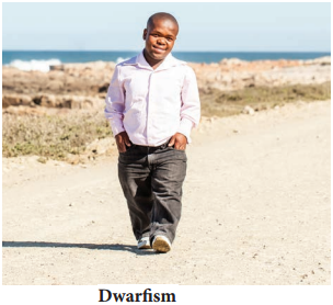
The hyper secretion and hypo secretion of hormones leads to several disorders. Dwarfism is due to hyposecretion of growth hormone (GH) in children, skeletal growth and sexual maturity is arrested. They attain a maximum height of 4 feet only (Figure 11.8).
Hypo and Hyper Activity of Endocrine Glands and Related Disorders img 1

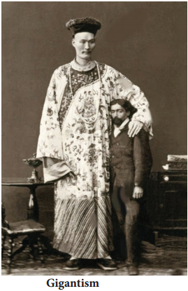
Gigantism is due to hypersecretion of growth hormone (GH) in children. Overgrowth of skeletal structure occurs (up to 8 feet) and the visceral growth is not appropriate with that of limbs. Figure 11.9.
Hypo and Hyper Activity of Endocrine Glands and Related Disorders img 2

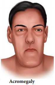
Acromegaly is due to excessive secretion of growth hormone in adults. Over growth of hand bones, feet bones, jaw bones, malfunctioning of gonads, enlargement of viscera, tongue, lungs, heart, liver, spleen and endocrine gland like thyroid, adrenal etc., are the symptoms of acromegaly. (Figure 11.10)
Hypo and Hyper Activity of Endocrine Glands and Related Disorders img 3

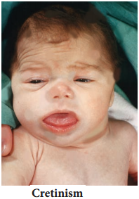
In infants, hypothyroidism causes cretinism. A cretin shows retarded skeletal growth, absence of sexual maturity, retarded mental ability, thick wrinkled skin, protruded enlarged tongue, bloated face, thick and short limbs occurs. The other symptoms are low BMR, slow pulse rate, subnormal body temperature and elevated blood cholesterol levels. (Figure 11.11)
Hypo and Hyper Activity of Endocrine Glands and Related Disorders img 4

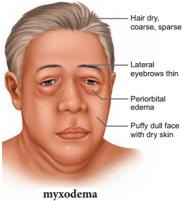
Hyposecretion of thyroid in adults causes myxodema. It is otherwise called Gull’s disease. This disease is characterised by decreased mental activity, memory loss, slowness of movement, speech, and general weakness of body, dry coarse skin, scarce hair, puffy appearance, disturbed sexual function, low BMR, poor appetite, and subnormal body temperature. (Figure 11.12)
Hypo and Hyper Activity of Endocrine Glands and Related Disorders img 5

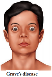
Grave’s disease also called as thyrotoxicosis or exophthalmic goitre. This disease is caused due to hyper secretion of thyroid. It is characterised by enlargement of thyroid gland, increased BMR (50% – 100%), elevated respiratory and excretory rates, increased heart beat, high BP, increased body temperature, protrusion of eyeball and weakness of eye muscles and weight loss. (Figure 11.13)
Hypo and Hyper Activity of Endocrine Glands and Related Disorders img 6

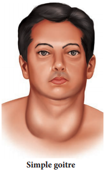
Simple goitre is also known asEndemic goitre. It is caused due to hyposecretion of thyroxine. The symptoms includes enlargement of thyroid gland, fall in serum thyroxine level, increased TSH secretion. (Figure 11.14)
Hypo and Hyper Activity of Endocrine Glands and Related Disorders img 7

Tetany is caused due to the hyposecretion of parathyroid hormone (PTH). Due to hyposecretion of PTH serum calcium level decreases (Hypocalcemia), as a result serum phosphate level increases. Calcium and phosphate excretion level decreases. Generalized convulsion, locking of jaws increased heart beat rate, increased body temperature, muscular spasm are the major symptoms of tetany.

Hyperparathyroidism is caused due to excess PTH in blood. Demineralisation of bone, cyst formation, softening of bone, loss of muscle tone, general weakness, renal disorders are the symptoms of hyperparathyroidism.

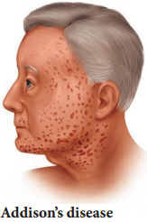
Addison’s disease is caused due to hyposecretion of glucocorticoids and mineralocorticoids from the adrenal cortex. Muscular weakness, low BP., loss of appetite, vomiting, hyper pigmentation of skin, low metabolic rate, subnormal temperature, reduced blood volume, weight loss are the symptoms that occur in Addison’s disease (Figure 11.15). Reduced aldosterone secretion increases urinary excretion of NaCl and water and decreases potassium excretion leading to dehydration.
Hypo and Hyper Activity of Endocrine Glands and Related Disorders img 8

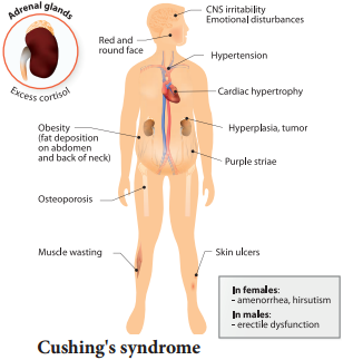
Cushing’s syndrome is caused due to excess secretion of cortisol. Obesity of the face and trunk, redness of face, hand, feet, thin skin, excessive hair growth, loss of minerals from bone (osteoporosis) systolic hypertension are features of Cushing’s syndrome. Suppression of sexual function like atrophy of gonads are the other symptoms of Cushing’s syndrome. (Figure 11.16)
Hypo and Hyper Activity of Endocrine Glands and Related Disorders img 9

Hypoglycaemia is due to increased secretion of insulin thereby blood glucose level decreases. In this disorder blood glucose level lowers than normal fasting index. Increased heartbeat, weakness, nervousness, headache, confusion, lack of co-ordination, slurred speech, serious brain defects like epilepsy and coma occurs.

Hyperglycaemia is otherwise known as Diabetes mellitus. It is caused due to reduced secretion of insulin. As the result, blood glucose level is elevated. Diabetes mellitus is of two types, Type I Diabetes and Type II Diabetes. Type I diabetes is also known Insulin dependent diabetes, caused by the lack of insulin secretion due to illness or viral infections.

Type II diabetes is also known as Non – Insulin dependent diabetes, caused due to reduced sensitivity to insulin, often called as insulin resistance. Symptoms of diabetes includes, polyurea (excessive urination), polyphagia (excessive intake of food), polydipsia (excessive consumption liquids due to thirst), ketosis (breakdown of fat into glucose results in accumulation of ketone bodies) in blood. Gluconeogenesis (Conversion of non – carbohydrate form like amino acids and fat into glucose) also occur in diabetes.

Diabates insipidus is caused due to hyposecretion of vasopressin (ADH) from neurohypophysis. The symptom includes frequent urination (polyurea) and excessive consumption of liquids due to thirst (polydipsia).

Human Endocrine System Various Types and its Function

Learninsta presents the core concepts of Biology with high-quality research papers and topical review articles.

Human Endocrine System Various Types and its Function

There are two glandular systems such as the exocrine glands and the endocrine glands. The exocrine glands secrete enzymes, saliva and sweat and have ducts that carry their substances to the membrane surfaces. Example: salivary gland and gastric gland. The endocrine glands, called ductless glands produce hormones and lack ducts; they release their hormone to the surrounding tissue fluid.

The hormones circulate around the body and eventually reach the target organs. Endocrine glands (Figure: 11. 1) include the pituitary, thyroid, parathyroid, pineal, adrenal, thymus and are also known as exclusive endocrine glands.

The hypothalamus along with its neural function also produces hormones and is considered as a neuro endocrine gland. In addition several organs such as pancreas, gastro intestinal tract epithelium, kidney, heart, gonads and placenta are also have endocrine tissues and are known as partial endocrine glands.
Human Endocrine System img 1
Table 11.1 Chemical Nature of Hormones
Human Endocrine System img 2

Hypothalamus

Hypothalamus is a small cone shaped structure that projects downward from the brain ending into the pituitary stalk. It interlinks both the nervous system and endocrine system. Though pituitary gland is known as master endocrine glands that controls the other endocrine glands, but it is, in turn controlled by the hypothalamus. Hypothalamus contains groups of neurosecretory cells. It produces neurotransmitters which regulate the secretions of the pituitary (Figure 11. 2). The hormones produced by the hypothalamus act either as a releasing hormone or as an inhibitory hormone.
Human Endocrine System img 3
Table 11.2 The major hypothalamic hormones and their functions
Human Endocrine System img 4

In the basal region of the brain, the hypothalamic hypophyseal portal blood vessel connects hypothalamus and anterior pituitary. It allows hypothalamic hormones to control anterior pituitary secretion. The posterior pituitary is connected with hypothalamus by a nerve bundle called hypothalamic hypophyseal axis. It produces nerve signal that control the posterior pituitary secretion. Hypothalamus maintains homeostasis, blood pressure, body temperature, cardio and fluid electrolyte balance of the body. As the part of limbic system it influences various emotional responses.

Pituitary Gland or Hypophysis

The pituitary gland (means to grow under) is ovoid in shape and is located in the sella turcica, a bony cavity of the sphenoid bone at the base of brain and connected to the hypothalamic region of the brain by a stalk called infundibulum. It is about one centimetre in diameter and 0.5 gm in weight. The pituitary consists of two lobes, anterior glandular adenohypophysis and posterior neural neurohypophysis.

The anterior lobe originates from the embryonic invagination of pharyngeal epithelium called Rathke’s pouch and the posterior lobe is originates from the base of the brain as an outgrowth of hypothalamus. Anatomically the adenohypophysis has three lobes or zones namely pars intermedia, pars distalis and pars tuberalis. The neurohypophysis is otherwise known as pars nervosa.

The anterior lobe of pituitary secretes six tropic hormones such as growth hormone (GH), thyroid stimulating hormone (TSH), adreno corticotropic hormone (ACTH), follicle stimulating hormone (FSH), luteinizing hormone (LH), luteotropic hormone (LTH) and melanocyte stimulating hormone (MSH) (in lower animals only). The posterior lobe of pituitary secretes the hormones namely vasopressin and oxytocin.

Hormones of Adenohypophysis

(i) Growth Hormone (GH):

It is also known as somatotropic hormone (STH) or Somatotropin. It is a peptide hormone. Growth hormone promotes growth of all the tissues and metabolic process of the body. It influences the metabolism of carbohydrates, proteins and lipids and increases the rate of protein biosynthesis in the cells.

It stimulates chondrogenesis (cartilage formation), osteogenesis (bone formation) and helps in the retention of minerals like nitrogen, potassium, phosphorus, sodium etc., in the body. GH increases the release of fatty acid from adipose tissue and decreases the rate of glucose utilization for energy by the cells. Thus it conserves glucose for glucose dependent tissues, such as the brain.

(ii) Throid Stimulating Hormone (TSH) or Thyrotropin:

TSH is a glycoprotein hormone, which stimulates the thyroid gland to secrete Tri-iodothyronine (T3) and thyroxine (T4). TSH secretion is regulated by negative feedback mechanism. It’s release from the anterior pituitary is induced by the thyrotropin releasing hormone (TRH). When thyroxine level in the blood increases, TRH acts on both the pituitary and hypothalamus to inhibit TSH secretion.

(iii) Adreno Cortico Tropic Hormone

(ACTH):

ACTH is a peptide hormone that stimulates the adrenal cortex to secrete glucocorticoids and mineralocorticoids. It stimulates melanin synthesis in melanocytes, induces the release of fatty acids from adipose tissues and stimulates insulin secretion. ACTH secretion is regulated by negative feedback mechanism.

(iv) Follicle Stimulating Hormone (FSH):

FSH is a glycoprotein hormone which regulates the functions of the gonads (ovary and testis). In males, FSH along with androgens acts on the germinal epithelium of seminiferous tubules and stimulates the production and release of sperms (spermatogenesis). In females, FSH acts on the ovaries and brings about the development and maturation of graffian follicles.

(v) Luteinizing Hormone (LH):

LH is a glycoprotein hormone which is also known as interstitial cell stimulating hormone (ICSH). In males, ICSH acts on the interstitial cells of testis to produce the male sex hormone, testosterone. In females, LH along with FSH matures the ovarian follicles.

LH independently induces ovulation, maintains the corpus luteum and promotes synthesis and release of ovarian hormones. FSH and LH are collectively referred as gonadotropins. FSH and LH are not produced during childhood. The secretion of FSH and LH starts only during pre pubertal period.

(vi) Luteotropic Hormone (LTH):

LTH is also called luteotropin or lactogenic hormone or prolactin or mammotropin. It is a protein hormone which stimulates milk secretion after the child birth in females. High prolactin secretion during lactation suppresses LH secretion and ovulation since it induces the corpus luteum hence named as luteo tropic hormone.

Hormones of Neurohypophysis

(i) Vasopressin or Antidiuretic Hormone (ADH) :

ADH is a peptide hormone which promotes reabsorption of water and electrolytes by distal tubules of nephron and thereby reduces loss of water through urine. Hence it is called as anti diuretic hormone. It also causes constriction of blood vessels when released in large amount and increases blood pressure. ADH deficiency causes Diabetes insipidus which induces the production of large amount of urine.

(ii) Oxytocin (Means Quick Birth):

It is a peptide hormone which stimulates vigorous contraction of the smooth muscles of uterus during child birth and ejection of milk from the mammary glands.

Pineal Gland

In human, the pineal gland or epiphysis cerebri or conarium is located behind the third ventricle of brain and is formed of parenchymal cells and interstitial cells. It secretes the hormone, melatonin, which plays a central role in the regulation of circadian rhythm of our body and maintains the normal sleep wake cycle. It also regulates the timing of sexual maturation of gonads. In addition melatonin also influences metabolism, pigmentation, menstrual cycle and defence mechanism of our body.

Thyroid Gland

The butterfly shaped thyroid gland is a bilobed gland located below the larynx on each side of upper trachea. It is the largest endocrine gland in the body. Its two lateral lobes are connected by a median tissue mass called isthmus.

Each lobe is made up of many lobules. The lobules consist of follicles called acini (acinus in singular). Each acinus is lined with glandular, cuboidal or squamous epithelial cells. The lumen of acinus is filled with colloid, a thick glycoprotein mixture consisting of thyroglobulin molecules.

Hormones of the thyroid gland are often called the major metabolic hormones. The follicular cells of thyroid gland secrete two hormones namely tri-iodothyronine (T3) and thyroxine or tetra – iodothyronine (T4). The parafollicular cells or ‘C’ cells of thyroid gland secrete a hormone called thyrocalcitonin. Iodine is essential for the normal synthesis of thyroid hormones.

Thyroid releasing hormone from the hypothalamus stimulates the adenohypophysis to secrete TSH, which inturn stimulates the thyroid gland to secrete the thyroid hormones. Thyroid hormones show a negative feedback effect on the hypothalamus and pituitary (Figure 11. 3).
Human Endocrine System img 5

Functions of Thyroxine or Tetraiodothyronine (T4):

Thyroxine regulates the basal metabolic rate (BMR) and body heat production. It stimulates protein synthesis and promotes growth. It is essential for the development of skeletal and nervous system. Thyroxine plays an important role in maintaining blood pressure. It reduces serum cholesterol levels, Optimum levels of thyroxine in blood is necessary for gonadial functions.

TCT is a polypeptide hormone, which regulates the blood calcium and phosphate levels. It reduces the blood calcium level and opposes the effects of parathyroid hormone.
Human Endocrine System img 6

Parathyroid Gland

In human, four tiny parathyroid glands are found in the posterior wall of the thyroid glands. This gland is composed of two types of cells, the chief cells and oxyphil cells. The chief cells secrete parathyroid hormone (PTH) and the functions of oxyphil cells are not known.

Parathyroid Hormone or Parathormone (PTH)

PTH is a hypercalcemic hormone. It is a peptide hormone involved in controlling the calcium and phosphate homeostasis. The secretion of PTH is controlled by calcium level in the blood. It increases the blood calcium level by stimulating osteoclasts to dissolve the bone matrix.

As a result calcium and phosphate are released into the blood. PTH enhances the reabsorption of calcium and excretion of phosphates by the renal tubules and promotes activation of vitamin D to increase calcium absorption by intestinal mucosal cells.

Thymus Gland

Thymus gland is partially an endocrine and partially a lymphoid organ. It is a bilobed structure located just above the heart and aorta, behind the sternum. It is covered by firous capsule and anatomically it is divisible into an outer cortex and an inner medulla.

It secretes four hormones such as thymulin, thymosin, thymopoietin and thymic humoral factor (THF). The primary function of thymus is the production of immuno competent ‘T’ lymphocytes which provides cell mediated immunity.
Human Endocrine System img 7

Adrenal Gland

A pair of adrenal glands are located at the anterior end of the kidneys, hence also called suprarenal glands. Anatomically the outer region is the cortex and the inner region is the medulla. Histologically the adrenal cortex has three distinct zones, zona glomerulosa, zona fasciculata and zona reticularis.

Zona glomerulosa an outer thin layer constitutes about 15% of adrenal cortex, and secretes mineralocorticoids. Zona fasciculata, the middle widest layer constitutes about 75% of adrenal cortex and secretes glucocorticoids such as cortisol, corticosterone and trace amounts of adrenal androgen and oestrogen. Zona reticularis, an inner zone of adrenal cortex constitute about 10% of adrenal cortex and secretes the adrenal androgen, trace amount of oestrogen and glucocorticoids.

Adrenal Medulla:

It is the central part of adrenal gland and is composed of ovoid and columnar cells, which are found around the network of blood capillaries. Adrenalin (epinephrine) and nor adrenalin (nor epinephrine) are the two hormones secreted by the adrenal medulla. Both adrenalin and nor adrenalin are catecholamines.

Function of Adrenal Hormones:

Glucocorticoids stimulate gluconeogensis, lipolysis and proteolysis (the life saving activity). Cortisol is a glucocorticoid involved in maintaining cardio vascular and kidney functions. It produces anti inflammatory reactions and suppresses the immune response.

It stimulates the RBC production. It is also known as stress combat hormone. Mineralocorticoids regulates water and electrolyte balance of our body. Aldosterone stimulates the reabsorption of sodium and water and eliminates potassium and phosphate ions through excretion, thus it helps in maintaining electrolytes, osmotic pressure and blood pressure. Adrenal androgen plays a role in hair growth in the axial region, pubis and face during puberty.

The adrenal medulla secretes the hormones adrenalin and noradrenalin and are referred as “3F hormone” (fight, flight and fright hormone). Adrenalin increases liver glycogen breakdown into glucose and increases the release of fatty acids from fat cells.

During emergency it increases heart beat rate and blood pressure. It stimulates the smooth muscles of cutaneous and visceral arteries to decrease blood flow. It increases blood flow to the skeletal muscles thereby increases the metabolic rate of skeletal muscles, cardiac muscles and nervous tissue.
Human Endocrine System img 8
Human Endocrine System img 9

Pancreas

Pancreas is a composite gland which performs both exocrine and endocrine functions. It is located just below the stomach as a leaf like structure. The pancreas is composed of two major tissues such the acini and islets of langerhans.

Acini secretes digestive enzymes and the islets of langerhans secretes hormones like insulin and glucagon. Human pancreas has one to two million islets of langerhans. In each islet about 60% cells are beta cells, 30% cells are alpha cells and 10% cells are delta cells. The alpha cells secrete glucagon, the beta cells secrete insulin and delta cells secrete somatostatin.

Insulin:

Insulin is a peptide hormone and plays an important role in glucose homeostasis. It’s main effect is to lower blood glucose levels by increasing the uptake of glucose into the body cells, especially muscle and fat cells. Insulin also inhibits the breakdown of glycogen to glucose, the conversion of amino acids or fats to glucose, so insulin is rightly called a hypoglycemic hormone.

Glucagon:

Glucagon is a polypeptide hormone. It is a potent hyperglycemic hormone that acts on the liver and promotes the breakdown of glycogen to glucose (Glygogenolysis), synthesis of glucose from lactic acid and from non-carbohydrate molecules (Gluconeogenesis).

Glucagon releases glucose from the liver cells, increasing the blood glucose levels. Since glucagon reduces the cellular uptake and utilisation of glucose it is called a hyperglycemic hormone. Prolonged hyperglycemia leads to the disorder called diabetes mellitus.

Gonads

Testis:

A pair of testis is present in the scrotal sac of males. The testis functions as a sex organ and also as an endocrine gland. The testis is composed of seminiferous tubules and interstitial cells or Leydig cells. The Leydig cells secrete several male sex hormones, collectively called androgens, mainly testosterone.

Functions of Testosterone:

Under the influence of FSH and LH, testosterone initiates maturation of male reproductive organs, and the appearance of secondary sexual characters, muscular growth, growth of facial and axillary hair, masculine voice and male sexual behaviour. It enhances the total bone matrix and plays a stimulating role in the process of spermatogenesis.

Ovary:

Females have a pair of ovaries located in the pelvic region of the abdomen. The ovary is composed of ovarian follicles and stromal tissues. It produces the eggs or ova. The ovaries secrete the steroid hormones oestrogen and progesterone. Oestrogen is responsible for the maturation of reproductive organs and the development of secondary sexual characters at puberty.

Along with progesterone, oestrogens promotes breast development and initiate the cyclic changes during menstrual cycle. Progesterone prepares the uterus for implantation of the fertilized ovum. It decreases the uterine contraction during pregnancy and stimulates the development of mammary glands and milk secretion. It is responsible for premenstrual changes in the uterus and is essential for the formation of placenta.

Hormones of Heart, Kidney and Gastro Intestinal Tract

Some tissues of the heart, kidney and gastro intestinal tract acts as partial endocrine glands. In the heart, cardiocytes on the atrial wall’s secretes an important peptide hormone called atrial natriuretic factor (ANF). When blood pressure is increased, ANF is secreted and causes dilation of the blood vessels to reduce the blood pressure.

In kidneys, hormones such as renin, erythropoietin and calcitriol are secreted. Renin is secreted by juxta glomerular cells (JGA), which increases blood pressure when angiotensin is formed in blood. Erythropoietin is also secreted by the JGA cells of the kidney and stimulates erythropoiesis (formation of RBC) in bone marrow. Calcitriol is sercreted by proximal tubules of nephron. It is an active form of vitamin D3 which promotes calcium and phosphorus absorption from intestine and accelerates bone formation.

Gastro Intestinal Tract Hormones

Group of specialized endocrine cells present in gastro-intestinal tract secretes hormones such as gastrin, cholecystokinin (CCK), secretin and gastric inhibitory peptides (GIP). Gastrin acts on the gastric glands and stimulates the secretion of HCl and pepsinogen.

Cholecystokinin (CCK) is secreted by duodenum in response to the presence of fat and acid in the diet. It acts on the gall bladder to release bile into duodenum and stimulates the secretion of pancreatic enzymes and its discharge. Secretin acts on acini cells of pancreas to secrete bicarbonate ions and water to neutralize the acidity. Gastric inhibitory peptide (GIP) inhibits gastric secretion and motility.

Endocrine Glands and Hormones

Learninsta presents the core concepts of Biology with high-quality research papers and topical review articles.

Endocrine Glands and Hormones

Physiological functions of our human body is regulated and coordinated by both neural and endocrine systems. The endocrine system influences the metabolic activities by means of hormones (hormone means to excite) which are chemical messengers released into the blood and circulated as chemical signals and acts specifically on certain organs or tissues called target organs or target tissues.

Hormones may speed up or slow down or alter the activity of the target organs. The hormones secreted do not remain permanently in the blood but are converted by the liver into inactive compounds and excreted by the kidneys.

Hormones are chemical messengers because they act as organic catalysts and coenzymes to perform specific functions in the target organs. The target organs contain receptor molecules either on the surface or within the cell.

Although different hormones come in contact with cells, only the cells that contain receptor molecules specific for the hormone are physiologically activated. A single hormone may have multiple effects on a single target tissue or on different target tissues.

Many hormones exhibit long term changes like growth, puberty and pregnancy. Hormones often influence many organs and organ systems at the same time. Serious deficiency or excess secretion of hormones leads to disorders. Hormones coordinate different physical, physiological, mental activities and maintain homeostasis. Hormones are composed of water soluble proteins or peptides or amines or fat soluble steroids.

While many parts of the body make hormones, the major glands that make up the endocrine system are the:

  • Hypothalamus
  • Pituitary
  • Thyroid
  • Parathyroids
  • Adrenals
  • Pineal body
  • The ovaries
  • The testes

Endocrine glands are ductless glands and release the substances that they make (hormones) directly into the bloodstream. These glands form part of the endocrine system and information on them is included in this website. There is another type of gland called an exocrine gland (e.g. sweat glands, lymph nodes).

The following are integral parts of the endocrine system:

Hypothalamus. The hypothalamus is located at the base of the brain, near the optic chiasm where the optic nerves behind each eye cross and meet.

  • Pineal body
  • Pituitary
  • Thyroid and parathyroid
  • Thymus
  • Adrenal gland
  • Pancreas
  • Ovary

The symptoms of an endocrine disorder vary widely and depend on the specific gland involved. However, most people with endocrine disease complain of fatigue and weakness. Blood and urine tests to check your hormone levels can help your doctors determine if you have an endocrine disorder.

When you are facing a diagnosis of a hormonal condition, like diabetes or thyroid disease, your doctor may suggest you see an endocrinologist. You may be wondering why you need to see a specialist instead of simply sticking with your primary doctor.

Endocrinologists are qualified to diagnose and treat conditions like diabetes, thyroid diseases, infertility, growth issues, metabolic disorders, osteoporosis, some cancers, and disorders in the hormone-producing adrenal glands and pituitary glands.

Beginning the examination with the hands establishes a personal connection with the patient and reinforces the trust between physician and patient that was initiated during the history. It opens the possibility for an equal exchange between doctor and patient as both observe and talk about the hands.
Endocrine Glands and Hormones img 1

Sensory Reception and Processing

Learninsta presents the core concepts of Biology with high-quality research papers and topical review articles.

Sensory Reception and Processing

Our senses make us aware of changes that occur in our surroundings and also within our body. Sensation [awareness of the stimulus] and perception [interpretation of the meaning of the stimulus] occur in the brain.

Receptors are Classified Based on Their Location:

1. Exteroceptors are located at or near the surface of the body. These are sensitive to external stimuli and receive sensory inputs for hearing, vision, touch, taste and smell.

2. Interoceptors are located in the visceral organs and blood vessels. They are sensitive to internal stimuli. Proprioceptors are also a kind of interoceptors. They provide information about position and movements of the body.

These are located in the skeletal muscles, tendons, joints, ligaments and in connective tissue coverings of bones and muscles. Receptors based on the type of stimulus are shown in Table 10.3.
Sensory Reception and Processing img 1

Photoreceptor – Eye

Eye is the organ of vision; located in the orbit of the skull and held in its position with the help of six extrinsic muscles. They are superior, inferior, lateral, median rectus muscles, superior oblique and inferior oblique muscles. These muscles aid in the movement of the eyes and they receive their nerve innervation from III, IV and VI cranial nerves.

Eyelids, eye lashes and eye brows are the accessory structures useful in protecting the eyes. The eye lids protect the eyes from excessive light and foreign objects and spread lubricating secretions over the eyeballs.

Eyelashes and the eyebrows help to protect the eyeballs from foreign objects, perspiration and also from the direct rays of sunlight. Sebaceous glands at the base of the eyelashes are called ciliary glands which secrete a lubricating fluid into the hair follicles.

Lacrymal glands, located in the upper lateral region of each orbit, secrete tears. Tears are secreted at the rate of 1mL/day and it contains salts, mucus and lysozyme enzyme to destroy bacteria. The conjunctiva is a thin, protective mucous membrane found lining the outer surface of the eyeball (Figure 10.13).
Sensory Reception and Processing img 2

The eye has two compartments, the anterior and posterior compartments. The anterior compartment has two chambers, first one lies between the cornea and iris and the second one lies between the iris and lens. These two chambers are filled with watery fluid called aqueous humor.

The posterior compartment lies between the lens and retina and it is filled with a jelly like fluid called vitreous humor that helps to retain the spherical nature of the eye. Eye lens is transparent and biconvex, made up of long columnar epithelial cells called lens fires. These cells are accumulated with the proteins called crystalline.

The Eye Ball

The eye ball is spherical in nature. The anterior one – sixth of the eyeball is exposed; the remaining region is fitted well into the orbit. The wall of the eye ball consists of three layers: firous Sclera, vascular Choroid and sensory Retina (Figure 10.14).
Sensory Reception and Processing img 3

The outer coat is composed of dense non-vascular connective tissue. It has two regions: the anterior cornea and the posterior sclera. Cornea is a non-vascular transparent coat formed of stratified squamous epithelium which helps the cornea to renew continuously as it is very vulnerable to damage from dust. Sclera forms the white of the eye and protects the eyeball.

Posteriorly the sclera is innervated by the optic nerve. At the junction of the sclera and the cornea, is a channel called ‘canal of schlemm’ which continuously drains out the excess of aqueous humor.

Choroid

Is highly vascularized pigmented layer that nourishes all the eye layers and its pigments absorb light to prevent internal reflection. Anteriorly the choroid thickens to form the ciliary body and iris. Iris is the coloured portion of the eye lying between the cornea and lens. The aperture at the centre of the iris is the pupil through which the light enters the inner chamber.

Iris is made of two types of muscles the dilator papillae (the radial muscle) and the sphincter papillae (the circular muscle). In the bright light, the circular muscle in the iris contract; so that the size of pupil decreases and less light enters the eye.

In dim light, the radial muscle in the iris contract; so that the pupil size increases and more light enters the eye. Smooth muscle present in the ciliary body is called the ciliary muscle which alters the convexity of the lens for near and far vision.

The ability of the eyes to focus objects at varying distances is called accommodation which is achieved by suspensory ligament, ciliary muscle and ciliary body. The suspensory ligament extends from the ciliary body and helps to hold the lens in its upright position. The ciliary body is provided with blood capillaries that secrete a watery fluid called aqueous humor that fills the anterior chamber.

Retina Forms the Inner Most Layer of the Eye and it Contains Two Regions:

A sheet of pigmented epithelium (non visual part) and neural visual regions. The neural retina layer contains three types of cells: photoreceptor cells – cones and rods (Figure 10.15 and Table 10.4), bipolar cells and ganglion cells.

The yellow flat spot at the centre of the posterior region of the retina is called macula lutea which is responsible for sharp detailed vision. A small depression present in the centre of the yellow spot is called fovea centralis which contains only cones.

The optic nerves and the retinal blood vessels enter the eye slightly below the posterior pole, which is devoid of photo receptors; hence this region is called blind spot.
Sensory Reception and Processing img 4
Differences between rod and cone cells
Sensory Reception and Processing img 5

Mechanism of Vision

When light enters the eyes, it gets refracted by the cornea, aqueous humor and lens and it is focused on the retina and excites the rod and cone cells. The photo pigment consists of Opsin, the protein part and Retinal, a derivative of vitamin A.

Light induces dissociation of retinal from opsin and causes the structural changes in opsin. This generates an action potential in the photoreceptor cells and is transmitted by the optic nerves to the visual cortex of the brain, via bipolar cells, ganglia and optic nerves, for the perception of vision.

Refractive Errors of Eye

Myopia (near sightedness):

The affected person can see the nearby objects but not the distant objects. This condition may result due to an elongated eyeball or thickened lens; so that the image of distant object is formed in front of the yellow spot. This error can be corrected using concave lens that diverge the entering light rays and focuses it on the retina.

Hypermetropia (Long Sightedness):

The affected person can see only the distant objects clearly but not the objects nearby. This condition results due to a shortened eyeball and thin lens; so the image of closest object is converged behind the retina. This defect can be overcome by using convex lens that converge the entering light rays on the retina.

Presbyopia:

Due to aging, the lens loses elasticity and the power of accommodation. Convex lenses are used to correct this defect.

Astigmatism

Is due to the rough (irregular) curvature of cornea or lens. Cylindrical glasses are used to correct this error (Figure 10.16).
Sensory Reception and Processing img 6

Cataract:

Due to the changes in nature of protein, the lens becomes opaque. It can be corrected by surgical procedures.

Phonoreceptor

The ear is the site of reception of two senses namely hearing and equilibrium. Anatomically, the ear is divided into three regions: the external ear, the middle ear and internal ear.

The external ear consists of pinna, external auditory meatus and ear drum. The pinna is flap of elastic cartilage covered by skin. It collects the sound waves. The external auditory meatus is a curved tube that extends up to the tympanic membrane [the ear drum]. The tympanic membrane is composed of connective tissues covered with skin outside and with mucus membrane inside.

There are very fine hairs and wax producing sebaceous glands called ceruminous glands in the external auditory meatus. The combination of hair and the ear wax [cerumen] helps in preventing dust and foreign particles from entering the ear.

The middle ear is a small air-filled cavity in the temporal bone. It is separated from the external ear by the eardrum and from the internal ear by a thin bony partition; the bony partition contains two small membrane covered openings called the oval window and the round window.

The Middle Ear Contains Three Ossicles:

Malleus [hammer bone], incus [anvil bone] and stapes [stirrup bone] which are attached to one another. The malleus is attached to the tympanic membrane and its head articulates with the incus which is the intermediate bone lying between the malleus and stapes.

The stapes is attached to the oval window in the inner ear. The ear ossicles transmit sound waves to the inner ear. A tube called Eustachian tube connects the middle ear cavity with the pharynx. This tube helps in equalizing the pressure of air on either sides of the ear drum.

Inner ear is the fluid filled cavity consisting of two parts, the bony labyrinth and the membranous labyrinths. The bony labyrinth consists of three areas: cochlea, vestibule and semicircular canals. The cochlea is a coiled portion consisting of 3 chambers namely: scala vestibuli and scala tympani – these two are filled with perilymph; and the scala media is filled with endolymph.

At the base of the cochlea, the scala vestibule ends at the ‘oval window’ whereas the scala tympani ends at the ‘round window’ of the middle ear. The chambers scala vestibuli and scala media are separated by a membrane called Reisner’s membrane whereas the scala media and scala tympani are separated by a membrane called Basilar membrane (Figure 10.17)
Sensory Reception and Processing img 7

Organ of Corti

The organ of Corti (Figure.10.18) is a sensory ridge located on the top of the Basilar membrane and it contains numerous hair cells that are arranged in four rows along the length of the basilar membrane. Protruding from the apical part of each hair cell is hair like structures known as stereocilia. During the conduction of sound wave, stereocilia makes a contact with the stiff gel membrane called tectorial membrane, a roof like structure overhanging the organ of corti throughout its length.

Mechanism of Hearing

Sound waves entering the external auditory meatus fall on the tympanic membrane. This causes the ear drum to vibrate, and these vibrations are transmitted to the oval window through the three auditory ossicles. Since the tympanic membrane is 17-20 times larger than the oval window, the pressure exerted on the oval window is about 20 times more than that on the tympanic membrane.

This increased pressure generates pressure waves in the fluid of perilymph. This pressure causes the round window to alternately bulge outward and inward meanwhile the basilar membrane along with the organ of Corti move up and down.

These movements of the hair alternately open and close the mechanically gated ion channels in the base of hair cells and the action potential is propagated to the brain as sound sensation through cochlear nerve.
Sensory Reception and Processing img 8

Defects of Ear

Deafness may be temporary or permanent. It can be further classified into conductive deafness and sensory-neural deafness. Possible causes for conductive deafness may be due to

  • The blockage of ear canal with earwax
  • Rupture of eardrum
  • Middle ear infection with fluid accumulation
  • Restriction of ossicular movement. In sensory-neural deafness, the defect may be in the organ of Corti or the auditory nerve or in the ascending auditory pathways or auditory cortex.

Organ of Equilibrium

Balance is part of a sense called proprioception, which is the ability to sense the position, orientation and movement of the body. The organ of balance is known as the vestibular system which is located in the inner ear next to the cochlea. The vestibular system is composed of a series of fluid filled sacs and tubules.

These sacs and tubules contain endolymph and are kept in the surrounding perilymph (Figure 10.19). These two fluids, perilymph and endolymph, respond to the mechanical forces, during changes occurring in body position and acceleration.
Sensory Reception and Processing img 8

The utricle and saccule are two membranous sacs, found nearest the cochlea and contain equilibrium receptor regions called maculae that are involved in detecting the linear movement of the head. The maculae contain the hair cells that act as mechanorecptors.

These hair cells are embeded in a gelatinous otolithic membrane that contains small calcareous particles called otoliths. This membrane adds weight to the top of the hair cells and increase the inertia.

The canals that lie posterior and lateral to the vestibule are semicircular canals; they are anterior, posterior and lateral canals oriented at right angles to each other. At one end of each semicircular canal, at its lower end has a swollen area called ampulla. Each ampulla has a sensory area known as crista ampullaris which is formed of sensory hair cells and supporting cells. The function of these canals is to detect rotational movement of the head.

Oldfactory Receptors

The receptors for taste and smell are the chemoreceptors. The smell receptors are excited by air borne chemicals that dissolve in fluids. The yellow coloured patches of oldfactory epithelium form the oldfactory organs that are located on the roof of the nasal cavity.

The oldfactory epithelium is covered by a thin coat of mucus layer below and oldfactory glands bounded connective tissues, above. It contains three types of cells: supporting cells, Basal cells and millions of pin shaped oldfactory receptor cells (which are unusual bipolar cells).

The oldfactory glands and the supporting cells secrete the mucus. The unmyelinated axons of the oldfactory receptor cells are gathered to form the filaments of oldfactory nerve [cranial nerve I] which synapse with cells of oldfactory bulb. The impulse, through the oldfactory nerves, is transmitted to the frontal lobe of the brain for identification of smell and the limbic system for the emotional responses to odour.

Gustatory Receptor:

The sense of taste is considered to be the most pleasurable of all senses. The tongue is provided with many small projections called papillae which give the tongue an abrasive feel. Taste buds are located mainly on the papillae which are scattered over the entire tongue surface.

Most taste buds are seen on the tongue (Figure 10.20) few are scattered on the sof palate, inner surface of the cheeks, pharynx and epiglottis of the larynx. Taste buds are flask-shaped and consist of 50 – 100 epithelial cells of two major types.
Sensory Reception and Processing img 10

Gustatory epithelial cells (taste cells) and Basal epithelial cells (Repairing cells) Long microvilli called gustatory hairs project from the tip of the gustatory cells and extends through a taste pore to the surface of the epithelium where they are bathed by saliva.

Gustatory hairs are the sensitive portion of the gustatory cells and they have sensory dendrites which send the signal to the brain. The basal cells that act as stem cells, divide and differentiate into new gustatory cells (Figure 10.20).
Sensory Reception and Processing img 11

Skin-Sense of Touch

Skin is the sensory organ of touch and is also the largest sense organ. This sensation comes from millions of microscopic sensory receptors located all over the skin and associated with the general sensations of contact, pressure, heat, cold and pain.

Some parts of the body, such as the finger tips have a large number of these receptors, making them more sensitive. Some of the sensory receptors present in the skin (Figure 10.21) are:

Tactile Merkel Disc

Is light touch receptor lying in the deeper layer of epidermis.

Hair Follicle Receptors

Are light touch receptors lying around the hair follicles.

Meissner’s Corpuscles

Are small light pressure receptors found just beneath the epidermis in the dermal papillae. They are numerous in hairless skin areas such as finger tips and soles of the feet.

Pacinian Corpuscles

Are the large egg shaped receptors found scattered deep in the dermis and monitoring vibration due to pressure. It allows to detect different textures, temperature, hardness and pain.

Ruffi Endings

Which lie in the dermis responds to continuous pressure.

Krause End Bulbs

Are thermoreceptors that sense temperature.

Reflex Action and Reflex Arc

Learninsta presents the core concepts of Biology with high-quality research papers and topical review articles.

Reflex Action and Reflex Arc

When dust falls in our eyes, the eyelids close immediately not waiting for our willingness; on touching a hot pan, the hand is withdrawn rapidly. Do you know how this happens?

The spinal cord remains as a connecting functional nervous structure in between the brain and effector organs. But sometimes when a very quick response is needed, the spinal cord can effect motor initiation as the brain and brings about an effect. This rapid action by spinal cord is called reflex action. It is a fast, involuntary, unplanned sequence of actions that occurs in response to a particular stimulus.

The nervous elements involved in carrying out the reflex action constitute a reflex arc or in other words the pathway followed by a nerve impulse to produce a reflex action is called a reflex arc (Figure 10.12).
Reflex Action and Reflex Arc img 1

Functional Components of a Reflex Arc

Sensory Receptor:
It is a sensory structure that responds to a specific stimulus.

Sensory Neuron:
This neuron takes the sensory impulse to the grey (afferent) matter of the spinal cord through the dorsal root of the spinal cord.

Interneurons:
One or two interneurons may serve to transmit the impulses from the sensory neuron to the motor neuron.

Motor Neuron:
It transmits impulse from CNS to the effector organ.

Effector Organs:
It may be a muscle or gland which responds to the impulse received. There are two types of reflexes. They are:-

(1) Unconditional Reflex:

Is an inborn reflex for an unconditioned stimulus. It does not need any past experience, knowledge or training to occur; Ex: blinking of an eye when a dust particle about to fall into it, sneezing and coughing due to foreign particle entering the nose or larynx.

(2) Conditioned Reflex:

Is a respone to a stimulus that has been acquired by learning. This does not naturally exists in animals. Only an experience makes it a part of the behaviour. Example: excitement of salivary gland on seeing and smelling a food. The conditioned reflex was first demonstrated by the Russian physiologist Pavlov in his classical conditioning experiment in a dog. The cerebral cortex controls the conditioned reflex.

Peripheral Neural System (PNS)

PNS consists of all nervous tissue outside the CNS. Components of PNS include nerves, ganglia, enteric plexuses and sensory receptors. A nerve is a chord like structure that encloses several neurons inside. Ganglia (singular-ganglion) are small masses of nervous tissue, consisting primarily of neuron cell bodies and are located outside the brain and spinal cord.

Enteric plexuses are extensive networks of neurons located in the walls of organs of the gastrointestinal tract. The neurons of these plexuses help in regulating the digestive system. The specialized structure that helps to respond to changes in the environment i.e. stimuli are called sensory receptor which triggers nerve impulses along the afferent fires to CNS. PNS comprises of cranial nerves arising from the brain and spinal nerves arising from the spinal cord.

(A) Cranial Nerves:

There are 12 pairs of cranial nerves, of which the first two pairs arise from the fore brain and the remaining 10 pairs from the mid brain. Other than the Vagus nerve, which extends into the abdomen, all cranial nerves serve the head and face.

(B) Spinal Nerves:

31 pairs of spinal nerves emerge out from the spinal cord through spaces called the intervertebral foramina found between the adjacent vertebrae. The spinal nerves are named according to the region of vertebral column from which they originate

  • Cervical nerves (8 pairs)
  • Thoracic nerves (12 pairs)
  • Lumbar nerves (5 pairs)
  • Sacral nerves (5 pairs)
  • Coccygeal nerves (1 pair)

Each spinal nerve is a mixed nerve containing both afferent (sensory) and efferent (motor) fibres. It originates as two roots:

  • A posterior dorsal root with a ganglion outside the spinal cord and
  • An anterior ventral root with no external ganglion.

Somatic Neural System (SNS)

The somatic neural system (SNS or voluntary neural system) is the part of the peripheral neural system associated with the voluntary control of body movements via skeletal muscles. The sensory and motor nerves that innervate striated muscles form the somatic neural system. Major functions of the somatic neural system include voluntary movement of the muscles and organs, and reflex movements.

Autonomic Neural System

The autonomic neural system is auto functioning and self governed. It is a part of peripheral neural system that innervates smooth muscles, glands and cardiac muscle. This system controls and coordinates the involuntary activities of various organs. ANS controlling centre is in the hypothalamus. Autonomic neural system comprises the following components:

Preganglionic Neuron

Whose cell body is in the brain or spinal cord; its myelinated axon exits the CNS as part of cranial or spinal nerve and ends in an autonomic ganglion.

Autonomic Ganglion

Consists of axon of preganglionic neuron and cell bodies of postganglionic neuron.

Postganglionic Neuron

Conveys nerve impulses from autonomic ganglia to visceral effector organs. The autonomic neural system consists of Sympathetic neural system and Parasympathetic neural system.

Central Neural System (CNS) | Brain | Spinal Cord

Learninsta presents the core concepts of Biology with high-quality research papers and topical review articles.

Central Neural System (CNS) | Brain | Spinal Cord

The CNS includes the brain and the spinal cord, which are protected by the bones of the skull and vertebral column. During its embryonic development, CNS develops from the ectoderm.

Brain

The brain acts as the command and control system. It is the site of information processing. It is located in the cranial cavity and is covered by three cranial meninges. The outer thick layer is Duramater which lines the inner surface of the cranial cavity; the median thin layer is Arachnoid mater which is separated from the duramater by a narrow subdural space.

The innermost layer is Piamater which is closely adhered to the brain but separated from the arachnoid mater by the subarachnoid space. The brain is divided into three major regions: Forebrain, Midbrain and Hindbrain.

Fore Brain

It Comprises the Following Regions:

Cerebrum and Diencephalon. Cerebrum is the ‘seat of intelligence’ and forms the major part of the brain. The cerebrum consists of an outer cortex, inner medulla and basal nuclei. The superficial region of the cerebrum is called cerebral cortex, which looks grey due to the presence of unmyelinated nerve cells. Cerebral cortex consists of neuronal cell body, dendrites, associated glial and blood vessels. The surface of the cerebrum shows many convolutions (folds) and grooves.

The folds are called gyri (singular gyrus); the shallow grooves between the gyri are called sulci (singular sulcus) and deep grooves are called fissures. These sulci and gyri increase the surface area of the cerebral cortex. Several sulci divide the cerebrum into eight lobes: a pair of frontals, parietals, temporals and occipital lobes (Figure 10.7 & Table 10.2).
Central Neural System (CNS) - Brain - Spinal Cord img 1
Central Neural System (CNS) - Brain - Spinal Cord img 2

A median longitudinal fissure divides the cerebrum longitudinally into two cerebral hemispheres (Figure 10.7). A transverse fissure separates the cerebral hemispheres from the cerebellum. The hemispheres are connected by a tract of nerve fires called corpus callosum. Cerebral cortex has three functional areas namely sensory areas occur in the parietal, temporal and occipital lobes of the cortex.
Central Neural System (CNS) - Brain - Spinal Cord img 1

They receive and interpret the sensory impulses. Motor area of the cortex which controls voluntary muscular movements lies in the posterior part of the frontal lobes. The areas other than sensory and motor areas are called Association areas that deal with integrative functions such as memory, communications, learning and reasoning. Inner to the cortex is medulla which is white in colour and acts as a nerve tract between the cortex and the diencephalon.

Diencephalon consists largely of following three paired structures. Epithalamus forms the roof of the diencephalon and it is a non-nervous tissue. The anterior part of epithalamus is vascular and folded to form the anterior choroid plexus. Just behind the choroid plexus, the epithalamus forms a short stalk which ends in a rounded body called pineal body which secretes the hormone, melatonin which regulates sleep and wake cycle.

Thalamus is composed of grey mater which serves as a relay centre for impulses between the spinal cord, brain stem and cerebrum. Within the thalamus, information is sorted and edited and plays a key role inlearning and memory. It is a major coordinating centre for sensory and motor signalling.
Central Neural System (CNS) - Brain - Spinal Cord img 3

Hypothalamus forms the floor of the diencephalon. The downward extension of the hypothalamus, the infundibulum connects the hypothalamus with the pituitary gland. The hypothalamus contains a pair of small rounded body called mammillary bodies that are involved in olfactory reflxes and emotional responses to odour.

Hypothalamus maintains homeostasis and has many centres which control the body temperature, urge for eating and drinking. It also contains a group of neurosecretory cells which secrete the hypothalamic hormones. Hypothalamus also acts as the satiety centre.

Limbic System

The inner part of the cerebral hemisphere constitutes the limbic system. The main components of limbic system are oldfactory bulbs, cingulate gyrus, mammillary body, amygdala, hippocampus and hypothalamus. The limbic system is called ‘emotional brain’ because it plays a primary role in the regulation of pleasure, pain, anger, fear, sexual feeling and affection. The hippocampus and amygdala also play a role in memory (Figure 10.9).
Central Neural System (CNS) - Brain - Spinal Cord img 4

Brain stem is the part of the brain between the spinal cord and the diencephalon. It consists of mid brain, pons varolii and medulla oblongata (Figure 10.10).
Central Neural System (CNS) - Brain - Spinal Cord img 5

Mid Brain

The mid brain is located between the diencephalon and the pons. The lower portion of the midbrain consists of a pair of longitudinal bands of nervous tissue called cerebral peduncles which relay impulses back and forth between cerebrum, cerebellum, pons and medulla. The dorsal portion of the midbrain consists of four rounded bodies called corpora quadrigemina which acts as a reflex centre for vision and hearing.

Hind Brain

Rhombencephalon forms the hind brain. It comprises of cerebellum, pons varolii and medulla oblongata. Cerebellum is the second largest part of the brain. It consists of two cerebellar hemispheres and central worm shaped part, the vermis. The cerebellum controls and coordinates muscular movements and body equilibrium. Any damage to cerebellum often results in uncoordinated voluntary muscle movements.

Pons varoli lies infront of the cerebellum between the midbrain and the medulla oblongata. The nerve fires in the pons varolii form a bridge between the two cerebellar hemispheres and connect the medulla oblongata with the other region of the brain. The respiratory nuclei found in the pons cooperate with the medulla to control respiration.

Medulla oblongata forms the posterior most part of the brain. It connects the spinal cord with various parts of the brain. It receives and integrates signals from spinal cord and sends it to the cerebellum and thalamus. Medulla contains vital centres that control cardio vascular reflexes, respiration and gastric secretions.

Ventricles of the Brain

The brain has four hollow, fluid filled spaces. The C – shaped space found inside each cerebral hemisphere forms the lateral ventricles I and II which are separated from each other by a thin membrane called theseptum pellucidum.

Each lateral ventricle communicates with the narrow III ventricle in the diencephalon through an opening called interventricular foramen (foramen of Monro). The ventricle III is continuous with the ventricle IV in the hind brain through a canal called aqueduct of Sylvius (cerebral aqueduct).

Choroid plexus is a network of blood capillaries found in the roof of the ventricles and forms cerebro spinal fluid (CSF) from the blood. CSF provides buoyancy to the CNS structures; CSF acts as a shock absorber for the brain and spinal cord; it nourishes the brain cells by transporting constant supply of food and oxygen; it carries harmful metabolic wastes from the brain to the blood; and maintains a constant pressure inside the cranial vessels.

Spinal Cord

The spinal cord is a long, slender, cylindrical nervous tissue. It is protected by the vertebral column and surrounded by the three membranes as in the brain. The spinal cord that extends from the brain stem into the vertebral canal of the vertebral column up to the level of 1st or 22nd lumbar vertebra.

So the nerve roots of the remaining nerves are greatly elongated to exit the vertebral column at their appropriate space. The thick bundle of elongated nerve roots within the lower vertebral canal is called the cauda equina (horse’s tail) because of its appearance.

In the cross section of spinal cord (Figure 10.11), there are two indentations: the posterior median sulcus and the anterior median fissure. Although there might be slight variations, the cross section of spinal cord is generally the same throughout its length. In contrast to the brain, the grey matter in the spinal cord forms an inner butterfly shaped region surrounded by the outer white matter.
Central Neural System (CNS) - Brain - Spinal Cord img 6

The grey matter consists of neuronal cell bodies and their dendrites, interneurons and glial cells. White matter consists of bundles of nerve fibres. In the center of the grey matter there is a central canal which is filled with CSF. Each half of the grey matter is divided into a dorsal horn, a ventral horn and a lateral horn.

The dorsal horn contains cell bodies of interneurons on which afferent neurons terminate. The ventral horn contains cell bodies of the efferent motor neurons supplying the skeletal muscle. Autonomic nerve fires, supplying cardiac and smooth muscles and exocrine glands, originate from the cell bodies found in the lateral horn.

In the white matter, the bundles of nerve fires form two types of tracts namely ascending tracts which carry sensory impulses to the brain and descending tracts which carry motor impulses from the brain to the spinal nerves at various levels of the spinal cord. The spinal cord shows two enlargements, one in the cervical region and another one in the lumbosacral region. The cervical enlargement serves the upper limb and lumbar enlargement serves the lower limbs.

Neuron as a Structural and Functional Unit of Neural System

Learninsta presents the core concepts of Biology with high-quality research papers and topical review articles.

Neuron as a Structural and Functional Unit of Neural System

A neuron is a microscopic structure composed of three major parts namely cell body (soma), dendrites and axon. The cell body is the spherical part of the neuron that contains all the cellular organelles as a typical cell (except centriole). The plasma membrane covering the neuron is called neurilemma and the axon is axolemma.

The repeatedly branched short fibres coming out of the cell body are called dendrites, which transmit impulses towards the cell body. The cell body and the dendrites contain cytoplasm and granulated endoplasmic reticulum called Nissl’s granules.

An axon is a long fibre that arises from a cone shaped area of the cell body called the Axon hillock and ends at the branched distal end. Axon hillock is the place where the nerve impulse is generated in the motor neurons. The axon of one-neuron branches and forms connections with many other neurons. An axon contains the same organelles found in the dendrites and cell body but lacks Nissl’s granules and Golgi apparatus.

The axon, particularly of peripheral nerves is surrounded by Schwann cells (a type of glial cell) to form myelin sheath, which act as an insulator. Myelin sheath is associated only with the axon; dendrites are always non-myelinated.

Schwann cells are not continuous along the axon; so there are gaps in the myelin sheath between adjacent Schwann cells. These gaps are called Nodes of Ranvier. Large myelinated nerve fibres conduct impulses rapidly, whereas nonmyelinated fibres conduct impulses quite slowly (Figure 10.1).
Neuron as a Structural and Functional Unit of Neural System img 1

Each branch at the distal end of the axon terminates into a bulb like structure called synaptic knob which possesses synaptic vesicles filled with neurotransmitters. The axon transmits nerve impulses away from the cell body to an inter neural space or to a neuro-muscular junction. The neurons are divided into three types based on number of axon and dendrites they possess (Figure 10.2).
Neuron as a Structural and Functional Unit of Neural System img 2

1. Multipolar Neurons

Have many processes with one axon and two or more dendrites. They are mostly interneurons.

2. Bipolar Neurons

Have two processes with one axon and one dendrite. These are found in the retina of the eye, inner ear and the oldfactory area of the brain.

3. Unipolar Neurons

Have a single short process and one axon. Unipolar neurons are located in the ganglia of cranial and spinal nerves.

Generation and Conduction of Nerve Impulses

This section deals with how the nerve impulses are produced and conducted in our body. Sensation felt in the sensory organs are carried by the nerve fibres in the form of electrical impulses. A nerve impulse is a series of electrical impulses, which travel along the nerve fibre.

Inner to the axolemma, the cytoplasm contains the intracellular fluid (ICF) with large amounts of potassium and magnesium phosphate along with negatively charged proteins and other organic molecules.

The extra cellular fluid (ECF) found outside the axolemma contains large amounts of sodium chloride, bicarbonates, nutrients and oxygen for the cell; and carbon dioxide and metabolic wastes released by the neuronal cells. The ECF and ICF (cytosol) contains negatively charged particles (anions) and positively charged particles (cations). These charged particles are involved in the conduction of impulses.

The neurons maintain an uneven distribution of various inorganic ions across their axolemma for transmission of impulses. This unequal distribution of ions establishes the membrane potential across the axolemma. The axolemma contains a variety of membrane proteins that act as ionic channels and regulates the movement of ions across the axolemma. (Shown in Table 10.1).
Neuron as a Structural and Functional Unit of Neural System img 3

Transmission of Impulses

The transmission of impulse involves two main phases; Resting membrane potential and Action membrane potential.

Resting Membrane Potential:

The electrical potential difference across the plasma membrane of a resting neuron is called the resting potential during which the interior of the cell is negative due to greater efflux of K+ outside the cell than Na+ influx into the cell.

When the axon is not conducting any impulses i.e. in resting condition, the axon membrane is more permeable to K+ and less permeable to Na+ ions, whereas it remains impermeable to negatively charged protein ions.
Neuron as a Structural and Functional Unit of Neural System img 4

The axoplasm contains high concentration of K+ and negatively charged proteins and low concentration of Na+ ions. In contrast, fluid outside the axon (ECF) contains low concentration of K+ and high concentration of Na+, and this forms a concentration gradient. This ionic gradient across the resting membrane is maintained by ATP driven Sodium Potassium pump, which exchanges 3Na+ outwards for 2K+ into the cells.

In this state, the cell membrane is said to be polarized. In neuron, the resting membrane potential ranges from -40mV to -90mV, and its normal value is -70mV. The minus sign indicates that the inside of the cell is negative with respect to the Figure 10.3 Ionic channels outside (Figure 10.4).
Neuron as a Structural and Functional Unit of Neural System img 5

Action Membrane Potential

An action potential occurs when a neuron sends information down an axon, away from the cell body. It includes following phases, depolarization, repolarisation and hypo polarization.

Depolarization – Reversal of Polarity

When a nerve fire is stimulated, sodium voltage-gate opens and makes the axolemma permeable to Na+ ions; meanwhile the potassium voltage gate closes. As a result, the rate of flow of Na+ ions into the axoplasm exceeds the rate of flow of K+ ions to the outside fluid [ECF].

Therefore, the axolemma becomes positively charged inside and negatively charged outside. This reversal of electrical charge is called Depolarization. During depolarization, when enough Na+ ions enter the cell, the action potential reaches a certain level, called threshold potential [-55 mV]. The particular stimulus which is able to bring the membrane potential to threshold is called threshold stimulus.

The action potential occurs in response to a threshold stimulus but does not occur at subthreshold stimuli. This is called all or none principle. Due to the rapid influx of Na+ ions, the membrane potential shoots rapidly up to +45mV which is called the Spike potential.

Repolarisation [Falling Phase]

When the membrane reaches the spike potential, the sodium voltage-gate closes and potassium voltage-gate opens. It checks influx of Na+ ions and initiates the efflux of K+ ions which lowers the number of positive ions within the cell. This, the potential falls back towards the resting potential. The reversal of membrane potential inside the axolemma to negative occurs due to the efflux of K+ ions. This is called Repolarisation.

Hyperpolarization

If repolarization becomes more negative than the resting potential -70 mV to about -90 mV, it is called Hyperpolarization. During this, K+ ion gates are more permeable to K+ even after reaching the threshold level as it closes slowly; hence called Lazy gates. The membrane potential returns to its original resting state when K+ ion channels close completely. During hyperpolarization the Na+ voltage gate remains closed (Figure 10.5).
Neuron as a Structural and Functional Unit of Neural System img 6

Conduction Speed of a Nerve Impulse

The conduction speed of a nerve impulse depends on the diameter of axon. The greater the axon’s diameter, the faster is the conduction. The myelinated axon conducts the impulse faster than the non-myelinated axon. The voltage-gated Na+ and K+ channels are concentrated at the nodes of Ranvier.

As a result, the impulse jumps node to node, rather than travelling the entire length of the nerve fire. This mechanism of conduction is called Saltatory Conduction. Nerve impulses travel at the speed of 1-300 m/s.

Synaptic Transmission

The junction between two neurons is called a Synapse through which a nerve impulse is transmitted. The first neuron involved in the synapse forms the presynaptic neuron and the second neuron is the post synaptic neuron. A small gap between the pre and postsynaptic membranes is called Synaptic Cleft that forms a structural gap and a functional bridge between neurons.

The axon terminals contain synaptic vesicles filled with neurotransmitters. When an impulse [action potential] arrives at the axon terminals, it depolarizes the presynaptic membrane, opening the voltage gated calcium channels. Influx of calcium ions stimulates the synaptic vesicles towards the pre-synaptic membrane and fuses with it.

In the neurilemma, the vesicles release their neurotransmitters into the synaptic cleft by exocytosis. The released neurotransmitters bind to their specific receptors on the post-synaptic membrane, responding to chemical signals.

The entry of the ions can generate a new potential in the post-synaptic neuron, which may be either excitatory or inhibitory. Excitatory post-synaptic potential causes depolarization whereas inhibitory post synaptic potential causes hyperpolarization of post-synaptic membrane (Figure 10.6).
Neuron as a Structural and Functional Unit of Neural System img 7

Human Neural System Definition and its Function

Learninsta presents the core concepts of Biology with high-quality research papers and topical review articles.

Human Neural System Definition and its Function

The human neural system is divided into two, the central neural system (CNS) and the peripheral neural system (PNS). The structural and functional units of the neural system are neurons that transmit nerve impulses. The non-nervous special cells called neuroglia form the supporting cells of the nervous tissue.

There are three functional classes of neurons. They are the afferent neurons that take sensory impulses to the Central Neural system (CNS) from the sensory organs; the efferent neurons that carry motor impulses from the CNS to the effector organs; and interneurons that lie entirely within the CNS between the afferent and efferent neurons.

The central neural system lacks connective tissue, so the interneuron space is filled by neuroglia. They perform several functions such as providing nourishment to the surrounding neurons; involving the memory process; repairing the injured tissues due to their dividing and regenerating capacity; and acting as phagocyte cells to engulf the foreign particles at the time of any injury to the brain.

The human nervous system consists of two main parts: the central nervous system (CNS) and the peripheral nervous system (PNS). The CNS contains the brain and spinal cord. The PNS consists mainly of nerves, which are long fibers that connect the CNS to every other part of the body.

The central nervous system is made up of the brain and spinal cord, and the peripheral nervous system is made up of the Somatic and the Autonomic nervous systems.

The nervous system of vertebrates (including humans) is divided into the central nervous system (CNS) and the peripheral nervous system (PNS). The (CNS) is the major division, and consists of the brain and the spinal cord. The spinal canal contains the spinal cord, while the cranial cavity contains the brain.

The nervous system is the major controlling, regulatory, and communicating system in the body. It is the center of all mental activity including thought, learning, and memory. Together with the endocrine system, the nervous system is responsible for regulating and maintaining homeostasis.

The Four Main Functions of the Nervous System are:

Control of body’s internal environment to maintain ‘homeostasis’ An example of this is the regulation of body temperature. Programming of spinal cord reflexes. An example of this is the stretch reflex. Memory and learning. Voluntary control of movement.

The Nervous System has two main Parts:

The central nervous system is made up of the brain and spinal cord. The peripheral nervous system is made up of nerves that branch off from the spinal cord and extend to all parts of the body.

The nervous system includes the brain, nerves and spinal cord. It is the communication center for the body, sending and receiving messages, regulating body functions and serving as the control center for the five senses and for emotions, speech, coordination, balance, and learning.

The 11 organ systems include the integumentary system, skeletal system, muscular system, lymphatic system, respiratory system, digestive system, nervous system, endocrine system, cardiovascular system, urinary system, and reproductive systems.
Human Neural System img 1

Neural System Definition, Function, Structure and its Types

Learninsta presents the core concepts of Biology with high-quality research papers and topical review articles.

Neural System Definition, Function, Structure and its Types

The neural system comprises of highly specialized cells called neurons, which can detect, receive, process and transmit different kinds of stimuli. Simple form of neural system as nerve net is seen in lower invertebrates. The neural system of higher animals are well developed and performs the following basic functions:

Sensory Functions:
It receives sensory input from internal and external environment.

Motor Functions:
It transmits motor commands from the brain to the skeletal and muscular system.

Autonomic Functions:
Reflex actions.

The nervous system is the part of an animal’s body that coordinates its behavior and transmits signals between different body areas. In vertebrates it consists of two main parts, called the central nervous system (CNS) and the peripheral nervous system (PNS). The CNS contains the brain and spinal cord.

The nervous system takes in information through our senses, processes the information and triggers reactions, such as making your muscles move or causing you to feel pain. For example, if you touch a hot plate, you reflexively pull back your hand and your nerves simultaneously send pain signals to your brain.

The central nervous system is made up of the brain and spinal cord, and the peripheral nervous system is made up of the Somatic and the Autonomic nervous systems.

The nervous system consists of the brain, spinal cord, sensory organs, and all of the nerves that connect these organs with the rest of the body.

The nervous system in a human is made of the brain, spinal cord, sensory organs and all the neurons that serve as communication channels between the various organs of the body.

The peripheral nervous system carries messages to and from the central nervous system. It sends information to the brain and carries out orders from the brain. Messages travel through the cranial nerves, those which branch out from the brain and go to many places in the head such as the ears, eyes and face.

The nervous system has three broad functions: sensory input, information processing, and motor output. In the PNS, sensory receptor neurons respond to physical stimuli in our environment, like touch or temperature, and send signals that inform the CNS of the state of the body and the external environment.

The neural or nervous system is a complex network of nerve cells or neurons. The nervous system is specialized to carry messages while the endocrine system provides chemical integration through hormones. To better understand the nervous system, one must realize the difference between a neuron and a nerve.

The Structure of a Neuron:

The above image shows the basic structural components of an average neuron, including the dendrite, cell body, nucleus, Node of Ranvier, myelin sheath, Schwann cell, and axon terminal.

The gap between two neurons called synapse, helps in quick transmission of impulses from one neuron to another. Always one-way communication i.e. unidirectional, transmitting from pre-synaptic to post-synaptic neurons. Can be used to calsculate timing of sensory inputs. Greater plasticity.

Neurons have specialized projections called dendrites and axons. The synapse contains a small gap separating neurons. The synapse consists of: a presynaptic ending that contains neurotransmitters, mitochondria and other cell organelles.
Neural Tissue img 1

Benefits of Regular Exercise

Learninsta presents the core concepts of Biology with high-quality research papers and topical review articles.

Benefits of Regular Exercise

Exercise and physical activity fall into four basic categories. Endurance, Strength, Balance and Flexibility. Endurance or aerobic activities increase the breathing and heart rate. They keep the circulatory system healthy and improve overall fitness.

Strength exercises make the muscles stronger. They help to stay independent and carry out everyday activities such as climbing stairs and carrying bags.

Balance exercises help to prevent falls which is a common problem in older adults. Many strengthening exercises also improves balance.

Flexibility exercises help to stretch body muscles for more freedom of joint movements. Regular exercises can produce the following beneficial physiological changes:

  • The muscles used in exercise grow larger and stronger.
  • The resting heart rate goes down.
  • More enzymes are synthesized in the muscle fibre.
  • Ligaments and tendons become stronger.
  • Joints become more flexible.
  • Protection from heart attack.
  • Influences hormonal activity.
  • Improves cognitive functions.
  • Prevents Obesity.
  • Promotes confidence, esteem.
  • Aesthetically better with good physique.
  • Over all well-being with good quality of life.
  • Prevents depression, stress and anxiety.

During muscular exercise, there is an increase in metabolism. The O2 need of the muscles is increased. This requirement is met with more oxygen rich RBCs available to the active sites. There is an increase in heart rate and cardiac output. Along with balanced diet, physical activity plays a significant role in strengthening the muscles and bones.
Benefits of Regular Exercise img 1

Disorders of Muscular and Skeletal System

Learninsta presents the core concepts of Biology with high-quality research papers and topical review articles.

Disorders of Muscular and Skeletal System

(a) Disorders of Muscular System

Myasthenia Gravis:

An autoimmune disorder affecting the action of acetylcholine at neuromuscular junction leading to fatigue, weakening and paralysis of skeletal muscles. Acetylcholine receptors on the sarcolemma are blocked by antibodies leading to weakness of muscles. When the disease progresses, it can make chewing, swallowing, talking and even breathing difficult.

Tetany:

Rapid muscle spasms occur in the muscles due to deficiency of parathyroid hormone resulting in reduced calcium levels in the body.

Muscle Fatigue:

Muscle fatigue is the inability of a muscle to contract after repeated muscle contractions. This is due to lack of ATP and accumulation of lactic acid by anaerobic breakdown of glucose.

Atrophy:

A decline or cessation of muscular activity results in the condition called atrophy which results in the reduction in the size of the muscle and makes the muscle to become weak, which occurs with lack of usage as in chronic bedridden patients.

Muscle Pull:

Muscle pull is actually a muscle tear. A traumatic pulling of the fibres produces a tear known as sprain. This can occur due to sudden stretching of muscle beyond the point of elasticity. Back pain is a common problem caused by muscle pull due to improper posture with static sitting for long hours.

Muscular Dystrophy:

The group of diseases collectively called the muscular dystrophy are associated with the progressive degeneration and weakening of skeletal muscle fires, leading to death from lung or heart failure. The most common form of muscular dystrophy is called Duchene Muscular Dystrophy (DMD).

(b) Disorders of Skeletal System

Arthritis and osteoporosis are the major disorders of skeletal system.

1. Arthritis:

Arthritis is an inflammatory (or) degenerative disease that damages the joints. There are several types of arthritis.

(i) Osteoarthritis:

The bone ends of the knees and other freely movable joints wear away as a person ages. The joints of knees, hip, fingers and vertebral column are affected.

(ii) Rheumatoid Arthritis:

The synovial membranes become inflamed and there is an accumulation of fluid in the joints. The joints swell and become extremely painful. It can begin at any age but symptoms usually emerge before the age of fifty.

(iii) Gouty Arthritis or Gout:

Inflammation of joints due to accumulation of uric acid crystals or inability to excrete it. It gets deposited in synovial joints.

2. Osteoporosis:

It occurs due to deficiency of vitamin D and hormonal imbalance. The bone becomes sof and fragile. It causes rickets in children and osteomalacia in adult females. It can be minimized with adequate calcium intake, vitamin D intake and regular physical activities.
Disorders of Muscular and Skeletal System img 1