Role of Other Organs in Excretion

Learninsta presents the core concepts of Biology with high-quality research papers and topical review articles.

Role of Other Organs in Excretion

Apart from kidneys, organs such as lungs, liver and skin help to remove wastes. Our lungs remove large quantities of carbon dioxide (18 L/day) and significant quantities of water every day. Liver secretes bile containing substances like, bilirubin and biliverdin, cholesterol, steroid hormones, vitamins and drugs which are excreted out along with the digestive wastes.

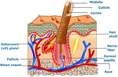
Sweat and sebaceous glands in the skin eliminate certain wastes through their secretions. Sweat produced by the sweat glands primarily helps to cool the body and secondarily excretes Na+ and Cl, small quantities of urea and lactate.

Sebaceous glands eliminate certain substances like sterols, hydrocarbons and waxes through sebum that provides a protective oily covering for the skin. Small quantities of nitrogenous wastes are also excreted through saliva.

Kidneys play a major role in the process of excretion in humans. Kidneys help in the elimination of wastes from the body in the form of urine. Apart from kidneys, organs like lungs, liver, skin and sebaceous glands help in excretion.

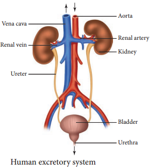
The excretory system in humans consists mainly of the kidneys and bladder. The kidneys filter urea and other waste products from the blood, which are then added to the urine within the bladder. Other organs, such as the liver, process toxins but put their wastes back into the blood.

Excretory Organs. Organs of excretion include the skin, liver, large intestine, lungs, and kidneys (see the figure below). Together, these organs make up the excretory system. They all excrete wastes, but they don’t work together in the same way that organs do in most other body systems. The appendix is a vestigial organ that has no role to play in excretion.

The liver regulates most chemical levels in the blood and excretes a product called bile. This helps carry away waste products from the liver. Production of bile, which helps carry away waste and break down fats in the small intestine during digestion. Production of certain proteins for blood plasma.

These chemical reactions produce waste products such as carbon dioxide, water, salts, urea and uric acid. Accumulation of these wastes beyond a level inside the body is harmful to the body. The excretory organs remove these wastes. This process of removal of metabolic waste from the body is known as excretion.

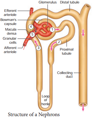
Humans have two kidneys and each kidney is supplied with blood from the renal artery. The kidneys remove from the blood the nitrogenous wastes such as urea, as well as salts and excess water, and excrete them in the form of urine.

Role of Liver in Excretion:

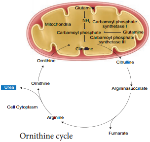
Liver converts the amino acids present in blood into ammonia and pyruvic acid. Pyruvic acid gets oxidized to release energy and ammonia gets converted into urea. Kidney helps in the filtration of the urea and urea gets excreted in the form of urine.

Certain waste and harmful substances are formed during the functioning of body cells. When these toxic materials are not removed from the body, they get mixed with blood and can damage the cells of the body. The removal of such poisonous waste materials is therefore necessary.

Skin has an important role in excretion in man . So Skin is important to clean our body by the process of excretion. Lungs release Carbon dioxide (CO2) which helps in the process of respiration and purifies blood. Intestine helps in excretion of food digested in stomach and in duodenum.

Excretion, the process by which animals rid themselves of waste products and of the nitrogenous by-products of metabolism. Through excretion organisms control osmotic pressure the balance between inorganic ions and water and maintain acid-base balance.
Role of Other Organs in Excretion img 1

The Physiology of Micturition Definition and its Uses

Learninsta presents the core concepts of Biology with high-quality research papers and topical review articles.

The Physiology of Micturition Definition and its Uses

The process of release of urine from the bladder is called micturition or urination. Urine formed by the nephrons is ultimately carried to the urinary bladder where it is stored till it receives a voluntary signal from the central nervous system. The stretch receptors present in the urinary bladder are stimulated when it gets filled with urine.

Stretching of the urinary bladder stimulates the CNS via the sensory neurons of the parasympathetic nervous system and brings about contraction of the bladder. Simultaneously, somatic motor neurons induce the sphincters to close. Smooth muscles contracts resulting in the opening of the internal sphincters passively and relaxing the external sphincter.

When the stimulatory and inhibitory controls exceed the threshold, the sphincter opens and the urine is expelled out. An adult human on an average excretes 1 to 1.5 L of urine per day. The urine formed is a yellow coloured watery fluid which is slightly acidic in nature (pH 6.0), Changes in diet may cause pH to vary between 4.5 to 8.0 and has a characteristic odour. The yellow colour of the urine is due to the presence of a pigment, urochrome.

On an average, 25-30 gms of urea is excreted per day. Various metabolic disorders can affect the composition of urine. Analysis of urine helps in clinical diagnosis of various metabolic disorders and the malfunctioning of the kidneys. For example the presence of glucose (glucosuria) and ketone bodies (ketonuria) in the urine are indications of diabetes mellitus.

The exact cause of micturition syncope isn’t fully understood. But it may be related to opening (vasodilation) of the blood vessels that occurs when getting up and standing at the toilet or that occurs at the rapid emptying of a full bladder. This is thought to result in a sudden drop in blood pressure.

Micturition involves the simultaneous coordinated contraction of the bladder detrusor muscle, which is controlled by parasympathetic (cholinergic) nerves, and the relaxation of the bladder neck and sphincter, which are controlled by sympathetic (α-adrenergic) nerves.

Micturition syncope causes more than 8 percent of all episodes of fainting. People who experience it are more prone to fainting under other circumstances, too. Micturition syncope occurs more often in men. It often happens after using the bathroom in the middle of the night or first thing in the morning.

The pons is a major relay center between the brain and the bladder. The mechanical process of urination is coordinated by the pons in the area known as the pontine micturition center (PMC). The conscious sensations associated with bladder activity are transmitted to the pons from the cerebral cortex.

Introduction. Micturition is the process of eliminating water and electrolytes from the urinary system, commonly known as urinating. It has two discrete phases: the storage/continence phase, when urine is stored in the bladder; and the voiding phase, where urine is released through the urethra.

Micturition is the process by which the urine from the urinary bladder is excreted. This reflex stimulates the urge to pass out urine. To discharge urine, the urethral sphincter relaxes and the smooth muscles of the bladder contract. This forces the urine out from the bladder.
Micturition img 1

Regulation of Kidney Function

Learninsta presents the core concepts of Biology with high-quality research papers and topical review articles.

Regulation of Kidney Function

ADH and Diabetes Insipidus

The functioning of kidneys is efficiently monitored and regulated by hormonal feedback control mechanism involving the hypothalamus, juxta glomerular apparatus and to a certain extent the heart. Osmoreceptors in the hypothalamus are activated by changes in the blood volume, body fluid volume and ionic concentration.

When there is excessive loss of fluid from the body or when there is an increase in the blood pressure, the osmoreceptors of the hypothalamus respond by stimulating the neurohypophysis to secrete the antidiuretic hormone (ADH) or vasopressin (a positive feedback). ADH facilitates reabsorption of water by increasing the number of aquaporins on the cell surface membrane of the distal convoluted tubule and collecting duct.

This increase in aquaporins causes the movement of water from the lumen into the interstitial cells, thereby preventing excess loss of water by diuresis. When you drink excess amounts of your favourite juice, osmoreceptors of the hypothalamus is no longer stimulated and the release of ADH is suppressed from the neurohypophysis (negative feedback) and the aquaporins of the collecting ducts move into the cytoplasm.

This makes the collecting ducts impermeable to water and the excess fluid flows down the collecting duct without any water loss. Hence dilute urine is produced to maintain the blood volume. Vasopressin secretion is controlled by positive and negative feedback mechanism.

Defects in ADH receptors or inability to secrete ADH leads to a condition called diabetes insipidus, characterized by excessive thirst and excretion of large quantities of dilute urine resulting in dehydration and fall in blood pressure.

Renin Angiotensin

Juxta glomerular apparatus (JGA) is a specialized tissue in the afferent arteriole of the nephron that consists of macula densa and granular cells. The macula densa cells sense distal tubular flow and affect afferent arteriole diameter, whereas the granular cells secrete an enzyme called renin. A fall in glomerular blood flow, glomerular blood pressure and glomerular filtration rate, can atctivate JG cells to release renin which converts a plasma protein, angiotensinogen (synthesized in the liver) to angiotensin I.

Angiotensin converting enzyme (ACE) converts angiotensin I to angiotensin II. Angiotensin II stimulates Na+ reabsorption in the proximal convoluted tubule by vasoconstriction of the blood vessels and increases the glomerular blood pressure.

Angiotensin II acts at different sites such as heart, kidney, brain, adrenal cortex and blood vessels. It stimulates adrenal cortex to secrete aldosterone that causes reabsorption of Na+, K+ excretion and absorption of water from the distal convoluted tubule and collecting duct.

This increases the glomerular blood pressure and glomerular filtration rate. This complex mechanism is generally known as Renin-AngiotensinAldosterone System (RAAS). Figure 8.9 shows the schematic representation of the various hormones in the regulation of body fluid concentration.
Regulation of Kidney Function img 1

Atrial Natriuretic Factor

Excessive stretch of cardiac atrial cells cause an increase in blood flow to the atria of the heart and release Atrial Natriuretic Peptide or factor (ANF) travels to the kidney where it increases Na+ excretion and increases the blood flow to the glomerulus, acting on the afferent glomerular arterioles as a vasodilator or on efferent arterioles as a vasoconstrictor.

It decreases aldosterone release from the adrenal cortex and also decreases release of renin, thereby decreasing angiotensin II. ANF acts antagonistically to the renin – angiotensin system, aldosterone and vasopressin.

Mechanism of Urine Formation in Human

Learninsta presents the core concepts of Biology with high-quality research papers and topical review articles.

Mechanism of Urine Formation in Human

The nitrogenous waste formed as a result of breakdown of amino acids is converted to urea in the liver by the Ornithine cycle or urea cycle (Figure 8.7).
Mechanism of Urine Formation in Human img 1

Urine formation involves three main processes namely, glomerular fitration, tubular reabsorption and tubular secretion.

(i) Glomerular Filtration

Blood enters the kidney from the renal artery, into the glomerulus. Blood is composed of large quantities of water, colloidal proteins, sugars, salts and nitrogenous end product. The first step in urine formation is the filtration of blood that takes place in the glomerulus.

This is called glomerular filtration which is a passive process. The fluid that leaves the glomerular capillaries and enters the Bowman’s capsule is called the glomerular filtrate.

The glomerular membrane has a large surface area and is more permeable to water and small molecules present in the blood plasma. Blood enters the glomerulus faster with greater force through the afferent arteriole and leaves the glomerulus through the efferent arterioles, much slower.

This force is because of the difference in sizes between the afferent and efferent arteriole (afferent arteriole is wider than efferent arteriole) and glomerular hydrostatic pressure which is around 55mm Hg.

Kidneys produce about 180L of glomerular filtrate in 24 hours. The molecules such as water, glucose, amino acids and nitrogenous substances pass freely from the blood into the glomerulus. Molecules larger than 5nm are barred from entering the tubule.

Glomerular pressure is the chief force that pushes water and solutes out of the blood and across the filtration membrane. The glomerular blood pressure (approximately 55 mmHg) is much higher than in other capillary beds. The two opposing forces are contributed by the plasma proteins in the capillaries.

These includes, colloidal osmotic pressure (30 mmHg) and the capsular hydrostatic pressure (15 mmHg) due to the fluids in the glomerular capsule. The net filtration pressure of 10 mmHg is responsible for the renal filtration.

Net filtration Pressure = Glomerular
hydrostatic pressure – (Colloidal osmotic pressure + Capsular hydrostatic pressure).
Net filtration pressure = 55 mmHg – (30 mmHg + 15 mmHg) = 10mmHg

The effective glomerular pressure of 10 mmHg results in ultrafiltration. Glomerular filtration rate (GFR) is the volume of filtrate formed min-1 in all nephrons (glomerulus) of both the kidneys. In adults the GFR is approximately 120-125mL/min. Blood from the glomerulus is passed out through the efferent arteriole.

The smooth muscle of the efferent arteriole contract resulting in vasoconstriction. Table 8.1 shows the relative concentrations of substances in the blood plasma and the glomerular filtrate. The glomerular filtrate is similar to blood plasma except that there are no plasma proteins.

In cortical nephrons, blood from efferent arteriole flows into peritubular capillary beds and enters the venous system carrying with it recovered solutes and water from the interstitial fluid that surrounds the tubule.

Table 8.1 Concentration of substances in the blood plasma and in the glomerular filtrate
Mechanism of Urine Formation in Human img 2

(ii) Tubular Reabsorption

This involves movement of the filtrate back into the circulation. The volume of filtrate formed per day is around 170-180 L and the urine released is around 1.5 L per day, i.e., nearly 99% of the glomerular filtrate that has to be reabsorbed by the renal tubules as it contains certain substances needed by the body.

This process is called selective reabsorption. Reabsorption takes place by the tubular epithelial cells in different segments of the nephron either by active transport or passive transport, diffusion and osmosis.

Proximal Convoluted Tubule (PCT):

Glucose, lactate, amino acids, Na+ and water in the filtrate is reabsorbed in the PCT. Sodium is reabsorbed by active transport through sodium-potassium (Na+K+) pump in the PCT. Small amounts of urea and uric acid are also reabsorbed.

Descending Limb

Of Henle’s loop is permeable to water due the presence of aquaporins, but not permeable to salts. Water is lost in the descending limb, hence Na+ and Cl gets concentrated in the filtrate.

Ascending Limb of Henle’s Loop

Is impermeable to water but permeable to solutes such as Na+, Cl and K+.

The distal convoluted tubule recovers water and secretes potassium into the tubule. Na+, Cl and water remains in the filtrate of the DCT. Most of the reabsorption from this point is dependent on the body’s need and is regulated by hormones. Reabsorption of bicarbonate (HCO3) takes place to regulate the blood pH. Homeostasis of K+ and Na+ in the blood is also regulated in this region.

Collecting Duct

Is permeable to water, secretes K+ (potassium ions are actively transported into the tubule) and reabsorbs Na+ to produce concentrated urine. The change in permeability to water is due to the presence of number of waterpermeable channels called aquaporins.

Tubular Secretion:

Substances such as H+, K+, NH4+, creatinine and organic acids move into the filtrate from the peritubular capillaries into the tubular fluid. Most of the water is absorbed in the proximal convoluted tubule and Na+ is exchanged for water in the loop of Henle. Hypotonic fluid enters the distal convoluted tubule and substances such as urea and salts pass from peritubular blood into the cells of DCT.

The urine excreted contains both filtered and secreted substances. Once it enters the collecting duct, water is absorbed and concentrated hypertonic urine is formed. For every H+ secreted into the tubular filtrate, a Na+ is absorbed by the tubular cell.

The H+ secreted combines with HCO3, HPO3 and NH3and gets fixed as H2CO4+, H2PO4+ and NH4+ respectively. Since H+ gets fixed in the fluid, reabsorption of H+ is prevented.

Formation of Concentrated Urine

Formation of concentrated urine is accomplished by kidneys using counter current mechanisms. The major function of Henle’s loop is to concentrate Na+ and Cl. There is low osmolarity near the cortex and high osmolarity towards the medulla.

This osmolarity in the medulla is due to the presence of the solute transporters and is maintained by the arrangement of the loop of Henle, collecting duct and vasa recta. This arrangement allows movement of solutes from the filtrate to the interstitial fluid. At the transition between the proximal convoluted tubule and the descending loop of Henle the osmolarity of the interstitial fluid is similar to that of the blood – about 300mOsm.

Ascending and Descending Limbs of Henle, Create a Counter Current Multiplier

(Interaction between flow of filtrate through the limbs of Henle’s and JMN) by active transport. Figure 8.8 (a) shows the counter current multiplier created by the long loops of Henle of the JM nephrons which creates medullary osmotic gradient.
Mechanism of Urine Formation in Human img 3

As the fluid enters the descending limb, water moves from the lumen into the interstitial fluid and the osmolarity of interstitial fluid decreases. To counteract this dilution the region of the ascending limb actively pumps solutes from the lumen into the interstitial fluid and the osmolarity increases to about 1200mOsm in medulla. This mismatch between water and salts creates osmotic gradient in the medulla. The osmotic gradient is also due to the permeability of the collecting duct to urea.

The vasa recta, maintains the medullary osmotic gradient via counter current exchanger (the flow of blood through the ascending and descending vasa recta blood vessels) by passive transport. Figure 8.8 (b) shows counter current exchanger where the vasa recta preserves the medullary gradient while removing reabsorbed water and solutes.

This system does not produce an osmotic gradient, but protects the medulla by removal of excess salts from the interstitial fluid and removing reabsorbed water. The vasa recta leave the kidney at the junction between the cortex and medulla. The interstitial fluid at this point is iso-osmotic to the blood.

When the blood leaves the efferent arteriole and enters vasa recta the osmolarity in the medulla increases (1200mOsm) and results in passive uptake of solutes and loss of water in descending vasa recta. As the blood enters the cortex, the osmolarity in the blood decreases (300mOsm) and the blood loses solutes and gains water.

At the final stage in collecting duct to form concentrated urine (hypertonic). Human kidneys can produce urine nearly four times concentrated than the initial filtrate formed.
Mechanism of Urine Formation in Human img 4

Human Excretory System – Structure of Kidney, Nephron

Learninsta presents the core concepts of Biology with high-quality research papers and topical review articles.

Human Excretory System – Structure of Kidney, Nephron

Structure of kidney

Excretory system in human consists of a pair of kidneys, a pair of ureters, urinary bladder and urethra (Figure. 8.2). Kidneys are reddish brown, bean shaped structures that lie in the superior lumbar region between the levels of the last thoracic and third lumber vertebra close to the dorsal inner wall of the abdominal cavity.

The right kidney is placed slightly lower than the left kidney. Each kidney weighs an average of 120-170 grams. The outer layer of the kidney is covered by three layers of supportive tissues namely, renal fascia, perirenal fat capsule and fibrous capsule.
Human Excretory System - Structure of Kidney, Nephron img 1

The longitudinal section of kidney (Figure. 8.3) shows, an outer cortex, inner medulla and pelvis. The medulla is divided into a few conical tissue masses called medullary pyramids or renal pyramids. The part of cortex that extends in between the medullary pyramids is the renal columns of Bertini.
Human Excretory System - Structure of Kidney, Nephron img 2

The centre of the inner concave surface of the kidney has a notch called the renal hilum, through which ureter, blood vessels and nerves innervate. Inner to the hilum is a broad funnel shaped space called the renal pelvis with projection called calyces.

The renal pelvis is continuous with the ureter once it leaves the hilum. The walls of the calyces, pelvis and ureter have smooth muscles which contracts rhythmically. The calyces collect the urine and empties into the ureter, which is stored in the urinary bladder temporarily. The urinary bladder opens into the urethra through which urine is expelled out.

Structure of a Nephron

Each kidney has nearly one million complex tubular structures called nephron (Figure 8.4). Each nephron consists of a filtering corpuscle called renal corpuscle (malpighian body) and a renal tubule. The renal tubule opens into a longer tubule called the collecting duct. The renal tubule begins with a double walled cup shaped structure called the Bowman’s capsule, which encloses a ball of capillaries that delivers fluid to the tubules, called the glomerulus.

The Bowman’s capsule and the glomerulus together constitute the renal corpuscle. The endothelium of glomerulus has many pores (fenestrae). The external parietal layer of the Bowman’s capsule is made up of simple squamous epithelium and the visceral layer is made of epithelial cells called podocytes. The podocytes end in foot processes which cling to the basement membrane of the glomerulus. The openings between the foot processes are called filtration slits.
Human Excretory System - Structure of Kidney, Nephron img 3

The renal tubule continues further to form the proximal convoluted tubule [PCT] followed by a U-shaped loop of Henle (Henle’s loop) that has a thin descending and a thick ascending limb. The ascending limb continues as a highly coiled tubular region called the distal convoluted tubule [DCT].

The DCT of many nephrons open into a straight tube called collecting duct. The collecting duct runs through the medullary pyramids in the region of the pelvis. Several collecting ducts fuse to form papillary duct that delivers urine into the calyces, which opens into the renal pelvis.

In the renal tubules, PCT and DCT of the nephron are situated in the cortical region of the kidney whereas the loop of Henle is in the medullary region. In majority of nephrons, the loop of Henle is too short and extends only very little into the medulla and are called cortical nephrons. Some nephrons have very long loop of Henle that run deep into the medulla and are called juxta medullary nephrons (JMN) (Figure 8.5 a and b)
Human Excretory System - Structure of Kidney, Nephron img 4

The capillary bed of the nephrons. First capillary bed of the nephron is the glomerulus and the other is the peritubular capillaries. The glomerular capillary bed is different from other capillary beds in that it is supplied by the afferent and drained by the efferent arteriole.

The efferent arteriole that comes out of the glomerulus forms a fine capillary network around the renal tubule called the peritubular capillaries. The efferent arteriole serving the juxta medullary nephron forms bundles of long straight vessel called vasa recta and runs parallel to the loop of Henle. Vasa recta is absent or reduced in cortical nephrons (Figure 8.6).
Human Excretory System - Structure of Kidney, Nephron img 5

Models of Excretion Definition and Its Explanation

Learninsta presents the core concepts of Biology with high-quality research papers and topical review articles.

Models of Excretion Definition and Its Explanation

Excretory system helps in collecting nitrogenous waste and expelling it into the external environment. Animals have evolved different strategies to get rid of these nitrogenous wastes. Ammonia produced during amino acid breakdown is toxic hence must be excreted either as ammonia, urea or uric acid.

The type of nitrogenous end product an animal excretes depends upon the habitat of the animal. Ammonia requires large amount of water for its elimination, whereas uric acid, being the least toxic can be removed with the minimum loss of water, and urea can be stored in the body for considerable periods of time, as it is less toxic and less soluble in water than ammonia.

Animals that excrete most of its nitrogen in the form of ammonia are called ammonoteles. Many fishes, aquatic amphibians and aquatic insects are ammonotelic. In bony fishes, ammonia diffuses out across the body surface or through gill surface as ammonium ions.

Reptiles, birds, land snails and insects excrete uric acid crystals, with a minimum loss of water and are called uricoteles. In terrestrial animals, less toxic urea and uric acid are produced to conserve water. Mammals and terrestrial amphibians mainly excrete urea and are called ureoteles. Earthworms while in soil are ureoteles and when in water are ammonoteles. Figure 8.1 shows the excretory products in different groups of animals.
Models of Excretion img 1

The animal kingdom presents a wide variety of excretory structures. Most invertebrates have a simple tubular structure in the form of primitive kidneys called protonephridia and metanephridia. Vertebrates have complex tubular organs called kidneys.

Protonephridia are excretory structures with specialized cells in the form of flame cells (cilia) in Platyhelminthes (example tapeworm) and Solenocytes (flagella) in Amphioxus. Nematodes have rennette cells, Metanephridia are the tubular excretory structures in annelids and molluscs.

Malpighian tubules are the excretory structures in most insects. Antennal glands or green glands perform excretory function in crustaceans like prawns. Vertebrate kidney differs among taxa in relation to the environmental conditions.

Nephron is the structural and functional unit of kidneys. Reptiles have reduced glomerulus or lack glomerulus and Henle’s loop and hence produce very little hypotonic urine, whereas mammalian kidneys produce concentrated (hyperosmotic) urine due to the presence of long Henle’s loop.

The Loop of Henle of the nephron has evolved to form hypertonic urine. Aglomerular kidneys of marine fishes produce little urine that is isoosmotic to the body fluid. Amphibians and fresh water fish lack Henle’s loop hence produce dilute urine (hypoosmotic).

Cardio Pulmonary Resuscitation (CPR)

Learninsta presents the core concepts of Biology with high-quality research papers and topical review articles.

Cardio Pulmonary Resuscitation (CPR)

In 1956, James Elam and Peter Safar were the first to use mouth to mouth resuscitation. CPR is a life saving procedure that is done at the time of emergency conditions such as when a person’s breath or heart beat has stopped abruptly in case of drowning, electric shock or heart attack.

CPR includes rescue of breath, which is achieved by mouth to mouth breathing, to deliver oxygen to the victim’s lungs by external chest compressions which helps to circulate blood to the vital organs.

CPR must be performed within 4 to 6 minutes after cessation of breath to prevent brain damage or death. Along with CPR, defibrillation is also done. Defibrillation means a brief electric shock is given to the heart to recover the function of the heart.

Cardiopulmonary resuscitation (CPR) is an emergency procedure that combines chest compressions often with artificial ventilation in an effort to manually preserve intact brain function until further measures are taken to restore spontaneous blood circulation and breathing in a person who is in cardiac arrest.

5 Steps for Performing CPR

  • Check the patient’s responsiveness.
  • Shake the unresponsive person by the shoulders and speak loudly to them in an attempt to rouse them.
  • Check their breathing and pulse.
  • Administer chest compressions.
  • Recheck breathing and pulse.

After every 30 chest compressions at a rate of 100 to 120 a minute, give 2 breaths. Continue with cycles of 30 chest compressions and 2 rescue breaths until they begin to recover or emergency help arrives.

Cardiopulmonary resuscitation (CPR) is a lifesaving technique. It aims to keep blood and oxygen flowing through the body when a person’s heart and breathing have stopped. CPR can be performed by any trained person. It involves external chest compressions and rescue breathing.

Types of CPR

High-Frequency Chest Compressions. This technique involves imitating hear beats by giving more chest compressions at intervals of time in high frequency. Open-Chest CPR. Open chest CPR is a procedure in which the heart is retrieved through thoracotomy. Interposed Abdominal Compression CPR.

How is CPR Performed? There are two commonly known versions of CPR: For healthcare providers and those trained: conventional CPR using chest compressions and mouth-to-mouth breathing at a ratio of 30:2 compressions-to-breaths.

CPR stands for cardiopulmonary resuscitation. It is an emergency life-saving procedure that is done when someone’s breathing or heartbeat has stopped.

The three basic parts of CPR are easily remembered as “CAB”: C for compressions, A for airway, and B for breathing. C is for compressions. Chest compressions can help the flow of blood to the heart, brain, and other organs.
Cardio Pulmonary Resuscitation (CPR) img 1

Disorders of Circulatory System – Types, Causes and Risk

Learninsta presents the core concepts of Biology with high-quality research papers and topical review articles.

Disorders of the Circulatory System – Types, Causes and Risk

Hypertension

Hypertension is the most common circulatory disease. The normal blood pressure in man is 120/80 mmHg. In cases when the diastolic pressure exceeds 90 mm Hg and the systolic pressure exceeds 150 mm Hg persistently, the condition is called hypertension. Uncontrolled hypertension may damage the heart, brain and kidneys.

Coronary Heart Disease

Coronary heart disease occurs when the arteries are lined by atheroma. The build up of atheroma contains cholesterol, fires, dead muscle and platelets and is termed Atherosclerosis. The cholesterol rich atheroma forms plaques in the inner lining of the arteries making them less elastic and reduces the blood flow. Plaque grows within the artery and tends to form blood clots, forming coronary thrombus. Thombus in a coronary artery results in heart attack.

Stroke

Stroke is a condition when the blood vessels in the brain bursts, (Brain haemorrhage) or when there is a block in the artery that supplies the brain, (atherosclerosis) or thrombus. The part of the brain tissue that is supplied by this damaged artery dies due to lack of oxygen (cerebral infarction).

Angina Pectoris

Angina pectoris (ischemic pain in the heart muscles) is experienced during early stages of coronary heart disease. Atheroma may partially block the coronary artery and reduce the blood supply to the heart. As a result, there is tightness or choking with difficulty in breathing. This leads to angina or chest pain. Usually it lasts for a short duration of time.

Myocardial Infarction (Heart Failure)

The prime defect in heart failure is a decrease in cardiac muscle contractility. The Frank – Starling curve shifts downwards and towards the right such that for a given EDV, a failing heart pumps out a smaller stroke volume than a normal healthy heart.

When the blood supply to the heart muscle or myocardium is remarkably reduced it leads to death of the muscle fires. This condition is called heart attack or myocardial infarction. The blood clot or thrombosis blocks the blood supply to the heart and weakens the muscle fires.

It is also called Ischemic heart disease due to lack of oxygen supply to the heart muscles. If this persists it leads to chest pain or angina. Prolonged angina leads to death of the heart muscle resulting in heart failure.

Rheumatoid Heart Disease

Rheumatic fever is an autoimmune disease which occurs 2-4 weeks after throat infection usually a streptococcal infection. The antibodies developed to combat the infection cause damage to the heart. Effects include firous nodules on the mitral valve, firosis of the connective tissue and accumulation of fluid in the pericardial cavity.
Disorders of the Circulatory System img 1

Cardiac Cycle and Regulation of Cardiac Activity

Learninsta presents the core concepts of Biology with high-quality research papers and topical review articles.

Cardiac Cycle and Regulation of Cardiac Activity

The type of heart in human is myogenic because the heart beat originates from the muscles of the heart. The nervous and endocrine systems work together with paracrine signals (metabolic activity) to influence the diameter of the arterioles and alter the blood flow. The neuronal control is achieved through autonomic nervous system (sympathetic and parasympathetic).

Sympathetic neurons release norepinephrine and adrenal medulla releases epinephrine. The two hormones bind to β – adrenergic receptors and increase the heart rate. The parasympathetic neurons secrete acetylcholine that binds to muscarinic receptors and decreases the heart beat.

Vasopressin and angiotensin II, involved in the regulation of the kidneys, results in vasoconstriction while natriuretic peptide promotes vasodilation. Vagus nerve is a parasympathetic nerve that supplies the atrium especially the SA and the AV nodes.

Cardiac cycle helps in the circulation of blood. The cardiac cycle is a normal activity of the human heart and is regulated automatically by the nodal tissues – sinoatrial node (SA node) and atrioventricular node (AV node). The variation in the cardiac cycle results in an increase or decrease in the cardiac output

There are two primary modes by which the blood volume pumped by the heart, at any given moment, is regulated:

  • Intrinsic cardiac regulation, in response to changes in the volume of blood flowing into the heart; and
  • Control of heart rate and cardiac contractility by the autonomic nervous system.

The failure of the pumping action of the heart, resulting in loss of consciousness and absence of pulse and breathing: a medical emergency requiring immediate resuscitative treatment. cardiac arrest, cardiac pacemaker, cardialgic, Caria. cardiac arrest. n. cardiac inefficacity by cardiac tachycardia.

The rhythmic control of the cardiac cycle and its accompanying heartbeat relies on the regulation of impulses generated and conducted within the heart. Systole occurs when the ventricles of the heart contract and diastole occurs between ventricular contractions when the right and left ventricles relax and fill.

The principal functions of the heart are regulated by the sympathetic and parasympathetic divisions of the autonomic nervous system. In general, the sympathetic nerves to the heart are facilitatory, whereas the parasympathetic (vagus) nerves are inhibitory.

The main purpose of the heart is to pump blood through the body; it does so in a repeating sequence called the cardiac cycle. The cardiac cycle is the coordination of the filling and emptying of the heart of blood by electrical signals that cause the heart muscles to contract and relax.

It induces the force of contraction of the heart and its heart rate. In addition, it controls the peripheral resistance of blood vessels. The ANS has both sympathetic and parasympathetic divisions that work together to maintain balance.

One part of the autonomic nervous system is a pair of nerves called the vagus nerves, which run up either side of the neck. These nerves connect the brain with some of our internal organs, including the heart.

Sympathetic efferent nerves are present throughout the atria, ventricles (including the conduction system), and myocytes in the heart and also the sinoatrial (SA) and atrioventricular (AV) nodes.
Regulation of Cardiac Activity img 1

Double Circulation – Blood Circulation in Humans

Learninsta presents the core concepts of Biology with high-quality research papers and topical review articles.

Double Circulation – Blood Circulation in Humans

Circulation of the blood was first described by William Harvey (1628). There are two types of blood circulation in vertebrates, single circulation and double circulation which is shown in Figure 7.10 (a and b) and 7.11.
Double Circulation - Blood Circulation in Humans img 1
Double Circulation - Blood Circulation in Humans img 2

The blood circulates twice through the heart first on the right side then on the left side to complete one cardiac cycle. The complete double blood circulation is more prominent in mammals because of the complete partition of all the chambers (Auricles and ventricles) in the heart.

In systemic circulation, the oxygenated blood entering the aorta from the left ventricle is carried by a network of arteries, arterioles and capillaries to the tissues. The deoxygenated blood from the tissue is collected by venules, veins and vena cava and emptied into the right atrium.

In pulmonary circulation, the blood from heart (right ventricle) is taken to the lungs by pulmonary artery and the oxygenated blood from the lungs is emptied into the left auricle by the pulmonary vein.

Completely separated circuits have an important advantage. Different pressures are maintained in the pulmonary and systemic circulation. Why is this advantageous? In the lungs the capillaries must be very thin to allow gas exchange, but if the blood flows through these thin capillaries under high pressure the fluid can leak through or ruptures the capillary walls and can accumulate in the tissues.

This increases the diffusion distance and reduces the efficiency of the gas exchange. In contrast high pressure is required to force blood through the long systemic circuits. Hence the arteries close to the heart have increased pressure than the arteries away from the heart. Completely separated circuits (pulmonary and systemic) allow these two different demands to be met with.

Human Circulatory System

Learninsta presents the core concepts of Biology with high-quality research papers and topical review articles.

Human Circulatory System

The structure of the heart was described by Raymond de viessens, in 1706. Human heart is made of special type of muscle called the cardiac muscle. It is situated in the thoracic cavity and its apex portion is slightly tilted towards left. It weighs about 300g in an adult. The size of our heart is roughly equal to a closed fist. Heart is divided into four chambers, upper two small auricles or atrium and lower two large ventricles.

The walls of the ventricles are thicker than the auricles due to the presence of papillary muscles. The heart wall is made up of three layers, the outer epicardium, middle myocardium and inner endocardium. The space present between the membranes is called pericardial space and is filled with pericardial fluid.

The two auricles are separated by inter auricular septum and the two ventricles are separated by inter ventricular septum. The separation of chambers avoids mixing of oxygenated and deoxygenated blood. The auricle communicates with the ventricle through an opening called auriculo ventricular aperture which is guarded by the auriculo ventricular valves.

The opening between the right atrium and the right ventricle is guarded by the tricuspid valve (three flaps or cusps), whereas a bicuspid (two flaps or cusps) or mitral valve guards the opening between the left atrium and left ventricle (Figure 7.6). The valves of the heart allows the blood to flow only in one direction, i.e., from the atria to the ventricles and from the ventricles to the pulmonary artery or the aorta.
Human Circulatory System img 1

The opening of right and left ventricles into the pulmonary artery and aorta are guarded by aortic and pulmonary valves and are called semilunar valves.

Each semilunar valve is made of three half – moon shaped cusps. The myocardium of the ventricle is thrown into irregular muscular ridges called trabeculae corneae. The trabeculae corneae are modified into chordae tendinae.

The opening and closing of the semilunar valves are achieved by the chordae tendinae. The chordae tendinae are attached to the lower end of the heart by papillary muscles. Heart receives deoxygenated blood from various parts of the body through the inferior venacava and superior venacava which open into the right auricle. Oxygenated blood from lungs is drained into the left auricle through four pulmonary veins.

Origin and Conduction of Heart Beat

The heart in human is myogenic (cardiomyocytes can produce spontaneous rhythmic depolarisation that initiates contractions). The sequence of electrical conduction of heart is shown in Figure 7.7. The cardiac cells with fastest rhythm are called the Pacemaker cells, since they determine the contraction rate of the entire heart.

These cells are located in the right sinuatrial (SA) node / Pacemaker. On the left side of the right atrium is a node called auriculo ventricular node (AV node). Two special cardiac muscle fibres originate from the auriculo ventricular node and are called the bundle of His which runs down into the interventricular septum and the fibres spread into the ventricles. These fibres are called the Purkinje fibres.
Human Circulatory System img 2

Pacemaker cells produce excitation through depolarisation of their cell membrane. Early depolarisation is slow and takes place by sodium influx and reduction in potassium efflux. Minimum potential is required to activate voltage gated calcium (Ca+) channels that causes rapid depolarisation which results in action potential. The pace maker cells repolarise slowly via K+ efflux.

HEART BEAT:

Rhythmic contraction and expansion of heart is called heart beat. The contraction of the heart is called systole and the relaxation of the heart is called diastole. The heart normally beats 70-72 times per min in a human adult. During each cardiac cycle two sounds are produced that can be heard through a stethoscope.

The first heart sound (lub) is associated with the closure of the tricuspid and bicuspid valves whereas second heart sound (dub) is associated with the closure of the semilunar valves. These sounds are of clinical diagnostic significance. An increased heart rate is called tachycardia and decreased heart rate is called bradycardia.

Cardiac Cycle

The events that occur at the beginning of heart beat and lasts until the beginning of next beat is called cardiac cycle. It lasts for 0.8 seconds. The series of events that takes place in a cardiac cycle.

PHASE 1:

Ventricular Diastole:

The pressure in the auricles increases than that of the ventricular pressure. AV valves are open while the semi lunar valves are closed. Blood flows from the auricles into the ventricles passively.

PHASE 2:

Atrial Systole:

The atria contracts while the ventricles are still relaxed. The contraction of the auricles pushes maximum volume of blood to the ventricles until they reach the end diastolic volume (EDV). EDV is related to the length of the cardiac muscle fibre. More the muscle is stretched, greater the EDV and the stroke volume.

PHASE 3:

Ventricular Systole (isovolumetric contraction):

The ventricular contraction forces the AV valves to close and increases the pressure inside the ventricles. The blood is then pumped from the ventricles into the aorta without change in the size of the muscle fibre length and ventricular chamber volume (isovolumetric contraction).

PHASE 4:

Ventricular Systole (ventricular ejection):

Increased ventricular pressure forces the semilunar valves to open and blood is ejected out of the ventricles without backflow of blood. This point is the end of systolic volume (ESV).

PHASE 5:

(Ventricular Diastole):

The ventricles begins to relax, pressure in the arteries exceeds ventricular pressure, resulting in the closure of the semilunar valves. The heart returns to phase 1 of the cardiac cycle.

Cardiac Output

The amount of blood pumped out by each ventricle per minute is called cardiac output(CO). It is a product of heart rate (HR) and stroke volume (SV). Heart rate or pulse is the number of beats per minute. Pulse pressure = systolic pressure – diastolic pressure.

Stroke volume (SV) is the volume of blood pumped out by one ventricle with each beat. SV depends on ventricular contraction. CO = HR X SV. SV represents the difference between EDV (amount of blood that collects in a ventricle during diastole) and ESV (volume of blood remaining in the ventricle after contraction).

SV = EDV – ESV. According to Frank – Starling law of the heart, the critical factor controlling SV is the degree to which the cardiac muscle cells are stretched just before they contract. The most important factor stretching cardiac muscle is the amount of blood returning to the heart and distending its ventricles, venous return.

During vigorous exercise, SV may double as a result of venous return. Heart’s pumping action normally maintains a balance between cardiac output and venous return. Because the heart is a double pump, each
side can fail independently of the other. If the left side of the heart fails, it results in pulmonary congestion and if the right side fails, it results in peripheral congestion. Frank – Starling effect protects the heart from abnormal increase in blood volume.

Blood Pressure

Blood pressure is the pressure exerted on the surface of blood vessels by the blood. This pressure circulates the blood through arteries, veins and capillaries. There are two types of pressure, the systolic pressure and the diastolic pressure. Systolic pressure is the pressure in the arteries as the chambers of the heart contracts. Diastolic pressure is the pressure in the arteries when the heart chambers relax.

Blood pressure is measured using a sphygmomanometer (BP apparatus). It is expressed as systolic pressure/diastolic pressure. Normal blood pressure in man is about 120/80mm Hg. Mean arterial pressure is a function of cardiac output and resistance in the arterioles.

The primary reflex pathway for homeostatic control of mean arterial pressure is the baroreceptor reflex. The baroreceptor reflex functions every morning when you get out of bed. When you are lying flat the gravitational force is evenly distributed.

When you stand up, gravity causes blood to pool in the lower extremities. The decrease in blood pressure upon standing is known as orthostatic hypotension. Orthostatic reflex normally triggers baroreceptor reflex. This results in increased cardiac output and increased peripheral resistance which together increase the mean arterial pressure.

Electrocardiogram (ECG)

An electrocardiogram (ECG) records the electrical activity of the heart over a period of time using electrodes placed on the skin, arms, legs and chest. It records the changes in electrical potential across the heart during one cardiac cycle. The special flap of muscle which initiates the heart beat is called as sinu-auricular node or SA node in the right atrium.

It spreads as a wave of contraction in the heart. The waves of the ECG are due to depolarization and not due to contraction of the heart. This wave of depolarisation occurs before the beginning of contraction of the cardiac muscle. A normal ECG shows 3 waves designated as P wave, QRS complex and T wave as shown in Figure 7.8 and the stages of the ECG graph are shown in Figure 7.9.
Human Circulatory System img 3
Human Circulatory System img 4

P Wave (Atrial depolarisation)

It is a small upward wave and indicates the depolarisation of the atria. This is the time taken for the excitation to spread through atria from SA node. Contraction of both atria lasts for around 0.8-1.0 sec.

PQ Interval (AV node delay)

It is the onset of P wave to the onset of QRS complex. This is from the start of depolarisation of the atria to the beginning of ventricular depolarisation. It is the time taken for the impulse to travel from the atria to the ventricles (0.12-0.21sec). It is the measure of AV conduction time.

QRS Complex (Ventricular depolarisation)

No separate wave for atrial depolarisation in the ECG is visible. Atrial depolarisation occurs simultaneously with the ventricular depolarisation. The normal QRS complex lasts for 0.06-0.09 sec. QRS complex is shorter than the P wave, because depolarisation spreads through the Purkinjie fires. Prolonged QRS wave indicates delayed conduction through the ventricle, often caused due to ventricular hypertrophy or due to a block in the branches of the bundle of His.

ST Segment

It lies between the QRS complex and T wave. It is the time during which all regions of the ventricles are completely depolarised and reflects the long plateau phase before repolarisation. In the heart muscle, the prolonged depolarisation is due to retardation of K+ efflux and is responsible for the plateau. The ST segment lasts for 0.09 sec.

T Wave (Ventricular repolarisation)

It represents ventricular repolarisation. The duration of the T wave is longer than QRS complex because repolarisation takes place simultaneously throughout the ventricular depolarisation.