Apomixis Definition and its Types

Learninsta presents the core concepts of Biology with high-quality research papers and topical review articles.

Apomixis Definition and its Types

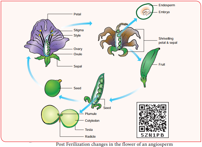
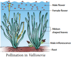
Reproduction involving fertilization in flowering plants is called amphimixis and wherever reproduction does not involve union of male and female gametes is called apomixis.

The term Apomixis was introduced by Winkler in the year 1908. It is defied as the substitution of the usual sexual system (Amphimixis) by a form of reproduction which does not involve meiosis and syngamy. Maheswari (1950) classifid Apomixis into two types – Recurrent and Non recurrent

Recurrent apomixis:
It includes vegetative reproduction and agamospermy.

Non recurrent apomixis:
Haploid embryo sac developed aftr meiosis, develops into a embryo without fertilization. The outline classifiation of Recurrent apomixis is given below.
Apomixis Definition and its Types img 1

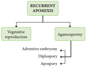
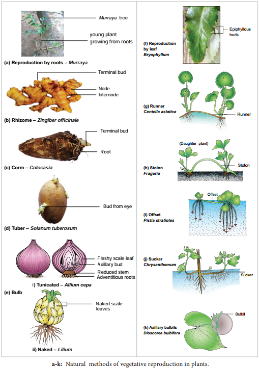
Vegetative reproduction:
Plants propagate by any part other than seeds

Bulbils – Fritillaria imperialis; Bulbs – Allium; Runner – Mentha arvensis; Sucker Chrysanthemum

Agamospermy:
It refers to processes by which Embryos are formed by eliminating meiosis and syngamy.

Adventive embryony:
An Embryo arises directly from the diploid sporophytic cells either from nucellus or integument. It is also called sporophytic budding because gametophytic phase is completely absent. Adventive embryos are found in Citrus and Mangifera.

Diplospory (Generative apospory):
A diploid embryo sac is formed from megaspore mother cell without a regular meiotic division Examples. Eupatorium and Aerva.

Apospory:
Megaspore mother cell (MMC) undergoes the normal meiosis and four megaspores formed gradually disappear. A nucellar cell becomes activated and develops into a diploid embryo sac. This type of apospory is also called somatic apospory. Examples Hieracium and Parthenium.

Post Fertilization Structure and Events

Learninsta presents the core concepts of Biology with high-quality research papers and topical review articles.

Post Fertilization Structure and Events

After fertilization, several changes take place in the floral parts up to the formation of the seed (Figure 1.20).
Post Fertilization Structure and Events img 1

The events after fertilization (endosperm, embryo development, formation of seed, fruits) are called post fertilization changes.
Post Fertilization Structure and Events img 2

Endosperm

The primary endosperm nucleus (PEN) divides immediately after fertilization but before the zygote starts to divide, to form the endosperm. The primary endosperm nucleus is the result of triple fusion (two polar nuclei and one sperm nucleus) and thus has 3n number of chromosomes. It is a nutritive tissue and regulatory structure that nourishes the developing embryo.

Depending upon the mode of development three types of endosperm are recognized in angiosperms. They are nuclear endosperm, cellular endosperm and helobial endosperm (Figure 1.21).
Post Fertilization Structure and Events img 3

Nuclear endosperm:
Primary Endosperm Nucleus undergoes several mitotic divisions without cell wall formation thus a free nuclear condition exists in the endosperm. Examples: Coccinia, Capsella and Arachis

Cellular endosperm:
Primary endosperm nucleus divides into 2 nuclei and it is immediately followed by wall formation. Subsequent divisions also follow cell wall formation. Examples: Adoxa, Helianthus and Scoparia

Helobial endosperm:
Primary Endosperm Nucleus moves towards base of embryo sac and divides into two nuclei. Cell wall formation takes place leading to the formation of a large micropylar and small chalazal chamber. The nucleus of the micropylar chamber undergoes several free nuclear division whereas that of chalazal chamber may or may not divide. Examples: Hydrilla and Vallisneria.

The endosperms may either be completely consumed by the developing embryo or it may persist in the mature seeds. These seeds without endosperms are called non-endospermous or ex-albuminous seeds. Examples: Pea, Groundnut and Beans. These seeds with endosperms are called endospermous or albuminous seeds. The endosperms in these seeds supply nutrition to the embryo during seed germination.
Examples: Paddy, Coconut and Castor.

Ruminate endosperm:
The endosperm with irregularity and unevenness in its surface forms ruminate endosperm. Examples: Areca catechu, Passiflra and Myristica

Functions of endosperm:

  • It is the nutritive tissue for the developing embryo.
  • In majority of angiosperms, the zygote divides only after the development of endosperm.
  • Endosperm regulates the precise mode of embryo development.

Development of Dicot embryo

The Stages involved in the development of Dicot embryo (Capsella bursa-pastoris – Onagrad or crucifer type) is given in Figure 1.22. The embryo develops at micropylar end of embryo sac. The zygote undergoes transverse division to form upper or terminal cell and lower or basal cell.

Further divisions in the zygote during the development lead to the formation of embryo. Embryo undergoes globular, heart shaped stages before reaching a mature stage. Th mature embryo has a radicle, two cotyledons and a plumule.
Post Fertilization Structure and Events img 4

Seed

The fertilized ovule is called seed and possesses an embryo, endosperm and a protective coat. Seeds may be endospermous (wheat, maize, barley and sunflower) or non endospermous. (Bean, Mango, Orchids and cucurbits).

Cicer seed (example for Dicot seed)

The mature seeds are attached to the fruit wall by a stalk called funiculus. The funiculus disappears leaving a scar called hilum. Below the hilum a small pore called micropyle is present. It facilitates entry of oxygen and water into the seeds during germination.

Each seed has a thick outer covering called seed coat. The seed coat is developed from integuments of the ovule. The outer coat is called testa and is hard whereas the inner coat is thin, membranous and is called tegmen.

In Pea plant the tegmen and testa are fused. Two cotyledons laterally attached to the embryonic axis and store the food materials in pea whereas in other seeds like castor the endosperm contains reserve food and the Cotyledons are thin. The portion of embryonal axis projecting beyond the cotyledons is called radicle or embryonic root.

The other end of the axis called embryonic shoot is the plumule. Embryonal axis above the level of cotyledon is called epicotyl whereas the cylindical region between the level of cotyledon is called hypocotyl (Figure 1.23 a).
Post Fertilization Structure and Events img 5

Oryza seed (example for Monocot seed)

The seed of paddy is one seeded and is called Caryopsis. Each seed remains enclosed by a brownish husk which consists of glumes arranged in two rows. The seed coat is a brownish, membranous layer closely adhered to the grain.

Endosperm forms the bulk of the grain and is the storage tissue. It is separated from embryo by a defiite layer called epithelium. The embryo is small and consists of one shieldshaped cotyledon known as scutellum present towards lateral side of embryonal axis.

A short axis with plumule and radicle protected by the root cap is present. The plumule is surrounded by a protective sheath called coleoptile. The radicle including root cap is also covered by a protective sheath called coleorhiza. The scutellum supplies the growing embryo with food material absorbed from the endosperm with the help of the epithelium (Figure 1.23 b).
Post Fertilization Structure and Events img 6

Fertilization of Asexual and sexual Reproduction in Plants

Learninsta presents the core concepts of Biology with high-quality research papers and topical review articles.

Fertilization of Asexual and sexual Reproduction in Plants

The fusion of male and female gamete is called fertilization. Double fertilization is seen in angiosperms.

Events of fertilization

The stages involved in double fertilization are:- germination of pollen to form pollen tube in the stigma; growth of pollen tube in the style; direction of pollen tube towards the micropyle of the ovule; entry of the pollen tube into one of the synergids of the embryo sac, discharge of male gametes; syngamy and triple fusion.

The events from pollen deposition on the stigma to the entry of pollen tube in to the ovule is called pollen – pistil interaction. It is a dynamic process which involves recognition of pollen and to promote or inhibit its germination and growth.
Fertilization of Asexual and sexual Reproduction in Plants img 1

Pollen on the stigma

In nature, a variety of pollens fall on the receptive stigma, but all of them do not germinate and bring out fertilization. The receptive surface of the stigma receives the pollen. If the pollen is compatible with the stigma it germinates to form a tube. This is facilitated by the stigmatic fluid in wet stigma and pellicle in dry stigma.

These two also decide the incompatibility and compatibility of the pollen through recognition rejection protein reaction between the pollen and stigma surface. Sexual incompatibility may exist between different species (interspecific) or between members of the same species (intraspecific). The latter is called self-incompatibility. The first visible change in the pollen, soon after it lands on stigma is hydration.

The pollen wall proteins are released from the surface. During the germination of pollen its entire content moves into the pollen tube. The growth is restricted to the tip of the tube and all the cytoplasmic contents move to the tip region.

The remaining part of the pollen tube is occupied by a vacuole which is cut of from the tip by callose plug. The extreme tip of pollen tube appears hemispherical and transparent when viewed through the microscope. This is called cap block. As soon as the cap block disappear the growth of the pollen tube stops.

Pollen tube in the style

After the germination the pollen tube enters into the style from the stigma. The growth of the pollen tube in the style depends on the type of style.

Types of style

There are three types of style

  1. Hollow or open style
  2. Solid style or closed style
  3. Semisolid or half closed style.

1. Hollow style (Open style):

It is common among monocots. A hollow canal running from the stigma to the base of the style is present. The canal is lined by a single layer of glandular canal cells (Transmitting tissue). They secrete mucilaginous substances. The pollen tube grows on the surface of the cells lining the stylar canal.

The canal is filed with secretions which serve as nutrition for growing pollen tubes and also controlling incompatibility reaction between the style and pollen tube. The secretions contain carbohydrates, lipids and some enzymes like esterases, acid phosphatases as well as compatibility controlling proteins.

2. Solid style (Closed type):

It is common among dicots. It is characterized by the presence of central core of elongated, highly specialised cells called transmitting tissue. This is equivalent to the lining cells of hollow style and does the same function. Its contents are also similar to the content of those cells. The pollen tube grows through the intercellular spaces of the transmitting tissue.

3. Semi-solid style (half closed type):

This is intermediate between solid and open type. There is a difference of opinion on the nature of transmitting tissue. Some authors consider that it is found only in solid styles while others consider the lining cells of hollow style also has transmitting tissue.

Entry of pollen tube into the ovule:-
There are three types of pollen tube entry into the ovule (Figure 1.18).
Fertilization of Asexual and sexual Reproduction in Plants img 2

Porogamy:-
when the pollen tube enters through the micropyle.

Chalazogamy:-
When the pollen tube enters through the integument.

Mesogamy:-
when the pollen tube enters through the integument.

Entry of pollen tube into embryo sac:

Irrespective of the place of entry of pollen tube into ovule, it enters the embryo sac at the micropylar end. Th pollen enters into embryo sac directly into one of the synergids. The growth of pollen tube towards the ovary, ovule and embryo sac is due to the presence of chemotropic substances.

The pollen tube after travelling the whole length of the style enters into the ovary locule where it is guided towards the micropyle of the ovule by a structure called obturator (See Do you know). After reaching the embryo sac, a pore is formed in pollen tube wall at its apex or just behind the apex.

The content of the pollen tube (two male gametes, vegetative nucleus and cytoplasm) are discharged into the synergids into which pollen tube enters. The pollen tube does not grow beyond it, in the embryo sac. The tube nucleus disorganizes.

Double fertilization and triple fusion

S.G. Nawaschin and L.Guignard in 1898 and 1899, observed in Lilium and Fritillaria that both the male gametes released from a male gametophyte are involved in the fertilization. They fertilize two diffrent components of the embryo sac.

Since both the male gametes are involved in fertilization, the phenomenon is called double fertilization and is unique to angiosperms. One of the male gametes fuses with the egg nucleus (syngamy) to form Zygote (Figure 1.19).
Fertilization of Asexual and sexual Reproduction in Plants img 3

The second gamete migrates to the central cell where it fuses with the polar nuclei or their fusion product, the secondary nucleus and forms the primary endosperm nucleus (PEN). Since this involves the fusion of three nuclei, this phenomenon is called triple fusion. This act results in endosperm formation which forms the nutritive tissue for the embryo.

Pre-Fertilization: Structures and Events

Learninsta presents the core concepts of Biology with high-quality research papers and topical review articles.

Pre-Fertilization: Structures and Events

The hormonal and structural changes in plant lead to the differentiation and development of floral primordium. The structures and events involved in prefertilization are given below.

Male Reproductive part Androecium

Androecium is made up of stamens. Each stamen possesses an anther and a fiament. Anther bears pollen grains which represent the male gametophyte. In this chapter we shall discuss the structure and development of anther in detail.

Development of anther:

A very young anther develops as a homogenous mass of cells surrounded by an epidermis. During its development, the anther assumes a fourlobed structure. In each lobe, a row or a few rows of hypodermal cells becomes enlarged with conspicuous nuclei. This functions as archesporium.

The archesporial cells divide by periclinal divisions to form primary parietal cells towards the epidermis and primary sporogenous cells towards the inner side of the anther. The primary parietal cells undergo a series of periclinal and anticlinal division and form 2-5 layers of anther walls composed of endothecium, middle layers and tapetum, from periphery to centre.

Microsporogenesis:

Th stages involved in the formation of haploid microspores from diploid microspore mother cell through meiosis is called Microsporogenesis. The primary sporogeneous cells directly, or may undergo a few mitotic divisions to form sporogenous tissue. The last generation of sporogenous tissue functions as microspore mother cells. Each microspore mother cell divides meiotically to form a tetrad of four haploid microspores
(microspore tetrad).

Microspores soon separate from one another and remain free in the anther locule and develop into pollen grains. The stages in the development of microsporangia is given in Figure 1.4. In some plants, all the microspores in a microsporangium remain held together called pollinium.

Example: Calotropis. Pollinia are attached to a clamp or clip like sticky structure called corpusculum. The fiamentous or thread like part arising from each pollinium is called retinaculum. The whole structure looks like inverted letter ‘Y’ and is called translator.
Pre-fertilization Structures and events img 1

T.S. of Mature anther

Transverse section of mature anther reveals the presence of anther cavity surrounded by an anther wall. It is bilobed, each lobe having 2 theca (dithecous). A typical anther is tetrasporangiate. The T.S. of Mature anther is given in Figure 1.5.
Pre-fertilization Structures and events img 2

1. Anther wall

The mature anther wall consists of the following layers

a. Epidermis
b. Endothecium
c. Middle layers
d. Tapetum.

a. Epidermis:

It is single layered and protective in function. The cells undergo repeated anticlinal divisions to cope up with the rapidly enlarging internal tissues.

b.Endothecium:

It is generally a single layer of radially elongated cells found below the epidermis. The inner tangential wall develops bands (sometimes radial walls also) of α cellulose (sometimes also slightly lignified). The cells are hygroscopic. In the anthers of aquatic plants, saprophytes, cleistogamous flowers and extreme parasites endothecial diffrentiation is absent.

The cells along the junction of the two sporangia of an anther lobe lack these thickenings. Ths region is called stomium. Ths region along with the hygroscopic nature of endothecium helps in the dehiscence of anther at maturity.

c. Middle layers:

Two to three layers of cells next to endothecium constitute middle layers. They are generally ephemeral. They disintegrate or get crushed during maturity.

d. Tapetum:

It is the innermost layer of anther wall and attains its maximum development at the tetrad stage of microsporogenesis. It is derived partly from the peripheral wall layer and partly from the connective tissue of the anther lining the anther locule. Thus, the tapetum is dual in origin.

It nourishes the developing sporogenous tissue, microspore mother cells and microspores. The cells of the tapetum may remain uninucleate or may contain more than one nucleus or the nucleus may become polyploid.

It also contributes to the wall materials, sporopollenin, pollenkitt, tryphine and number of proteins that control incompatibility reaction .Tapetum also controls the fertility or sterility of the microspores or pollen grains. There are two types of tapetum based on its behaviour. They are:

Secretory tapetum (parietal/glandular/cellular):
The tapetum retains the original position and cellular integrity and nourishes the developing microspores.

Invasive tapetum (periplasmodial):
The cells loose their inner tangential and radial walls and the protoplast of all tapetal cells coalesces to form a periplasmodium.

Functions of Tapetum:

  • It supplies nutrition to the developing microspores.
  • It contributes sporopollenin through ubisch bodies thus plays an important role in pollen wall formation.
  • The pollenkitt material is contributed by tapetal cells and is later transferred to the pollen surface.
  • Exine proteins responsible for ‘rejection reaction’ of the stigma are present in the cavities of the exine. Thse proteins are derived from tapetal cells.

2. Anther Cavity:

The anther cavity is filed with microspores in young stages or with pollen grains at maturity. Th meiotic division of microspore mother cells gives rise to microspores which are haploid in nature.

3. Connective:

It is the column of sterile tissue surrounded by the anther lobe. It possesses vascular tissues. It also contributes to the inner tapetum.

Microspores and pollen grains

Microspores are the immediate product of meiosis of the microspore mother cell whereas the pollen grain is derived from the microspore. The microspores have protoplast surrounded by a wall which is yet to be fully developed. The pollen protoplast consists of dense cytoplasm with a centrally located nucleus. The wall is diffrentiated into two layers, namely, inner layer called intine and outer layer called exine.

Intine is thin, uniform and is made up of pectin, hemicellulose, cellulose and callose together with proteins. Exine is thick and is made up of cellulose, sporopollenin and pollenkitt. The exine is not uniform and is thin at certain areas.

When these thin areas are small and round it is called germ pores or when elongated it is called furrows. It is associated with germination of pollen grains. The sporopollenin is generally absent in germ pores. The surface of the exine is either smooth or sculptured in various patterns (rod like, grooved, warty, punctuate
etc.) Th sculpturing pattern is used in the plant identifiation and classifiation.

Shape of a pollen grain varies from species to species. It may be globose, ellipsoid, fusiform, lobed, angular or crescent shaped. The size of the pollen varies from 10 micrometers in Myosotis to 200 micrometers in members of the family Cucurbitaceae and Nyctaginaceae.

Pollenkitt is contributed by the tapetum and coloured yellow or orange and is chiefly made of carotenoids or flvonoids. It is an oily layer forming a thick viscous coating over pollen surface. It attracts insects and protects damage from UV radiation.

Development of Male gametophyte:

The microspore is the first cell of the male gametophyte and is haploid. The development of male gametophyte takes place while they are still in the microsporangium. The nucleus of the microspore divides mitotically to form a vegetative and a generative nucleus.

A wall is laid around the generative nucleus resulting in the formation of two unequal cells, a large irregular nucleus bearing with abundant food reserve called vegetative cell and a small generative cell.

Generally at this 2 celled stage, the pollens are liberated from the anther. In some plants the generative cell again undergoes a division to form two male gametes. The pollen is liberated at 2 celled stage. In 60% of the angiosperms pollen is liberated in 2 celled stage.

Further, the growth of the male gametophyte occurs only if the pollen reaches the right stigma. The pollen on reaching the stigma absorbs moisture and swells.

The intine grows as pollen tube through the germ pore. In case the pollen is liberated at 2 celled stage the generative cell divides in the pollen into 2 male cells (sperms) aftr reaching the stigma or in the pollen tube before reaching the embryo sac. The stages in the development of male gametophyte is given in Figure 1.6.
Pre-fertilization Structures and events img 3

Female reproductive part Gynoecium

The gynoecium represents the female reproductive part of the flower. The word gynoecium represents one or more pistils of a flower. The word pistil refers to the ovary, style and stigma. A pistil is derived from a carpel. The word ovary represents the part that contains the ovules. The stigma serves as a landing platform for pollen grains. The style is an elongated slender part beneath the stigma. The basal swollen part of the pistil is the ovary. The ovules are present inside the ovary cavity (locule) on the placenta.

Gynoecium (carpel) arises as a small papillate outgrowth of meristematic tissue from the growing tip of the flral primordium. It grows actively and soon gets diffrentiated into ovary, style and stigma. The ovules or megasporangia arise from the placenta. The number of ovules in an ovary may be one (paddy, wheat and mango) or many (papaya, water melon and orchids).

Structure of ovule (Megasporangium):

Ovule is also called megasporangium and is protected by one or two covering called integuments. A mature ovule consists of a stalk and a body. The stalk or the funiculus (also called funicle) is present at the base and it attaches the ovule to the placenta.

The point of attachment of funicle to the body of the ovule is known as hilum. It represents the junction between ovule and funicle. In an inverted ovule, the funicle is adnate to the body of the ovule forming a ridge called raphe. The body of the ovule is made up of a central mass of parenchymatous tissue called nucellus which has large reserve food materials.

The nucellus is enveloped by one or two protective coverings called integuments. Integument encloses the nucellus completely except at the top where it is free and forms a pore called micropyle. The ovule with one or two integuments are said to be unitegmic or bitegmic ovules respectively.

The basal region of the body of the ovule where the nucellus, the integument and the funicle meet or merge is called as chalaza.

There is a large, oval, sac-like structure in the nucellus toward the micropylar end called embryo sac or female gametophyte. It develops from the functional megaspore formed within the nucellus. In some species (unitegmic tenuinucellate) the inner layer of the integument may become specialized to perform the nutritive function for the embryo sac and is called as endothelium or integumentary tapetum (Example: Asteraceae).

There are two types of ovule based on the position of the sporogenous cell. If the sporogenous cell is hypodermal with a single layer of nucellar tissue around it is called tenuinucellate type. Normally tenuinucellate ovules have very small nucellus.

Ovules with subhypodermal sporogenous cell is called crassinucellate type. Normally these ovules have fairly large nucellus. Group of cells found at the base of the ovule between the chalaza and embryo sac is called hypostase and the thick-walled cells found above the micropylar end above the embryo sac is called epistase. The structure of ovule is given in Figure 1.7.
Pre-fertilization Structures and events img 4

Types of Ovules

The ovules are classifid into six main types based on the orientation, form and position of the micropyle with respect to funicle and chalaza. Most important ovule types are orthotropous, anatropous, hemianatropous and campylotropous. The types of ovule is given in Figure 1.8.
Pre-fertilization Structures and events img 5

Orthotropous:
In this type of ovule, the micropyle is at the distal end and the micropyle, the funicle and the chalaza lie in one straight vertical line. Examples: Piperaceae, Polygonaceae.

Anatropous:
The body of the ovule becomes completely inverted so that the micropyle and funiculus come to lie very close to each other. This is the common type of ovules found in dicots and monocots.

Hemianatropous:
In this, the body of the ovule is placed transversely and at right angles to the funicle. Example: Primulaceae.

Campylotropous:
The body of the ovule at the micropylar end is curved and more or less bean shaped. The embryo sac is slightly curved. All the three, hilum, micropyle and chalaza are adjacent to one another, with the micropyle oriented towards the placenta. Example: Leguminosae In addition to the above main types there are two more types of ovules they are,

Amphitropous:

The distance between hilum and chalaza is less. The curvature of the ovule leads to horse-shoe shaped nucellus. Example: some Alismataceae.

Circinotropous:

Funiculus is very long and surrounds the ovule. Example: Cactaceae

Megasporogenesis

The process of development of a megaspore from a megaspore mother cell is called megasporogenesis.

As the ovule develops, a single hypodermal cell in the nucellus becomes enlarged and functions as archesporium. In some plants, the archesporial cell may directly function as megaspore mother cell. In others, it may undergo a transverse division to form outer primary parietal cell and inner primary sporogenous cell. The parietal cell may remain undivided or divide by few periclinal and anticlinal divisions
to embed the primary sporogenous cell deep into the nucellus.

The primary sporogenous cell functions as a megaspore mother cell. The megaspore mother cell (MMO) undergoes meiotic division to form four haploid megaspores. Based on the number of megaspores that develop into the Embryo sac, we have three basic types of development: monosporic, bisporic and
tetrasporic.

The megaspores are usually arranged in a linear tetrad. Of the four megaspores formed, usually the chalazal one is functional and other three megaspores degenerate. The functional megaspore forms the female gametophyte or embryo sac.

This type of development is called monosporic development (Example: Polygonum). Of the four megaspores formed if two are involved in Embryo sac formation the development is called bisporic (Example: Allium). If all the four megaspores are involved in Embryo sac formation the development is called tetrasporic (Example: Peperomia). An ovule generally has a single embryo sac. The development of monosporic embryo sac (Polygonum type) is given in Figure 1.9.
Pre-fertilization Structures and events img 6

Development of Monosporic embryo sac.

To describe the stages in embryo sac development and organization the simplest monosporic type of development is given below. The functional megaspore is the first cell of the embryo sac or female gametophyte. The megaspore elongates along micropylar-chalazal axis.

The nucleus undergoes a mitotic division. Wall formation does not follow the nuclear division. A large central vacuole now appears between the two daughter nuclei. The vacuole expands and pushes the nuclei towards the opposite poles of the embryo sac.

Both the nuclei divide twice mitotically, forming four nuclei at each pole. At this stage all the eight nuclei are present in a common cytoplasm (free nuclear division). After the last nuclear division the cell undergoes appreciable elongation, assuming a sac-like appearance.

This is followed by cellular organization of the embryo sac. Of the four nuclei at the micropylar end of the embryo sac, three organize into an egg apparatus, the fourth one is lef free in the cytoplasm of the central cell as the upper polar nucleus.

Three nuclei of the chalazal end form three antipodal cells whereas the fourth one functions as the lower polar nucleus. Depending on the plant the 2 polar nuclei may remain free or may fuse to form a secondary nucleus (central cell).

The egg apparatus is made up of a central egg cell and two synergids, one on each side of the egg cell. Synergids secrete chemotropic substances that help to attract the pollen tube. The special cellular thickening called fiiform apparatus of synergids help in the absorption, conduction of nutrients from the nucellus to embryo sac. It also guides the pollen tube into the egg. Thus, a7 celled with 8 nuclei embryo sac is formed. The structure of embryo sac is given in Figure 1.10.
Pre-fertilization Structures and events img 7

Pollination

Pollination is a wonderful mechanism which provides food, shelter etc., for the pollinating animals. Many plants are pollinated by a particular animal species and the flowers are modifid accordingly and thus there exists a co-evolution between plants and animals.

Let us imagine if pollination fails. Do you think there will be any seed and fruit formation? If not what happens to pollinating organisms and those that depend on these pollinating organism for the food? Here lies the signifiance of the process of pollination.

The pollen grains produced in the another will germinate only when they reach the stigma of the pistil. The reproductive organs, stamens and pistil of the flower are spatially separated, a mechanism which is essential for pollen grains to reach the stigma is needed. This process of transfer of pollen grains from the anther to a stigma of a flower is called pollination.

Pollination is a characteristic feature of spermatophyte (Gymnosperms and Angiosperms). Pollination in gymnosperms is said to be direct as the pollens are deposited directly on the exposed ovules, whereas in angiosperms it is said to be indirect, as the pollens are deposited on the stigma of the pistil.

In majority of angiosperms, the flower opens and exposes its mature anthers and stigma for pollination. Such flowers are called chasmogamous and the phenomenon is chasmogamy. In other plants, pollination occurs without opening and exposing their sex organs.

Such flowers are called cleistogamous and the phenomenon is cleistogamy. Based upon the flower on which the pollen of a flower reaches, the pollination is classified into two kinds, namely, self-pollination (Autogamy) and cross-pollination(Allogamy).
Pre-fertilization Structures and events img 8

A. Self-pollination or Autogamy (Greek Auto = self, gamos = marriage):

According to a majority of Botanists, the transfer of pollen on the stigma of the same flower is called self-pollination or Autogamy. Self-pollination is possible only in those plants which bear bisexual flowers. In order to promote self-pollination the flowers of the plants have several adaptations or mechanisms. They are:

1. Cleistogamy:

In cleistogamy (Greek Kleisto = closed. Gamos = marriage) flowers never open and expose the reproductive organs and thus the pollination is carried out within the closed flower. Commelina, Viola, Oxalis are some examples for cleistogamous flowers. In Commelina benghalensis, two types of flowers are producedaerial and underground flowers.

The aerial flowers are brightly coloured, chasmogamous and insect pollinated. Th underground flowers are borne on the subterranean branches of the rhizome that are dull, cleistogamous and self pollinated and are not dependent on pollinators for pollination. (Figure 1.11).
Pre-fertilization Structures and events img 9

2. Homogamy:

When the stamens and stigma of a flower mature at the same time it is said to be homogamy. It favours self pollination to occur. Example: Mirabilis jalapa, Catharanthus roseus.

3. Incomplete dichogamy:

In dichogamous flowers the stamen and stigma of a flower mature at different time. Sometimes, the time of maturation of these essential organs overlap so that it becomes favourable for self-pollination.

B. Cross – pollination

It refers to the transfer of pollens on the stigma of another flower. The cross-pollination is of two types:

(i) Geitonogamy:
When the pollen deposits on another flower of the same individual plant, it is said to be geitonogamy. It usually occurs in plants which show monoecious condition. It is functionally cross-pollination but is generally similar to autogamy because the pollen comes from same plant.

ii. Xenogamy:
When the pollen (genetically different) deposits on another flower of a different plant of the same species, it is called as xenogamy.

Contrivances of cross-pollination

The flowers have several mechanisms that promote cross-pollination which are also called contrivances of cross-pollination or outbreeding devices. It includes the following.

1. Dicliny or Unisexuality

When the flowers are unisexual only crosspollination is possible. Thre are two types.

(i) Monoecious:
Male and female flowers on the same plant. Coconut, Bitter gourd. In plants like castor and maize, autogamy is prevented but geitonogamy takes place.

(ii) Dioecious:
Male and female flowers on different plants. Borassus, Carica and phoenix. Here both autogamy and geitonogamy are prevented.

2. Monocliny or Bisexuality

Flowers are bisexual and the special adaptation of the flowers prevents self-pollination.

(i) Dichogamy:
In bisexual flowers anthers and stigmas mature at different times, thus checking self-pollination. It is of two types.

a. Protandry:
The stamens mature earlier than the stigmas of the flowers. Examples: Helianthus, Clerodendrum (Figure 1.12 a).
Pre-fertilization Structures and events img 10

b. Protogyny:
The stigmas mature earlier than the stamens of the flower. Examples: Scrophularia nodosa and Aristolochia bracteata (Figure 1.12 b).
Pre-fertilization Structures and events img 11

(ii) Herkogamy:
In bisexual flowers the essential organs, the stamens and stigmas, are arranged in such a way that self-pollination becomes impossible. For example in Gloriosa superba, the style is reflxed away from the stamens and in Hibiscus the stigmas project far above the stamens (Figure 1.13).
Pre-fertilization Structures and events img 12

(iii) Heterostyly:
Some plants produce two or three different forms of flowers that are different in their length of stamens and style. Pollination will take place only between organs of the same length. (Figure 1.14)
Pre-fertilization Structures and events img 13

a. Distyly:

The plant produces two forms of flowers, Pin or long style, long stigmatic papillae, short stamens and small pollen grains; Thum-eyed or short style, small stigmatic papillae, long stamens and large pollen grains. Example: Primula (Figure 1.14 a).

The stigma of the Thum-eyed flowers and the another of the pin lie in same level to bring out pollination. Similarly the anther of Thum-eyed and stigma of pin ones is found in same height. This helps in effective pollination.

b. Tristyly:

The plant produces three kinds of flowers, with respect to the length of the style and stamens. Here,the pollen from flowers of one type can pollinate only the other two types but not their own type. Example: Lythrum (Figure 1.14 b).
Pre-fertilization Structures and events img 14

(iv) Self sterility/Self- incompatibility:
In some plants, when the pollen grain of a flower reaches the stigma of the same, it is unable to germinate or prevented to germinate on its own stigma. Examples: Abutilon, Passiflra. It is a genetic mechanism.

Agents of pollination

Pollination is effcted by many agents like wind, water, insects etc. On the basis of the agents that bring about pollination, the mode of pollination is divided into abiotic and biotic. The latter type is used by majority of plants.

Abiotic agents

  1. Anemophily – pollination by Wind
  2. Hydrophily – pollination by Water

Biotic agents

Zoophily:
Zoophily refers to pollination through animals and pollination through insects is called Entomophily.

1. Anemophily:

Pollination by wind. The wind pollinated flowers are called anemophilous. The wind pollinated plants are generally situated in wind exposed regions. Anemophily is a chance event. Therefore, the pollen may not reach the target flower effctively and are wasted during the transit from one flower to another. The common examples of wind pollinated flwers are – grasses, sugarcane, bamboo, coconut, palm, maize etc.,

Anemophilous plants have the following characteristic features:

  1. The flowers are produced in pendulous, catkin-like or spike inflrescence.
  2. The axis of inflrescence elongates so that the flowers are brought well above the leaves.
  3. The perianth is absent or highly reduced.
  4. The flowers are small, inconspicuous, colourless, not scented, do not secrete nectar.
  5. The stamens are numerous, fiaments are long, exerted and versatile.
  6. Anthers produce enormous quantities of pollen grains compared to number of ovules available for pollination. They are minute, light and dry so that they can be carried to long distances by wind.
  7. In some plants anthers burst violently and release the pollen into the air. Example: Urtica.
  8. Stigmas are comparatively large, protruding, sometimes branched and feathery, adapted to catch the pollen grains. Generally single ovule is present.
  9. Plant produces flwers before the new leaves appear, so the pollen can be carried without hindrance of leaves.

Pollination in Maize (Zea mays):

The maize is monoecious and unisexual. The male inflorescence (tassel) is borne terminally and female inflrescence (cob) laterally at lower levels. Maize pollens are large and heavy and cannot be carried by light breeze.

However, the mild wind shakes the male inflorescence to release the pollen which falls vertically below. The female inflorescence has long stigma (silk) measuring upto 23 cm in length, which projects beyond leaves. The pollens drop from the tassel is caught by the stigma (Figure 1.15).
Pre-fertilization Structures and events img 15

Hydrophily:

Pollination by water is called hydrophily and the flowers pollinated by water are said to be hydrophilous (Example: Vallisneria, Hydrilla). Though there are a number of aquatic plants, only in few plants pollination takes place by water. The floral envelop of hydrophilous plants are reduced or absent. In water plants like Eichhornia and water lilly pollination takes place through wind or by insects.

There are two types of hydrophily, Epihydrophily and Hypohydrophily. In most of the hydrophilous flowers, the pollen grains possesses mucilage covering which protects them from wetting.

a. Epihydrophily:
Pollination occurs at the water level. Examples: Vallisneria spiralis, Elodea.

Pollination in Vallisneria spiralis:

It is a dioecious, submerged and rooted hydrophyte. The female plant bears solitary flowers which rise to the surface of water level using a long coiled stalk at the time of pollination. A small cup shaped depression is formed around the female flower on the surface of the water.

The male plant produces male flowers which get detached and flat on the surface of the water. As soon as a male flowers comes in contact with the female flower and pollination takes place, Stalk of the female flower coils and goes under water where fruits are produced. (Figure 1.16).
Pre-fertilization Structures and events img 16

b. Hypohydrophily:
Pollination occurs inside the water. Examples: Zostera marina and Ceratophyllum.

Zoophily:

Pollination by the agency of animals is called zoophily and flowers are said to be zoophilous. Animals that bring about pollination may be birds, bats, snails and insects. Of these, insects are well adapted to bring pollination. Larger animals like primates (lemurs), arboreal rodents, reptiles (gecko lizard and garden lizard) have also been reported as pollinators.

A. Ornithophily:

Pollination by birds is called Ornithophily. Some common plants that are pollinated by birds are Erythrina, Bombax, Syzygium, Bignonia, Sterlitzia etc., Humming birds, sun birds, and honey eaters are some of the birds which regularly visit flowers and bring about pollination.

The ornithophilous flowers have the following characteristic features:

  1. The flowers are usually large in size.
  2. The flowers are tubular, cup shaped or urnshaped.
  3. The flowers are brightly coloured, red, scarlet, pink, orange, blue and yellow which attracts the birds.
  4. The flowers are scentless and produce nectar in large quantities. Pollen and nectar form the floral rewards for the birds visiting the flowers.
  5. The floral parts are tough and leathery to withstand the powerful impact of the visitors.

B. Cheiropterophily:

Pollination carried out by bats is called cheiropterophily. Some of the common cheiropterophilous plants are Kigelia africana, Adansonia digitata, etc.,

C. Malacophily:

Pollination by slugs and snails is called malacophily. Some plants of Araceae are pollinated by snails. Water snails crawling among Lemna pollinate them.
Pre-fertilization Structures and events img 17

D. Entomophily:

Pollination by insects is called Entomophily. Pollination by ant is called myrmecophily. Insects that are well adapted to bring pollination are bees, moths, butterfles, fles, wasps and beetles. Of the insects, bees are the main flower visitors and dominant pollinators. Insects are chief pollinating agents and majority of angiosperms are adapted for insect pollination. It is the most common type of pollination.

The characteristic features of entomophilous flowers are as follows:

  1. Flowers are generally large or if small they are aggregated in dense inflrescence. Example: Asteraceae flowers.
  2. Flowers are brightly coloured. Th adjacent parts of the flwers may also be brightly coloured to attract insect. For example in Poinsettia and Bougainvillea the bracts become coloured.
  3. Flowers are scented and produce nectar.
  4. Flowers in which there is no secretion of nectar, the pollen is either consumed as food or used in building up of its hive by the honeybees. Pollen and nectar are the floral rewards for the visitors.
  5. Flowers pollinated by fles and beetles produce foul odour to attract pollinators.
  6. In some flwers juicy cells are present which are pierced and the contents are sucked by the insects.

Pollination in Salvia (Lever mechanism):

The flower is protandrous and the corolla is bilabiate with 2 stamens. A lever mechanism helps in pollination. Each anther has an upper fertile lobe and lower sterile lobe which is separated by a long connective which helps the anthers to swing freely. When a bee visits a flower, it sits on the lower lip which acts as a platform. It enters the flwer to suck the nectar by pushing its head into the corolla.

During the entry of the bee into the flower the body strikes against the sterile end of the connective. This makes the fertile part of the stamen to descend and strike at the back of the bee. The pollen gets deposited on the back of the bee. When it visits another flower, the pollen gets rubbed against the stigma and completes the act of pollination in Salvia (Figure 1.17).
Pre-fertilization Structures and events img 18

Some of the other interesting pollination mechanisms found in plants are a Trap mechanism (Aristolochia);Pit fall mechanism (Arum); Clip or translator mechanism.

Sexual Reproduction in Plants

Learninsta presents the core concepts of Biology with high-quality research papers and topical review articles.

Sexual Reproduction

In previous classes reproduction in lower plants like algae and bryophytes was discussed in detail. Sexual reproduction involves the production and fusion of male and female gametes. The former is called gametogenesis and the latter is the process of fertilization. Let us recall the sexual reproduction in algae and bryophytes.

They reproduce by the production of gametes which may be motile or non motile depending upon the species. The gametic fusion is of three types (Isogamy, Anisogamy and Oogamy). In algae external fertilization takes place whereas in higher plants internal fertilization occurs.

Flower

A flower is viewed in multidimensional perspectives from time immemorial. It is an inspirational tool for the poets. It is a decorative material for all the celebrations. In Tamil literature the fie lands are denoted by different flowers. The flags of some countries are embedded with flowers. Flowers are used in the preparation of perfumes.

For a Morphologist, a flower is a highly condensed shoot meant for reproduction. As you have already learned about the parts of a flower in Unit II of Class XI, let us recall the parts of a flower. A Flower possesses four whorls – Calyx, Corolla, Androecium and Gynoecium.

Androecium and Gynoecium are essential organs (Figure 1.3). The process or changes involved in sexual reproduction of higher plants include three stages. They are Prefertilization, Fertilization and Post fertilization changes. Let us discuss these events in detail.
Sexual Reproduction img 1

Vegetative Propagation – Definition, Types, Examples & Explanations

Learninsta presents the core concepts of Biology with high-quality research papers and topical review articles.

Vegetative Propagation – Definition, Types, Examples & Explanations

Natural methods

Natural vegetative reproduction is a form of asexual reproduction in which a bud grows and develops into a new plant. The buds may be formed in organs such as root, stem and leaf. At some stage, the new plant gets detached from the parent plant and starts to develop into a new plant.

Some of the organs involved in the vegetative reproduction also serve as the organs of storage and perennation. The unit of reproductive structure used in propagation is called reproductive propagules or diaspores. Some of the organs that help in vegetative reproduction are given in Figure 1.1.
Vegetative Reproduction img 1

A. Vegetative reproduction in root

The roots of some plants develop vegetative or adventitious buds on them. Example Murraya, Dalbergia and Millingtonia. Some tuberous adventitious roots apart from developing buds also store food. Example Ipomoea batatus and Dahlia. Roots possessing buds become detached from the parent plant and grow into independent plant under suitable condition.

B. Vegetative reproduction in stem

From the Unit 3 of class XI (Vegetative morphology) you are familiar with the structure of various underground stem and sub aerial stem modifiations. Thse include rhizome (Musa paradisiaca, Zingiber offinale and Curcuma longa); corm (Amorphophallus and Colocasia); tuber (Solanum tuberosum); bulb (Allium cepa and Lilium) runner (Centella asiatica); stolon (Mentha, and Fragaria); offet (Pistia, and
Eichhornia); sucker (Chrysanthemum) and bulbils (Dioscorea and Agave). The axillary buds from the nodes of rhizome and eyes of tuber give rise to new plants.

C. Vegetative reproduction in leaf

In some plants adventitious buds are developed on their leaves. When they are detached from the parent plant they grow into new individual plants. Examples: Bryophyllum, Scilla, and Begonia. In Bryophyllum, the leaf is succulent and notched on its margin.

Adventious buds develop at these notches and are called epiphyllous buds. Thy develop into new plants forming a root system and become independent plants when the leaf gets decayed. Scilla is a bulbous plant and grows in sandy soils. The foliage leaves are long and narrow and epiphyllous buds develop at their tips. Thse buds develop into new plants when they touch the soil.

Advantages of natural vegetative reproduction

  • Only one parent is required for propagation.
  • The new individual plants produced are genetically identical.
  • In some plants, this enables to spread rapidly. Example: Spinifex
  • Horticulturists and farmers utilize these organs of natural vegetative reproduction for cultivation and to harvest plants in large scale.

Disadvantage of natural vegetative reproduction

New plants produced have no genetic variation.

Artificial Methods

Apart from the above mentioned natural methods of vegetative reproduction, a number of methods are used in agriculture and horticulture to propagate plants from their parts. Such methods are said to be artifiial propagation.

Some of the artifiial propagation methods have been used by man for a long time and are called conventional methods. Now-a-days, technology is being used for propagation to produce large number of plants in a short period of time. Such methods are said to be modern methods.

A. Conventional methods

The common methods of conventional propagation are cutting, grafting and layering.

a. Cutting:

It is the method of producing a new plant by cutting the plant parts such as root, stem and leaf from the parent plant. The cut part is placed in a suitable medium for growth. It produces root and grows into a new plant.

Depending upon the part used it is called as root cutting (Malus), stem cutting (Hibiscus, Bougainvillea and Moringa) and leaf cutting (Begonia, Bryophyllum). Stem cutting is widely used for propagation.

b. Graftng:

In this, parts of two different plants are joined so that they continue to grow as one plant. Of the two plants, the plant which is in contact with the soil is called stock and the plant used for graftng is called scion (Figure 1.2 a). Examples are Citrus, Mango and Apple.

There are different types of graftng based on the method of uniting the scion and stock. Thy are bud graftng, approach graftng, tongue graftng, crown graftng and wedge graftng.
Vegetative Reproduction img 2

(i) Bud graftng:

A T – shaped incision is made in the stock and the bark is lifted. The scion bud with little wood is placed in the incision beneath the bark and properly bandaged with a tape.

(ii) Approach graftng:

In this method both the scion and stock remain rooted. The stock is grown in a pot and it is brought close to the scion. Both of them should have the same thickness. A small slice is cut from both and the cut surfaces are brought near and tied together and held by a tape. After 1-4 weeks the tip of the stock and base of the scion are cut of and detached and grown in a separate pot.

(iii) Tongue grafting:

A scion and stock having the same thickness is cut obliquely and the scion is fi into the stock and bound with a tape.

(iv) Crown grafting:

When the stock is large in size scions are cut into wedge shape and are inserted on the slits or clefts of the stock and fixed in position using graft wax.

(v) Wedge grafting:

In this method a slit is made in the stock or the bark is cut. A twig of scion is inserted and tightly bound so that the cambium of the two is joined.

c. Layering:

In this method, the stem of a parent plant is allowed to develop roots while still intact. When the root develops, the rooted part is cut and planted to grow as a new plant. Examples: Ixora and Jasminum. Mound layering and Air layering are few types of layering (Figure 1.2 b).
Vegetative Reproduction img 3

i. Mound layering:

The method is applied for the plants having flxible branches. The lower branch with leaves is bent to the ground and part of the stem is buried in the soil and tip of the branch is exposed above the soil. After the roots emerge from the part of the stem buried in the soil, a cut is made in parent plant so that
the buried part grow into a new plant.

ii. Air layering:

In this method the stem is girdled at nodal region and hormones are applied to this region which promotes rooting. The portion is covered with damp or moist soil using a polythene sheet. Roots emerge in these branches after 2-4 months. Such branches are removed from the parent plant and grown in a
separate pot or ground.

Advantages of conventional methods

  • The plants produced are genetically uniform.
  • Many plants can be produced quickly by this method.
  • Some plants produce little or no seeds; in others, the seeds produced do not germinate. In such cases, plants can be produced in a short period by this method.
  • Some plants can be propagated more economically by vegetative propagation. Example: Solanum tuberosum.
  • Two diffrent plants with desirable characters such as disease resistance and high yield can be grafted and grown as a new plant with the same desirable characters.

Disadvantages of conventional methods

  • Use of virus infected plants as parents produces viral infected new plants.
  • Vegetative structures used for propagation are bulky and so they are diffilt to handle and store.

Asexual Reproduction in Plants

Learninsta presents the core concepts of Biology with high-quality research papers and topical review articles.

Asexual Reproduction

The reproduction method which helps to perpetuate its own species without the involvement of gametes is referred to as asexual reproduction we know that reproduction is one of the attributes of living things and the diffrent types of reproduction have also been discussed.

Lower plants, fungi and animals show diffrent methods of asexual reproduction. Some of the methods include, formation of Conidia (Aspergillus and Penicillium); Budding (Yeast and Hydra); Fragmentation (Spirogyra); production of Gemma (Marchantia); Regeneration (Planaria) and Binary fision (Bacteria).

The individuals formed by this method is morphologically and genetically identical and are called clones.

Higher plants also reproduce asexually by diffrent methods which are given below:

Asexual reproduction is a type of reproduction that does not involve the fusion of gametes or change in the number of chromosomes. The offspring that arise by asexual reproduction from either unicellular or multicellular organisms inherit the full set of genes of their single parent.

In asexual reproduction, an individual can reproduce without involvement with another individual of that species. The division of a bacterial cell into two daughter cells is an example of asexual reproduction.
Asexual Reproduction image 1

7 types of Asexual Reproduction

  • Budding: A form of asexual reproduction of yeast in which a new cell grows out of the body of a parent.
  • Vegetative Reproduction: Plants budding which creates a runner hich sends a clone.
  • Parthenogenesis
  • Binary Fission.
  • Regeneration.
  • Fragmentation.
  • Spores.

Biology | Definition, History, Explanations and Examples of Biological Concepts

Learn Biology Online – Definitions, Topics and Importance of Biology

Bio Botany – Chapters with Concepts

Asexual and Sexual Reproduction in Plants

Classical Genetics

Chromosomal Basis of Inheritance

Principles and Processes of Biotechnology

Plant Tissue Culture

Principles of Ecology

Ecosystem

Environmental Issues

Plant Breeding

Economically Useful Plants and Entrepreneurial Botany

Diversity of Living World 

Plant Kingdom 

Vegatative Morphology 

Reproductive Morphology 

Taxonomy and Systematic Botany

Cell: The Unit of Life 

Cell Biology and Biomolecules 

Cell Biology and Biomolecules 

Tissue and Tissue System 

Secondary Growth 

Transport in Plants 

Mineral Nutrition 

Photosynthesis 

Respiration 

Plant Growth and Development 

Bio Zoology – Chapters with Concepts

Reproduction in Organisms

Human Reproduction

Reproductive Health

Principles of Inheritance and Variation

Molecular Genetics

Evolution

Human Health and Diseases

Microbes in Human Welfare

Applications of Biotechnology

Organisms and Population

Biodiversity and its Conservation

Environmental Issues

The Living World 

Kingdom Animalia 

Tissue Level of Organisation 

Organ and Organ Systems in Animals 

Digestion and Absorption 

Respiration

Body Fluids and Circulation 

Excretion 

Locomotion and Movement 

Neural Control And Coordination 

Chemical Coordination and Integration 

Trends in Economic Zoology 

Techniques in Genetic Engineering

Learninsta presents the core concepts of Microbiology with high-quality research papers and topical review articles.

Techniques in Genetic Engineering

There are several techniques used in recombinant DNA technology or gene manipulation. The most frequently used methods are agarose gel electrophoresis, isolation and purification of nucleic acids, nucleic acid blotting techniques, DNA sequencing, chemical synthesis of DNA, gene transfer methods, polymerase chain reaction, construction of gene library, radiolabeling of nucleic acids etc, few of them are discussed here.

Agarose Gel Electrophoresis

Electrophoresis refers to the movement of charged molecules in an electric field. The negatively charged molecules move towards the positive electrode while the positively charged molecules migrate towards the negative electrode. Gel electrophoresis is a routinely used analytical technique for the separation and purification of specific DNA fragments.

The gel is composed of either polyacrylamide or agarose. Polyacrylamide gel electrophoresis (PAGE) is used for the separation of smaller DNA fragments while agarose electrophoresis is convenient for the separation of DNA fragments ranging in size from 100 base pairs to 20 kilobase pairs.

Gel electrophoresis can also be used for the separation of RNA molecules. A diagramatic view of the agarose gel electrophoresis unit is shown in Figure 12.30 a.
Techniques in Genetic Engineering img 1

Steps

  1. Gel is set with wells on one end.
  2. The gel is placed in an electrophoresis apparatus and covered with buffer solution.
  3. The DNA samples along with tracer dye are placed in the wells of gel.
  4. Power supply is switched on and gel is run till the tracer dye reaches the end of the gel.

As the DNA is negatively charged, DNA fragments move through the gel towards the positive electrode. The rate of migration of DNA is dependent on the size and shape. In general, smaller linear fragments move faster than the larger ones.

Hence, gel electrophoresis can be conveniently used for the separation of a mixture of DNA fragments, based on their size. The bands of the DNA can be detected by soaking the gel in ethidium bromide solution (Ethidium bromide can also be added to molten agarose prior to setting the gel).

When activated by ultraviolet radiation, DNA base pairs in association with ethidium bromide, emit orange fluorescence. And in this way the DNA fragments separated in agarose electrophoresis can be identified (Figure 12.30b).
Techniques in Genetic Engineering img 2

PAGE is composed of chains of acryl amide monomers crosslinked with methylene bisacryalmide units. The pore size of the gel is dependent on the total concentration of monomers and the cross links. PAGE is used for the separation of single stranded DNA molecules that differ in length by just one nucleotide.

Agarose gels cannot be used for this purpose. This is because polyacrylamide gels have smaller pore sizes than agarose gels and allow precise separation of DNA molecule from 10-1500 bp.

Polymerase Chain Reaction (PCR)

The PCR technique has already proven exceptionally valuable in many areas of molecular biology, medicine, and biotechnology. PCR technique has great practical importance and impact on biotechnology. Between 1983 and 1985 American biochemist Kary Mullis developed PCR technique that made it possible to synthesize large quantities of a DNA fragment without cloning it.

Mullis received the 1993 Nobel Prize for Chemistry for his invention. PCR is a cell free amplification technique. Figure 12.31 outlines how PCR technique works. To amplify (make large quantities) a particular DNA sequence by PCR a reaction mixture (often 100μl or less in volume) containing the following are required.
Techniques in Genetic Engineering img 3

  • Target DNA
  • Two primers-These are synthetic oligonucleotides, usually about 20 nucleotides long. These are fragments with sequences identical to those flanking the targeted sequence.
  • Thermostable DNA polymerase-Two popular enzymes employed in the PCR technique are Taq polymerase from the thermophilic bacterium.
  • Thermus aquaticus and the vent polymerase from Theromococcus litoralis. These polymerases employed in PCR technique are able to function at high temperatures.
  • Four deoxyribonucleoside triphosphates (dNTPs) – dCTP, dATP, dGTP, dTTP

Steps in PCR

1. Denaturation:

The target DNA containing the sequence to be amplified is heat denatured to separate its complementary strands at temperature 94 °C-95 °C.

2. Annealing:

The temperature is lowered to 37 °C-55 °C so that the primers can hydrogen bond or anneal to the DNA on both sides of the target sequence. Because the primes are present in excess the targeted DNA strands normally anneal to the primers rather than to each other.

3. Extension:

Heat resistant DNA polymerase extends the primers and synthesizes copies of the target DNA sequence using the deoxyribonucleoside triphosphate’s at 70 °C-75 °C.

The three – step cycle (Figure 12.32) is repeated to obtain copies of target DNA in large numbers. At the end of one cylcle, the targeted sequences on both strands have been copied. When the three – step cycle is repeated, the four strands from the first cycle are copied to produce eight fragments.

The third cycle yields 16 products. Theoretically, 20 cycles will Figure 12.32: Three steps PCR cycle The PCR technique has now been automated and is carried out by a specially designed machine (Figure 12.33) PCR machines are now fully automated and microprocessor controlled.
Techniques in Genetic Engineering img 4

They can process up to 96 samples at a time. PCR machines can carry out 25 cycles and amplify DNA 105 times in as little as 57 minutes.

The PCR has many applications in research and in commercial arena, including generating specific DNA segments for cloning or sequencing, amplifying DNA to detect specific genetic defects, and amplifying DNA for fingerprinting in crime scene investigation.

PCR technology is improving continually. Various forms of PCR are available. RNA too can be efficiently used in PCR procedures. Cellular RNAs and RNA viruses may be studied even when the RNA is present in very small amounts (as few as 100 copies can be transcribed and amplified). Quantitative PCR is quite valuable in virology and gene impression studies.

PCR is modified as per the specific demands of the situation. Thus there are many variations in the original PCR Examples nested PCR, inverse PCR, reverse transcription PCR, time quantitative PCR, RAPD, RFLP, AFLP.
Techniques in Genetic Engineering img 5

Vectors – Types and Characteristics

Learninsta presents the core concepts of Microbiology with high-quality research papers and topical review articles.

Vectors – Types and Characteristics

Vectors are the DNA molecules, which carry a foreign DNA fragment to be cloned. They are cloning vehicles, examples of which are Plasmids, Bacteriophages, cosmids, phagemids and artificial chromosomes. The vector types differ in the molecular properties they have and in the maximum size of DNA that can be cloned into each.

Characteristics of an ideal vector.

  1. Should be small in size
  2. Should contain one or more restriction site
  3. Should be self replicating
  4. Should contain an origin of replication sequence (ori)
  5. Should possess genetic markers (to detect the presence of vectors in recipient cells)

Plasmid Cloning Vectors

Bacterial plasmids are extra chromosomal elements that replicate autonomously in cells. Their DNA is circular and double stranded and carries sequences required for plasmid replication (ori sequence) and for the plasmid’s other functions. (Note: A few bacteria contain linear plasmids. Example: Streptomyces species, Borellia burgdorferi). The size of plasmids varies from 1to 500 kb. Plasmids were the first cloning vectors.

DNA fragments of about 570kb are efficiently cloned in plasmid cloning vectors. Plasmids are the easiest to work with. They are easy to isolate and purify, and they can be reintroduced into a bacteria by transformation.

Naturally occurring plasmid vectors rarely possess all the characteristics of an ideal vector. Hence plasmid cloning vectors are derivatives of natural plasmids and are “engineered” to have features useful for cloning DNA.

Examples of plasmid cloning vectors: pBR 322 (plasmid discovered by Bolivar and Rodriguez 322) and pUC 19 (plasmid from University of California). Herbert Boyer and Stanley Cohen in 1973 showed it was possible to transplant DNA segments from a frog into a strain of Escherichia coli using pSC101, a genetically modified plasmid, as the vector. The work laid the foundation for the birth of Genetech, the first company dedicated to commercialization of recombinant DNA.
Vectors - Types and Characteristics img 1
Vectors - Types and Characteristics img 2

Figure 12.24 a and 12.24 b shows genetic maps of plasmid cloning vectors PUC19 and PBR322 respectively. Plasmid cloning vector PUC 19 has 2,686 – bp and has following features:

  1. It has a high copy number; so many copies of a cloned piece of DNA can be generated readily.
  2. It has amp R (ampicillin resistant) selective marker
  3. It has a number of unique restriction sites clustered in one region, called a multiple cloning site (MCS) or polylinker
  4. The MCS is inserted into part of the E.coli β – galactosidase (lac Z+) gene. Figure 12.25 illustrates how a piece of DNA can be inserted into a plasmid cloning vector such as pUC19.
    Vectors - Types and Characteristics img 3

Bacteriophage as Cloning Vectors

They are viruses that replicate within the bacteria. A phage can be employed as vector since a foreign DNA can be spliced into phage DNA, without causing harm to phage genes. The phage will reproduce (replicate the foreign DNA) when it infects bacterial cell.

Both single and double stranded phage vectors have been employed in recombinant DNA technology. Derivatives of phage can carry fragments up to about 45 kb in length. Example PI bacteriophage and phage λ.
Vectors - Types and Characteristics img 4

The main advantage of using phage vectors is that foreign DNA can be packed into the phage (invitro packaging), the latter in turn can be injected into the host cell very effectively (Note: no transformation is required). Figure 12.28 shows how a λ phage is used for cloning.
Vectors - Types and Characteristics img 5

Cosmids:

Cosmids are the vectors possessing the characteristics of both plasmid and bacteriophage. The advantage with cosmids is that they carry larger fragments of foreign DNA (35-45 kb) compared to plasmids.

Phagemids:

Phagemids are the combination of plasmid and phage and can function as either plasmid or phage. Since they posses functional origins of replication of both plasmid and phage λ they can be propagated (as plasmid or phage) in appropriate E.coli.

Artificial chromosome Vectors:

Artificial chromosomes are cloning vectors that can accommodate very large pieces of DNA, producing recombinant DNA molecules resembling small chromosomes. Example: Yeast Artificial Chromosome (YAC), Bacterial Artificial Chromosomes (BACs)

Plasmid shuttle Vectors:

The plasmid vectors that are specifically designed to replicate in two or more different host organisms (say in E.coli and yeast) are referred to as shuttle vectors. The origins of replication for two hosts are combined in one plasmid.

Expression vectors:

An expression vector is a cloning vector containing the regulatory sequences (promoter sequence) necessary to allow the transcription and translation of a cloned gene or genes.
Vectors - Types and Characteristics img 6

Recombinant DNA Technology

Learninsta presents the core concepts of Microbiology with high-quality research papers and topical review articles.

Recombinant DNA Technology

One of the practical applications of microbial genetics and the technology arising from it is the recombinant DNA technology. The deliberate modification of an organism’s genetic information by directly changing its nucleic acid genome is called genetic engineering and is accomplished by a collection of methods known as recombinant DNA technology.

Recombinant DNA technology opens up totally new areas of research and applied biology. Thus, it is an essential part of biotechnology, which is now experiencing a stage of exceptionally rapid growth and development. In general sense, recombination is the process in which one or more nucleic acids molecules are rearranged or combined to produce a new nucleotide sequence.

Usually genetic material from two parents is combined to produce a recombinant chromosome with a new, different genotype. Recombination results in a new arrangement of genes or parts of genes and normally is accompanied by a phenotypic change.

There are many diverse and complex techniques involved in gene manipulation. However, the basic principles of recombinant DNA technology are reasonably simple, and broadly involve the following stages (Figure 12.22).
Recombinant DNA Technology img 1

  1. Isolation of DNA from the source (Donor)
  2. Generation of DNA fragments and selection of the desired piece of DNA
  3. Insertion of the selected DNA into a cloning vector (Example: a plasmid) to create a recombinant DNA or chimeric DNA.
  4. Introduction of the recombinant vectors into host cells (Example: bacteria)
  5. Multiplication and selection of clones containing the recombinant molecules
  6. Expression of the gene to produce the desired product.

Cloning in the molecular biology sense (as opposed to cloning whole organisms) is the making of many copies of a segment of DNA, such as a gene. Cloning makes it possible to generate large amounts of pure DNA, such as genes, which can then be manipulated in various ways, including mapping, sequencing, mutating and transforming cells. An overview of cloning strategies in recombinant DNA technology is shown in Figure 12.23.
Recombinant DNA Technology img 2